Introduction
Vallecular cysts are uncommon in infants. The first case was reported in 1881.Reference Abercrombie1 They can cause respiratory distress, dysphagia, failure to thrive,Reference De, Devine and Weiland2–Reference Hsieh, Yang, Wong, Li, Wang and Yeh5 and sometimes life-threatening airway obstruction.Reference Lewison and Lim6 The differential diagnosis includes thyroglossal duct cyst, dermoid cyst, adipose tumour, lymphangioma, haemangioma and lingual thyroid.Reference Hsieh, Yang, Wong, Li, Wang and Yeh5, Reference Wong, Li and Huang7 Post-operative airway management is difficult if an invasive surgical procedure is required, because of the small respiratory tract in infants. Therefore, pre-operative diagnosis and planning of the treatment strategy are very important. Vallecular cyst in infants is associated with a low morbidity rate, so few clinical studies have been reported in recent years.Reference Hsieh, Yang, Wong, Li, Wang and Yeh5, Reference Gutierrez, Berkowitz and Robertson8 Consequently, the appropriate diagnostic procedure and treatment strategy continue to be discussed.
We treated a female infant with a vallecular cyst manifesting as congenital stridor and failure to thrive, with curious magnetic resonance imaging (MRI) findings. We describe this case, and also review 51 other cases of vallecular cyst in infants reported over the past 23 years,Reference Gluckman, Chu and van Hasselt4, Reference Hsieh, Yang, Wong, Li, Wang and Yeh5, Reference Wong, Li and Huang7–Reference Sands, Anand and Manoukian23 focussing on diagnostic methods and surgical procedures.
Case report
A three-month-old female infant presented with congenital inspiratory stridor and failure to thrive.
She had been born uneventfully at 40 weeks via normal spontaneous delivery, weighing 3.342 kg, and had had no perinatal problems. She had suffered inspiratory stridor since birth, but a misdiagnosis of laryngomalacia had been made, without a detailed examination. Feeding difficulty and failure to thrive had persisted since the age of five weeks. She was referred to our hospital at the age of three months for detailed investigation.
On admission, physical examination revealed the following: body temperature, 36.6°C; heart rate, 150/minute; respiratory rate, 40/minute; body weight, 5.018 kg; and body height, 60 cm.
Chest radiography and routine laboratory investigations (including thyroid hormone levels and blood gas analysis) showed no abnormalities.
Ultrasonography (US) revealed a round, low, echogenic mass at the base of the tongue, and a normal thyroid gland. Flexible laryngoscopy revealed a smooth, cystic mass in the vallecula, and excluded airway abnormalities.
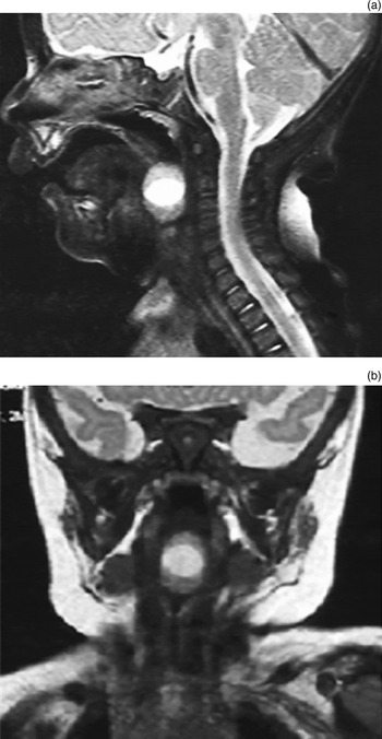
A T1-weighted MRI scan demonstrated a 15-mm diameter, well circumscribed, spherical mass in the vallecula. A T2-weighted MRI scan showed the mass contents as hyperintense and its margin as slightly hypointense (Figure 1).
(a) Sagittal and (b) coronal T2-weighted magnetic resonance images demonstrating a 15-mm diameter, well circumscribed mass in the vallecula, with hyperintense contents and a slightly hypointense margin.

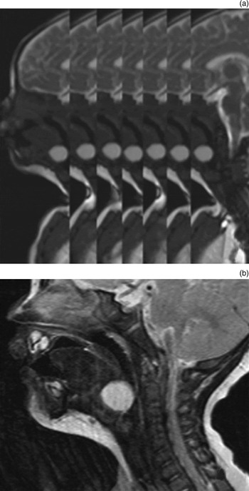
We had initially suspected a partly solid tumour. However, endoscopic and MRI findings were inconsistent, so we decided to repeat the MRI scan using a high-speed scan mode (utilising the Magnetom Avanto system; Siemens, Munich, Germany). Sagittal images indicated that the homogeneous lesion moved up and down slightly with breathing and swallowing, in the absence of larger body movements (Figure 2). This confirmed that the lesion was a homogeneous, cystic mass, and established the diagnosis of vallecular cyst.
(a) Sagittal, high-speed scan mode magnetic resonance image (MRI) scans demonstrating slight rising and falling movement of the lesion with breathing and swallowing, in the absence of large body movements. (b) Sagittal, T2-weighted MRI scan showing the lesion to be homogeneously hyperintense.

Marsupialisation of the cyst was performed under general anaesthesia, using electrical cautery under direct laryngoscopic visualisation with an operating microscope.
Histological examination of the operative specimen showed laryngeal epithelium with squamous metaplasia, connective tissue with mild inflammatory cell infiltration, and respiratory epithelium with squamous metaplasia. No thyroid tissue was found.
The post-operative course was uneventful. The patient's symptoms immediately resolved, and she was discharged 12 days after surgery.
Eight months after surgery, flexible laryngoscopic examination showed no abnormality in the larynx. The patient was in perfect health and had reached a normal weight.
Discussion
Congenital stridor is a relatively common symptom in infants, but vallecular cyst is a rare cause. The major differential diagnosis of congenital stridor includes laryngomalacia, subglottic stenosis and vocal fold paralysis;Reference Birch24, Reference Holinger25 in such cases, the incidence of vallecular cyst is only 0.9–2.0 per cent.Reference Birch24, Reference Holinger25 Vallecular cyst carries the risk of sudden airway obstruction and death, because of the anatomical location of the cyst and the small respiratory tract in infants.Reference Lewison and Lim6 Therefore, all otolaryngologists should consider this uncommon condition in the differential diagnosis of congenital stridor in infants.Reference Tuncer, Aydogan and Soylu14
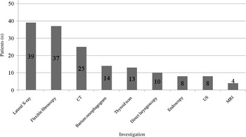
The diagnosis of vallecular cyst may be assisted by lateral radiography, flexible laryngoscopy, direct laryngoscopy, computed tomography (CT), MRI, US, barium oesophagography and thyroid scanning examinations. Figure 3 shows the use of diagnostic investigations in the 52 cases published to date. Radiography was used in 39 cases, but made no contribution to the diagnosis in 15 of these 39. Radiography is convenient and sometimes helpful,Reference Wong, Li and Huang7, Reference Oluwole12, Reference Ku13 but it is difficult to interpret and has many diagnostic pitfalls.Reference Wong, Li and Huang7, Reference Gutierrez, Berkowitz and Robertson8 These findings suggest that radiography is insufficient as the sole investigation for congenital stridor.
Use of diagnostic investigations in the 52 cases reviewed.

Flexible endoscopy (using a laryngoscope or bronchoscope) was used in 37 cases, and successfully identified the lesion in all cases. Flexible endoscopic examination is quick, easy and minimally invasive,Reference Gluckman, Chu and van Hasselt4 and allows dynamic assessment of the larynx, evaluation of swallowing and detection of congenital oropharyngeal lesions.Reference Ku13, Reference Tibesar and Thompson16 However, it cannot make fine distinctions between the various differential diagnoses of vallecular cyst.Reference Tibesar and Thompson16 Therefore, we propose that flexible endoscopy should be replaced by direct laryngoscopy, performed by a skilled operator under cardiopulmonary monitoring, to enable methodical examination of such cases.Reference Oluwole12, Reference Fu, Xue, Chen, Fan, Pan and Mao26 Flexible endoscopy is recommended for initial screening.
Computed tomography was used in 25 of the reviewed cases, and may be the most common type of radiographic imaging. Computed tomography may be indispensable for the diagnosis of lingual thyroglossal duct cyst in neonates;Reference Fu, Xue, Chen, Fan, Pan and Mao26 however, vallecular cyst, thyroglossal duct cyst, dermoid cyst, haemangioma and lymphangioma all have a similar, low density appearance on CT.Reference Lev and Lev27 In addition, CT involves a radiation risk for infants.
Magnetic resonance imaging is the diagnostic modality of choice,Reference Mahajan, Mathew, Singh, Gupta and Das20, Reference Grasso, Marchetti, Norbedo, Furlan, Schleef and Zocconi21 and detailed knowledge of the distinct MRI characteristics of common vallecular masses will enable correct diagnosis.Reference Tibesar and Thompson16 However, the use of MRI was described in only four of our reviewed cases. This modality requires sufficient sedation to exclude motion artefacts, together with cautious airway management in infants;Reference Weber, Kenna and Casselbrant28 it is also costly. Therefore, MRI is not widely used. Magnetic resonance imaging under sedation was performed in our patient. At first, we thought her lesion was partly solid, because of the above-described MRI findings in the apparent absence of motion artefacts. The interpretation of pharyngolaryngeal MRI in infants should consider the influence of breathing and swallowing, which cannot be prevented by sedation. High speed scanning should be used if findings are not logically consistent.
Ultrasonography is non-invasive, easy to perform, and familiar to otolaryngologists and paediatricians. It provides a good distinction between solid and cystic masses, as well as confirmation of a normal thyroid gland.Reference Ahrens, Lammert, Schmitt, Wahn, Paul and Niggemann18, Reference Breysem, Goosens, Vander Poorten, Vanhole, Proesmans and Smet22 Ultrasonography was used in only eight of our reviewed cases. We propose that US can provide a useful screening method for congenital stridor, especially for paediatricians who have no experience with flexible fibroscopy.
Lingual thyroid may present as a mass lesion at the base of the tongue, and thyroid scanning and thyroid function tests are necessary to confirm a normally located and functioning thyroid gland.Reference Hsieh, Yang, Wong, Li, Wang and Yeh5 Thyroid scanning was performed in 13 of our review cases. However, it carries a radiation risk and is difficult to administer in infants. We suggest that US and thyroid function tests, as used in our patient, are sufficient for confirmation of a normal thyroid gland.
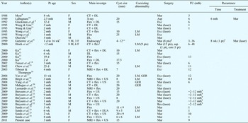
Table I summarises the 52 published cases of infantile vallecular cyst (including the present case). The mean age at presentation was 2.5 months (range, 2 days to nine months; 38 cases), and the female:male ratio was 1:1.3. The most common diagnostic strategy was a combination of CT, flexible fibroscopy and other techniques (22 cases); this strategy has become standard practice over the last 22 years. The use of MRI has various difficulties in infants, but has the greatest diagnostic effectiveness.Reference Tibesar and Thompson16 Hence, we recommend that a combination of MRI, flexible fibroscopy and US should be used to enable extensive examination of possible cases of vallecular cyst.
Reported cases of infantile vallecular cyst (past 23 years)

†Mean = 40 days. ‡All patients. **Mean = 8 mm. §With laser for 5 pts. #Forceps = this cyst was marsupialized by forceps at initial surgery. ¥For series. Pt = patient; investgn = diagnostic investigation; FU = follow up; wk = weeks; mth = months; d = days; F = female; M = male; CT = computed tomography; DL = direct laryngoscopy; flex = flexible laryngo(broncho)scopy; MRI = magnetic resonance imaging; US = ultrasonography; EUA = examination under anaesthesis; LM = laryngomalacia; GER = gastroesophageal reflux; mar = marsupialisation; asp = aspiration; exc = excision; con = conservative; ext = extirpation
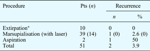
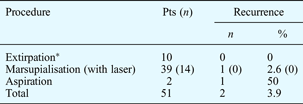
Surgical treatment for vallecular cyst in infants includes aspiration, marsupialisation (i.e. de-roofing) and extirpation (i.e. resection and excision). Surgery was performed in 51 of our 52 reviewed cases (Table II).
Surgery and recurrence rates: 51 previously published cases
*Including resection and excision. Pts = patients
In the reviewed cases, aspiration had a high rate of recurrence and was of limited use.Reference Mitchell, Irwin, Bailey and Evans3, Reference Tibesar and Thompson16 Small cysts can be aspirated,Reference Oluwole12 but aspiration should only be used as a palliative procedure, or as an initial manoeuvre if intubation is difficult.Reference Mitchell, Irwin, Bailey and Evans3, Reference LaBagnara10
• Vallecular cysts are uncommon in infants
• Magnetic resonance imaging presents various difficulties in infants, but has the highest diagnostic effectiveness for these lesions when used carefully
• The recurrence rate after marsupialisation is very low, so prompt marsupialisation is the recommended procedure for these lesions in infants
In general, marsupialisation or extirpation is used for definitive treatment.Reference Mitchell, Irwin, Bailey and Evans3–Reference Hsieh, Yang, Wong, Li, Wang and Yeh5, Reference Ku13, Reference Yao, Chiu, Wu, Wu and Huang17 Extirpation is the ideal treatment but sometimes requires an external approachReference Mitchell, Irwin, Bailey and Evans3 with or without tracheostomy. A published review of nine patients with congenital laryngeal cyst reported no recurrence in four after excision and in five after marsupialisation.Reference Pak, Woo and van Hasselt29 In our review of 52 cases (including the presented case), recurrence was encountered in none of 10 patients after extirpation, and in one of 39 patients after marsupialisation. In particular, recurrence occurred in none of 14 patients following marsupialisation with laser instruments. Therefore, our study suggests that marsupialisation and extirpation are equally effective, and that marsupialisation, especially with laser instruments, is to be recommended because of its minimal invasiveness.