Background
Lung cancer remains one of the most commonly diagnosed cancers in the world, as well as one of the commonest causes of cancer deaths. Reference Cheng, Cramb, Baade, Youlden, Nwogu and Reid1 In Singapore, it is the second and third most common cancer in males and females, respectively. 2 Significant advances have been made in the treatment modalities for early-stage and metastatic non-small-cell lung cancers (NSCLC), such as stereotactic ablative radiotherapy (SABR), targeted therapy and immunotherapy. However, the standard of care for advanced-stage non-metastatic NSCLC has not changed much over the past years; radical chemoradiotherapy remains the gold standard for stage III NSCLC as well as limited-stage SCLC. There have been iterative improvements in radiotherapy techniques, such as cone beam CT (CBCT), respiratory motion management, image-guided radiotherapy (IGRT) to name a few, which have at least increased the safety and accuracy of radiotherapy treatment, if not the overall survival rate.
One of the main challenges in treating lung cancer with radiotherapy is the fact that their position can vary greatly during respiration Reference Keall, Mageras and Balter3 ; this is especially so for tumours in the periphery and lower zones of the lung. Reference Liu, Balter and Tutt4 This can create significant uncertainty during contouring of target volumes as well as radiotherapy planning. In the Division of Radiation Oncology (DRO) at the National Cancer Centre Singapore (NCCS), 4D CT scans are routinely used for simulation when radical external beam radiotherapy is planned for lung cancer. The aims of 4D scans are to visualise and quantify the direction and magnitude of tumour movement as the patient breathes.
At the NCCS, we have been using the Varian Real-time Position Management™ (RPM) system (Varian Medical Systems, Palo Alto, CA, USA) for at least a decade. With this system, a marker block is positioned on the patient’s xiphoid region at simulation, and an infrared camera is used to track the anterior–posterior displacement of the block as the patient breathes. This generates a waveform, which is used as a surrogate for the patient’s respiratory pattern. A set of 4D CT scans are then acquired and sorted (or binned) into 10 discrete phases of respiration, based on the waveform acquired with the infrared camera. The software will then interpolate the scan images in each of these phases to generate a 3D ‘movie’ of the tumour and its motion during respiration. Such movements may be appreciated and accounted for during contouring and planning on our Eclipse™ (Varian Medical Systems) treatment planning system (TPS). Gross target volumes (GTV) for which internal motion has been accounted for are called internal target volumes (ITV). Reference Liu, Balter and Tutt4
However, this method of motion management is not without its limitations. Our experience shows that it is not always possible to reliably generate a surrogate respiratory waveform; factors affecting this include:
-
1. Exact position of the block on the xiphoid region
-
2. Thickness of the patient’s chest wall
-
3. Ability of the patient to breathe ‘regularly’
When this happens, the software has trouble ‘binning’ the 4D CT scan images into their respective respiratory phases, leading to tumours that appear as being ‘cut off’ or duplicated on the simulation scan. This raises concern about the accuracy and adequacy of the ITV generated with the scan. This is not helped by the fact that, due to time and resource limitations, patients are not routinely scheduled for pre-simulation ‘respiratory coaching’ sessions at our centre.
Presently at the NCCS, where the RPM™ system is unable to generate good-quality 4D simulation scans, or when the waiting time for 4D CT simulation is too long, planning is done with normal helical CT simulation scans. Generic planning target volume (PTV) expansions are then added to the GTV or clinical target volume (CTV) to account for potential internal tumour motions in the x (right–left), y (anterior–posterior) and z (superior–inferior) axes. In our current protocol, these are:
-
0·70 cm in the axial plane (0·20 + 0·50 cm setup uncertainty)
-
1·20 cm in the longitudinal plane (0·70 + 0·50 cm setup uncertainty)
and are applied uniformly to all lung tumours, regardless of the location. Reference Barrett, Dobbs, Morris and Roques5 However, these margins have never been validated at our centre, and it is common knowledge that tumour movements can vary greatly, depending on the location in the lung. Reference Onimaru, Shirato and Fujino6 In contrast, the ITV to PTV margin in the setting of 4D CT simulations is only 0·50 cm, in all axes, to account for setup uncertainties.
While the RPM™ system in its current use may not always give a representative tumour motion range for a given patient, it may be used to calculate a more useful and generalisable average motion range for a collective group of patients in our local setting. This data can be used to verify and assess the adequacy of our current generic margins. In addition, we sought to group these motion ranges by tumour location and derive new standard margins for each location (e.g. upper-zone tumours, central tumours, lower-zone tumours), which may be more realistic and applicable in the clinical setting.
Methods
Approval was obtained from the SingHealth Centralised Institutional Review Board (CIRB). Consecutive patients with early-stage lung cancer or lung metastases that were treated with SABR at the DRO, NCCS, between 2012 and 2016, and who had undergone 4D CT simulation scans were identified retrospectively. Only SABR cases were included because 4D CT simulation scans were not routinely used for conventional fractionated radiotherapy during the study period in our centre. In addition, smaller tumours were easier to group by location. All 4D CT scans were acquired on a GE Lightspeed™ (GE Healthcare Systems, Chicago, IL, USA) large-bore CT scanner at 2·5 mm slice thickness.
Grouping
Lung tumours were primarily grouped into three locations: upper zone (UZ), middle zone (MZ) and lower zone (LZ). For ease of clinical application, the lung was divided into equal thirds (without consideration of exact anatomical site) to derive UZ, MZ and LZ. The upper border of the lung was defined as the apex of the lung, while the lower border was defined as the midpoint between the dome of the diaphragm and the lowest part of the lung on a helical scan. Tumours with equivocal or overlapping locations or poorly defined margins (e.g. tumours adjacent to consolidation or collapsed lung) were excluded.
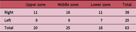
A total of 76 cases were identified. Eleven were excluded because of overlapping locations, and another two because of ill-defined volumes. Sixty-three cases (Table 1) from 41 male patients and 19 female patients were ultimately analysed (one patient had two lung lesions and another patient had three).
Tumours by location and laterality

Data analysis
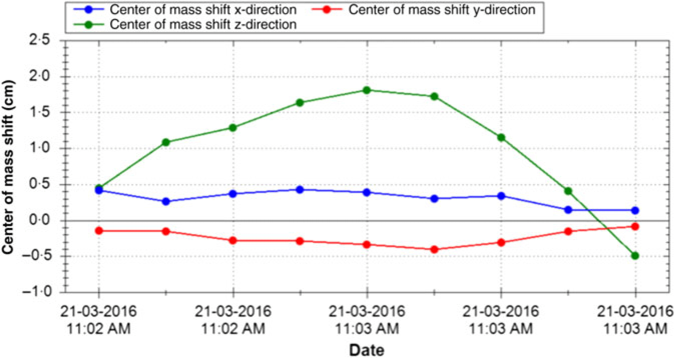
Analyses were done with the Eclipse™ TPS. To avoid biases and inconsistencies, the automated function of the TPS was used to contour the tumour volumes in each of the 10 phases of respiration. Displacements of the centre of mass (COM) of these volumes, in all three axes, were then generated by the TPS (Figure 1) and tabulated on Microsoft® Excel. These were then compared with the current generic margins and the number of cases that exceed them was recorded.
Example of COM shifts generated by the TPS.

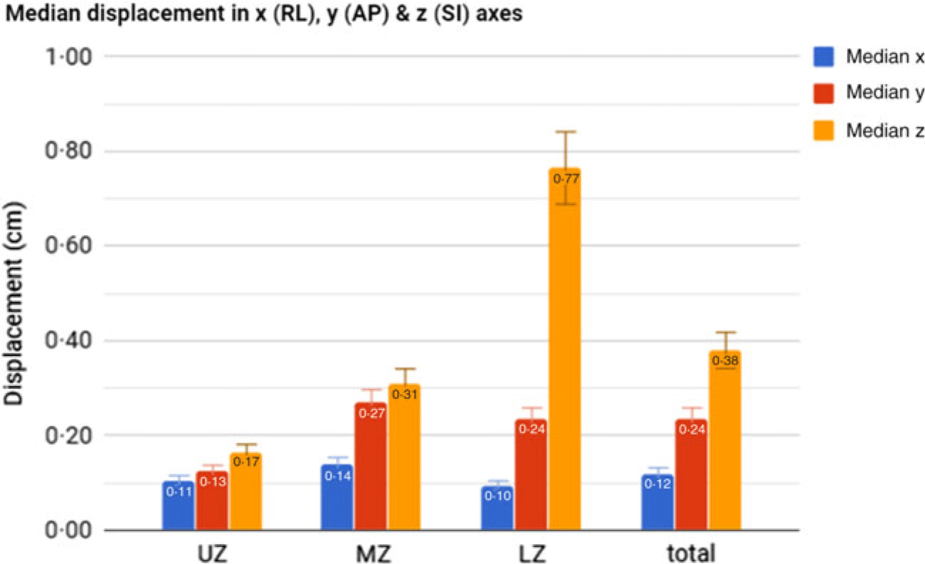
Median displacement of tumour by location.

The average displacements for each axis were also calculated and used to derive new standard margins for each tumour location according to the following formula:
 $${\rm{PTV}}\;{\rm{margin}} = {\rm{Median}}\;{\rm{displacement}} + 2{\rm{\;*\;Median}}\;{\rm{absolute}}\;{\rm{displacement}} + 0.50{\rm{\;cm\;}}\left( {{\rm{rounded}}\;{\rm{to}}\;{\rm{the}}\;{\rm{nearest\;}}0.10{\rm{\;cm}}} \right)$$
where 0·50 cm is the usual margin added for setup uncertainties. These new PTV margins should provide adequate coverage for 95% of cases.
Results
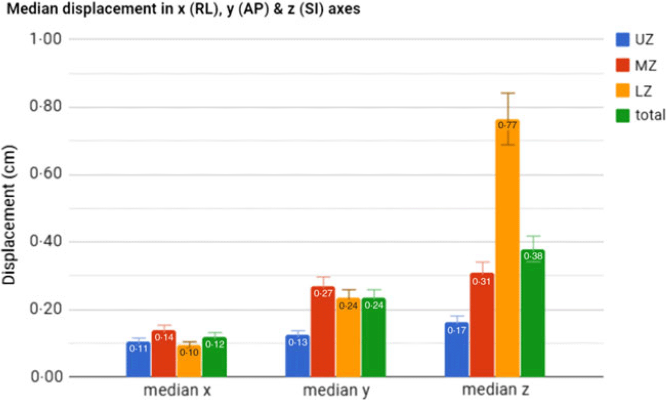
Unsurprisingly, the greatest motion was seen in the superior–inferior direction for all tumour locations. This ranges from a median of 0·17 cm (median absolute deviation (MAD) 0·12 cm) for tumours in UZ to 0·77 cm (MAD 0·27 cm) for those in LZ (Table 2 and Figure 2). Median tumour displacements in the right–left direction were <0·20 cm, and those in the anterior–posterior direction were <0·30 cm, regardless of the location (Figure 3).
Median displacement of tumour with respiration

Abbreviation: MAD, median absolute deviation.
Median displacement of tumour by axes.

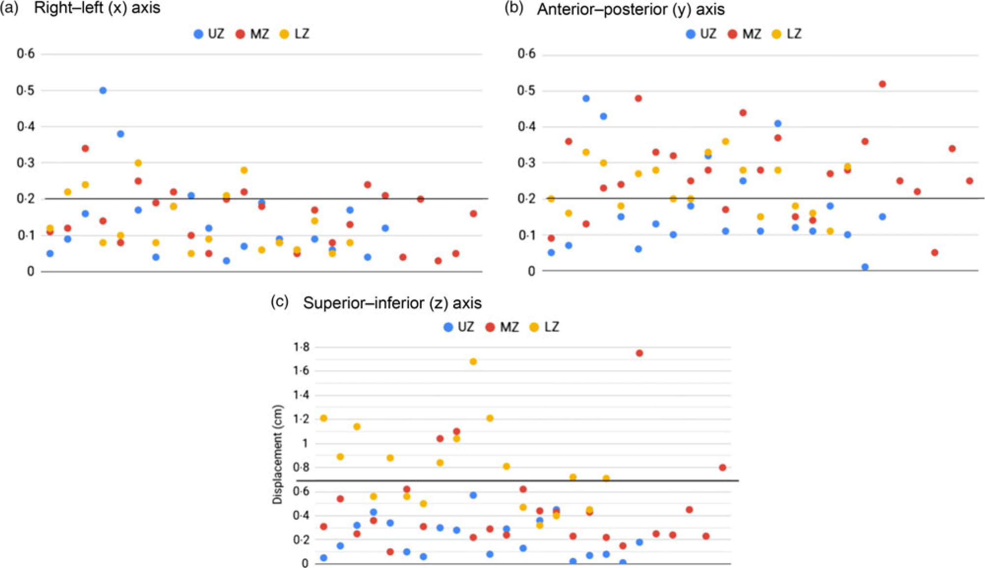
Tumour displacements in the (a) right–left axis, (b) anterior–posterior axis, (c) superior–inferior axis.

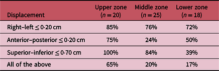
Applying our current default ‘ITV’ margins of 0·20 cm in the radial axis and 0·70 cm in the superior–inferior axis to these cases (Figures 4a–4c), they would be inadequate for 80% of MZ tumours and 83% of LZ tumours (Table 3). Of note, a large proportion of ‘failures’ was due to excessive displacements in the anterior–posterior axis; in particular, only 24% of MZ tumours had displacements that were less than the current default margin of 0·20 cm. When displacements in all three axes were considered together, the current margins were adequate for only one-third of the tumours in this study (65% of UZ tumours, 20% of MZ tumours and 17% of LZ tumours).
Percentage of cases with tumour displacements that fit current margins

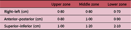
Applying the formula described previously, the following new standard PTV margins for the three tumour locations can be derived (Table 4). For tumours that straddle two or more locations, larger margins should be used.
New standard PTV margins by location

The major changes are an increase in the margin for the anterior–posterior axis for all locations and a significant increase in the superior–inferior margin for LZ tumours (from 1·20 to 2·10 cm).
Discussion
Over the years, tumour motion management has become increasingly important in the treatment of lung cancer. With the advent of techniques such as intensity-modulated radiotherapy and volumetric modulated arc therapy, doctors are seeking to treat ever larger volumes with radical or ablative intent. The NCCS is the largest cancer centre in this country (and possibly the region) and is even in the process of setting up a proton therapy unit. Our centre currently has three types of motion management systems: the RPM™ system, the SDX® System (Dyn’r Medical Systems, Aix-en-Provence, France) and the AlignRT® (Vision RT, London, UK) optical surface management system. However, SDX® has been used exclusively for breast treatments, and AlignRT® is yet to be fully integrated into the workflow. This leaves RPM™ as the only means with which to determine the respiratory pattern. The inherent limitations associated with this surrogate method are part of the reason why respiratory gating is not practiced at the NCCS. Instead, 4D CT simulation scans are primarily used to derive ITVs to ensure adequate coverage of tumour volumes throughout all phases of respiration.
Not infrequently, the RPM™ system fails to capture a nice respiratory waveform, particularly in patients with ‘thick’ chest walls, ‘shallow breathers’ or ‘irregular breathers’. When that happens, it may be prudent to err on the side of safety and apply generic PTV margins instead. In addition, such margins remain relevant when non-4D CT simulation scans are done in the interest of shorter waiting times or in the setting of more palliative treatments. However, the recommended generic PTV margins in our current protocol have not been validated, and it is not known whether they can be routinely applied.
The relationship between lung tumour location and respiration has been studied before. Reference Onimaru, Shirato and Fujino6–Reference Chen, Weinhous, Deibel, Ciezki and Macklis11 However, most of these studies involve small groups of subjects (around 20 patients) and used methods such as fluoroscopy, ultrafast CT scans and dynamic MRI to assess tumour motion. We found only one study which was comparable; Sarudis et al. Reference Sarudis, Karlsson Hauer, Nyman and Bäck12 evaluated 126 patients using 4D CT scans and the RPM™ system. They reported similar median shifts of 0·2 cm or less radially for tumours in all locations, while median shifts in the superior–inferior direction ranged from 0·2 cm for upper-lobe tumours to 0·9 cm for lower-lobe tumours (with a maximum shift of 5·3 cm). Our findings support their results and further highlight the magnitude and variability of lower-lobe tumour motion. Our current generic margins were only adequate for one-third of cases in this study, and as little as 17% of LZ tumours. For UZ and MZ tumours, this can be easily addressed by increasing the recommended margins by 1 or 2 mm. For LZ tumours, however, the required superior–inferior margin to avoid geographical misses is 2·10 cm; this far exceeds the current margin of 1·20 cm, and may be difficult to recommend or apply if organs-at-risk dose constraints are to be met. As such, motion mitigation (and not just motion tracking) strategies are strongly recommended for all LZ tumours, and especially large ones. These include respiratory gating, deep inspiration breath hold and abdominal compression; none of these are perfect or universally applicable, and there is much room for improvement. We also recommend that such tumour motion data be continually collected and reviewed and the standard margins be updated periodically.
A limitation of this study is that tumour motion was assessed at one time point, during simulation. Respiratory patterns and efforts can change during the course of radiotherapy (especially a long-course conventional fractionated treatment), and there may be further variations in tumour motion. At the moment, our centre does not routinely monitor respiratory patterns throughout the course of radiotherapy, but this is now the subject of another study.
Conclusion
Tumour motion is the greatest in LZ in the superior–inferior axis, and PTV margins as much as 2·10 cm may be required. Radial tumour motion is smaller and varies less with the location in the lungs; however, tumour displacement in the anterior–posterior axis is not insignificant and should not be overlooked. Motion mitigation strategies are essential for the treatment of LZ lung tumours, especially large ones.
Acknowledgements
None
Financial Support
None
Conflicts of Interest
None