

Introduction
The superfamily Hemiuroidea is a highly studied and rich group of trematodes. The 16 currently recognized hemiuroid families (Sokolov et al., Reference Sokolov, Atopkin, Urabe and Gordeev2019; World Register of Marine Species, 2022b) infect a wide range of marine and freshwater fishes, amphibians and aquatic reptiles (e.g. Thomas, Reference Thomas1939; Vercammen-Grandjean & Heyneman, Reference Vercammen-Grandjean and Heyneman1964; Blair, Reference Blair1984; Hafeezullah, Reference Hafeezullah1990; Shimazu et al., Reference Shimazu, Cribb, Miller, Urabe, Van Ha, Binh and Shed'ko2014). Although hemiuroids are highly diverse morphologically, all the families of this group with known life cycles but one (the Ptychogonimidae Dollfus, 1937) have cystophorous cercariae (Dollfus, Reference Dollfus1950). Cystophorous cercariae are unique to the Hemiuroidea and are characterized by a retractable cercarial body that is ejected from the caudal chamber through a delivery tube into the body cavity of the second intermediate host upon consumption of the cercaria (e.g. Køie, Reference Køie1995); the apparent secondary loss of the caudal appendage (and possibly of the delivery tube) in Ptychogonimus megastomum (Rudolphi, 1819) may be due to modifications in the life cycle of that trematode, wherein sporocysts containing the cercariae are trophically transmitted to the second-intermediate host (Palombi, Reference Palombi1941, Reference Palombi1942).
Hemiuroid life cycles are thought to typically include four hosts (Nikolaeva, Reference Nikolaeva1965; Kechemir, Reference Kechemir1978; Køie & Lester, Reference Køie and Lester1985). Known first intermediate hosts are molluscs (see review by Louvard et al. (Reference Louvard, Cutmore, Yong, Dang and Cribb2022)), principally gastropods and, much less frequently, bivalves (Wardle, Reference Wardle1975; Mateo et al., Reference Mateo, Peña, Guzmán and López1985; Castro, Reference Castro2019; Louvard et al., Reference Louvard, Cutmore, Yong, Dang and Cribb2022) and scaphopods (Palombi, Reference Palombi1941, Reference Palombi1942; Køie et al., Reference Køie, Karlsbakk and Nylund2002). Most hemiuroid asexual infections are found in benthic environments, with exceptions from holoplanktonic molluscs (Bonnevie, Reference Bonnevie1916; Vande Vusse, Reference Vande Vusse1980; Hochberg & Seapy, Reference Hochberg and Seapy1985; Lester & Newman, Reference Lester and Newman1986; Morales-Ávila et al., Reference Morales-Ávila, Saldierna-Martínez, Moreno-Alcántara and Violante-González2018). Fewer than half of the families have elucidated life cycles, however, and first intermediate hosts are unknown for most species. Especially glaring is the lack of knowledge about life cycles of the Didymozoidae Monticelli, 1888. Only two first intermediate hosts are known. Saccularina magnacetabula Louvard et al., 2022 infects the bivalve Anadara trapezia (Deshayes) (Arcoidea: Arcidae) (Louvard et al., Reference Louvard, Cutmore, Yong, Dang and Cribb2022), and two closely related but unidentified didymozoids infect the holoplanktonic gastropod Firoloida desmarestia Lesueur (Pterotracheoidea: Pterotracheidae) (Lozano-Cobo et al., Reference Lozano-Cobo, Oceguera-Figueroa, Silva-Segundo, Robinson and Gómez-Gutiérrez2022).
Opposite to host–parasite coevolution, speciation by host-switching is the main driver of host–parasite relationships (Araujo et al., Reference Araujo, Pires Braga, Brooks, Agosta, Hoberg, von Hartenthal and Boeger2015). Host-switching refers to the opportunistic establishment of a relationship between a parasite species and a previously unexploited host lineage (often not closely related to the usual hosts of that parasite) enabled by mutual encounter and compatibility (Agosta & Klemens, Reference Agosta and Klemens2008; Araujo et al., Reference Araujo, Pires Braga, Brooks, Agosta, Hoberg, von Hartenthal and Boeger2015). The exploitation of the new host is followed by the co-adaptation of both parasite and host to each other (Araujo et al., Reference Araujo, Pires Braga, Brooks, Agosta, Hoberg, von Hartenthal and Boeger2015). Host-switching often results in parasite adaptive radiation (Brooks et al., Reference Brooks, O'Grady and Glen1985). Although trematode speciation by host-switching is widespread at both definitive (e.g. Shoop, Reference Shoop1989; Jousson et al., Reference Jousson, Bartoli and Pawlowski2000; Cribb et al., Reference Cribb, Bray and Littlewood2001) and second intermediate host levels (e.g. Brooks et al., Reference Brooks, O'Grady and Glen1985; Cribb et al., Reference Cribb, Bray and Littlewood2001; Martin et al., Reference Martin, Sasal, Cutmore, Ward, Aeby and Cribb2018b, Reference Martin, Downie and Cribb2020), remarkably few digenean families infect more than one class of first intermediate hosts.
Didymozoids are known from holoplanktonic gastropods and benthic bivalves. Here, a didymozoid infection is reported from a dramatically different group of gastropods, the Vermetidae, on the basis of molecular phylogenetic analyses. The presence of didymozoid infections in arcid bivalves, holoplanktonic gastropods and sessile vermetids suggests that first intermediate host use within that family is driven by host ecology.
Materials and methods
Sample collection
Vermetid gastropods were chiselled by hand from emerged rocks as part of the study on trematodes infecting six species of vermetids conducted by Corner et al. (Reference Corner, Cribb and Cutmore2022) on Heron Island (HI), Lizard Island and Moreton Bay (MB) on the coast of Queensland, Australia. From all the locations and species sampled, a single hemiuroid infection was found in Polka Point, North Stradbroke Island (NSI), MB (27°29′46.3″S 153°23′54.6″E).
Adult hemiuroid trematodes were collected by the Marine Parasitology Laboratory (University of Queensland, Australia) from a wide range of fishes as part of a wider parasitological study. The search for didymozoids proceeded as per the protocol of Louvard et al. (Reference Louvard, Cutmore, Yong, Dang and Cribb2022).
All fishes and vermetids were dissected in saline. All digeneans, including larval stages, were killed in near-boiling saline and stored in 80% ethanol for morphological and molecular identifications. Anterior ends of infected vermetids were stored separately in 80% ethanol for morphological identification.
Morphological analyses
All procedures related to morphological analyses were performed on ethanol-preserved samples. For adult didymozoids, hologenophores sensu Pleijel et al. (Reference Pleijel, Jondelius, Norlinder, Nygren, Oxelman, Schander, Sundberg and Thollesson2008) were produced by removing a small portion of tissue from the side of the worms with a scalpel. The tissue sample was sequenced and the remainder preserved for morphological analyses; cercariae and sporocysts were left whole. Larvae were rinsed in fresh water, stained with Mayer's haematoxylin, destained in 1% hydrochloric acid, neutralized in 1% ammonium hydroxide, dehydrated in increasing ethanol concentrations from 50% to 100%, cleared in methyl salicylate and mounted in Canada balsam. Specimens were measured in micrometres using cellSens standard imaging software on an Olympus BX-53 compound microscope fitted with an Olympus SC50 digital camera. Specimens were drawn using a camera lucida and digitized with Adobe Illustrator CC 2018. Voucher specimens of both sporocysts and adult didymozoids were lodged in the Queensland Museum (QM), Brisbane, Queensland, Australia.
Vermetids were identified with both morphological and unpublished molecular data by Professor Timothy Rawlings (Cape Brenton University, Canada) using molecular sequences produced by Corner et al. (Reference Corner, Cribb and Cutmore2022). Morphological vouchers and molecular sequences of vermetid hosts were lodged by Corner et al. (Reference Corner, Cribb and Cutmore2022) in the QM and GenBank, respectively.
Adult didymozoids were identified on the basis of the work of Yamaguti (Reference Yamaguti1970) and Pozdnyakov & Gibson (Reference Pozdnyakov, Gibson, Bray, Gibson and Jones2008), as well as previous host–trematode–site records. Hologenophore specimens were lodged in the QM.
Molecular sequencing of trematodes and hosts
Total genomic DNA was extracted from individual sporocysts, adult trematodes and sections of vermetid tissue using a method derived from Sambrook & Russell (Reference Sambrook and Russell2001). Briefly, specimens were dried, then incubated thrice: at 37°C for >7 h in Tris (Merck, Australia)-ethylenediaminetetraacetic acid (Sigma Aldrich, UK); at 55°C for 2 h with added proteinase K (Invitrogen™, Australia; 10 mg/ml); and at 65°C for 10 min with added sodium chloride (Selby Biolab, Australia; 5 M) and cetyltrimethylammonium bromide (Sigma Aldrich, Australia). DNA was purified with chloroform (Merck, Australia) and phenol–chloroform–isoamyl alcohol (Ambion, Australia), precipitated with cold isopropanol (Sigma Aldrich, Australia) at room temperature, washed in 70% molecular-grade ethanol (Supelco, Germany), dried, rehydrated in 25 μl of Invitrogen™ ultraPURE™ (USA) distilled water at 4°C for >7 h and stored at −20°C.
Complete internal transcribed spacer 2 (ITS2) rDNA and a partial D1–D3 fragment of 28S rDNA regions, and the partial 16S mtDNA region, were amplified for parasites and vermetids, respectively. Reaction solutions comprised 5 μl of 5 × MyTaq™ Reaction Buffer (Bioline, Australia), 0.75 μl of each primer (10 μM), 0.25 μl of Taq polymerase (Bioline MyTaq™ DNA Polymerase, Australia) and 2 μl (ITS2 and 16S, approximately 10 ng) or 4 μl (28S, approximately 20 ng) of DNA template, completed to a total volume of 20 μl with Invitrogen™ ultraPURE™ distilled water. The ITS2 region was amplified using primers 3S (5′-GGT ACC GGT GGA TCA CGT GGC TAG TG-3′; Bowles et al., Reference Bowles, Hope, Tiu, Liu and McManus1993) and ITS2.2 (5′-CCT GGT TAG TTT CTT TTC CTC CGC-3′; Cribb et al., Reference Cribb, Anderson, Adlard and Bray1998) under the following denaturation–annealing–extension procedure: 1 × (3 min at 95°C, 2 min at 45°C, 90 s at 72°C); 4 × (45 s at 95°C, 45 s at 50°C, 90 s at 72°C); 30 × (20 s at 95°C, 20 s at 52°C; 90 s at 72°C); and 1 × (5 min extension only at 72°C). The 28S region was amplified using external primers LSU5 (5′-TAG GTC GAC CCG CTG AAY TTA AGC A-3′; Littlewood, Reference Littlewood1994) and 1200R (5′-GCA TAG TTC ACC ATC TTT CGG-3′; Lockyer et al., Reference Lockyer, Olson and Littlewood2003) under the following denaturation–annealing–extension procedure: 1 × (4 min denaturation only at 95°C); 30 × (1 min at 95°C, 1 min at 56°C, 2 min at 72°C); and 1 × (1 min at 95°C, 45 s at 55°C, 4 min at 72°C). The partial 16S region was amplified using primers 16SARL-CBU (5′-CGC CTG TWT ADC AAA AAC ATG-3′) and 16SBRH (5′-CCG GTC TGA ACT CAG ATC ACG-3′) or 16SBRH-ALT (5′-CCG GTC TGA ACT CAG ATC AYG T-3′) (all modified from Palumbi (Reference Palumbi1996)) under the denaturation–annealing–extension procedure described by Cribb et al. (Reference Cribb, Chapman, Cutmore and Huston2020).
Amplicons were run on 1% m/v agarose gels supplemented with 0.01% v/v SYBR™ Safe (Invitrogen, Australia). Amplified DNA was sequenced by Sanger cycle sequencing at the Australian Genome Research Facility (Brisbane) using the same primers as for amplification for ITS2 and 16S, and internal primers 300 F (5′-CAA GTA CCG TGA GGG AAA GTT-3′; Littlewood et al., Reference Littlewood, Curini-Galletti and Herniou2000) and ECD2 (5′-CTT GGT CCG TGT TTC AAG ACG GG-3′; Littlewood et al., Reference Littlewood, Rohde and Clough1997) for 28S. Sequences were checked for quality, contiged and trimmed in Geneious v.11.0.5. High-similarity Basic Local Alignment Search Tool searches were made against the United States National Center for Biotechnology Information nucleotide collection (nr/nt). Sequences produced in this study were lodged in GenBank.
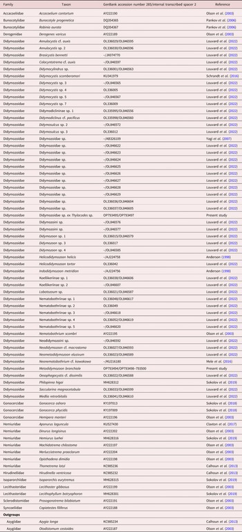
Sequences of the vermetid infections were aligned with selected GenBank sequences and sequences of didymozoid adults produced in this study (table 1) using Muscle v3.7 (Edgar, 2004) in MEGA7 (Kumar et al., 2016) for 28S (gap opening penalty = −400, gap extension penalty = −100) and on the Cyberinfrastructure for Phylogenetic Research (CIPRES) portal for ITS2, with unweighted pair group method with arithmetic mean clustering for iterations 1 and 2. Sequences of both alignments were trimmed in MEGA7 to the maximal length of the 50% shortest sequences. In-partition gaps were removed (Martin et al., Reference Martin, Cutmore and Cribb2018a) if affecting >25% of sequences. Alignments were further curated using Gblocks v.0.91b (Castresana, Reference Castresana2000; Dereeper et al., Reference Dereeper, Guignon and Blanc2008) with parameters of least-stringent selection (Kück et al., Reference Kück, Meusemann, Dambach, Thormann, von Reumont, Wägele and Misof2010). After these procedures, 31.1% of the original 28S alignment (representing 965 base pairs (bp) of 28S region) and 15.9% of the original ITS2 alignment (470 bp of ITS2 region) could be kept for phylogenetic analyses.
Molecular sequences used in the present study and Queensland Museum voucher identifiers for sequences produced in this study.

For Bayesian inference (BI) and maximum likelihood (ML) analyses, the best evolutionary model selected in jModelTest v.2.1.10 (Darriba et al., Reference Darriba, Taboada, Doallo and Posada2012) using a corrected Akaike information criterion (Akaike, Reference Akaike1974; Hurvich & Tsai, Reference Hurvich and Tsai1993) was the general time reversible model with estimates of invariant sites and gamma-distributed among-site variation (GTR + I + Γ) for the 28S dataset, and the TVM model with estimates of invariant sites and gamma-distributed among-site variation (TVM + I + Γ) for the ITS2 dataset. Alignments were converted into an appropriate format in Mesquite v.3.6 (Maddison, W.P., Maddison, D.R., 2018. Mesquite: A modular system for evolutionary analysis. Version 3.6. http://www.mesquiteproject.org). MrBayes 3.2.7a (Ronquist et al., Reference Ronquist, Teslenko and van der Mark2012) and RAxML-Blackbox (Stamatakis, Reference Stamatakis2014) were accessed through the CIPRES portal for BI and ML analyses, respectively. BI analyses used the values for amino-acid fixed substitution rate (revmatpr), gamma shape fixed parameter (shapepr), number of discrete categories to approximate the gamma distribution (ngammacat) and fixed proportion of invariable sites (prinvarpr) calculated in jModelTest. Other specified parameters were: ngen = 10,000,000; no discarding of sampled values as burn-in when calculating the convergence diagnostic; sumt burn-in = 3000; consensus tree with all compatible groups and tree probabilities; and sump burn-in = 3000. The trees were rooted in FigTree v1.4.3 (Rambaut, A., 2017. FigTree version 1.4.3, a graphical viewer of phylogenetic trees. Computer program distributed by the author, http://tree.bio.ed.ac.uk/software/figtree) and edited in Adobe Illustrator CC 2018.
Results
Morphological analyses
A single hemiuroid trematode infection was found from dissections of 612 specimens (0.16% prevalence) of a Thylacodes sp. (Gastropoda: Vermetidae) collected from MB (see Corner et al., Reference Corner, Cribb and Cutmore2022). The infected vermetid specimen was found at Polka Point, NSI, MB. Sporocysts were localized in the tissue at the base of the host's gills. Cystophorous cercariae characteristic of the Hemiuroidea were found in the sporocysts. Both cercariae and sporocysts are described below.
Molecular analyses
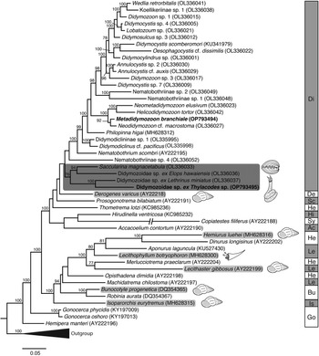
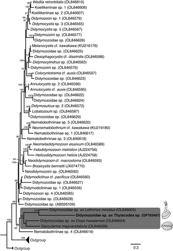
Forty-six representative 28S rDNA hemiuroid sequences from GenBank (table 1) and one novel 28S sequence produced in this study (that of Metadidymozoon branchiale Yamaguti, 1970 from Istiophorus platypterus (Shaw) (Carangiformes: Istiophoridae); GenBank number: OP793494, QM numbers: G240312–G240314) (table 1) were used to determine the family of the vermetid infection. The position of the vermetid taxon was the same in both ML and BI analyses, so phylogenetic trees are shown for the latter only (figs 1 and 2). In both BI (fig. 1) and ML (not shown) 28S analyses, the sequence from the vermetid infection was part of a clade within the Didymozoidae. Subsequently, 42 representative ITS2 sequences of didymozoid taxa were used to further explore the identity of the infection (table 1). In both BI and ML analyses of 28S and ITS2 datasets (figs 1 and 2; ML trees not shown), the sequence of the vermetid-infecting taxon formed a strongly supported clade with that of an uncharacterized didymozoid from the pharyngeal teeth area of Lethrinus miniatus (Forster) (Lethrinidae) from HI (Queensland, Australia) (see Louvard et al. (Reference Louvard, Cutmore, Yong, Dang and Cribb2022)). In all the analyses, the sequence of that uncharacterized taxon was itself part of a strongly supported clade comprising S. magnacetabula, described from the fin membrane of Elops hawaiensis Regan from MB, and another uncharacterized didymozoid from the same site, host and geographical location as S. magnacetabula (see Louvard et al. (Reference Louvard, Cutmore, Yong, Dang and Cribb2022)).
Phylogenetic relationships within the Hemiuroidea generated by Bayesian inference analysis of the partial 28S rDNA region from a 965 base pairs alignment. Sequences in boldface type were generated in this study. Numbers above nodes represent posterior probabilities (%); only values >75% are indicated. Vertical rectangular boxes represent hemiuroid families: Ac, Accacoeliidae; Bu, Bunocotylidae; De, Derogenidae; Di, Didymozoidae; Go, Gonocercidae; He, Hemiuridae; Hi, Hirudinellidae; Is, Isoparorchiidae; Le, Lecithasteridae; Sc, Sclerodistomidae; and Sn, Syncoeliidae. The clade formed by the vermetid infection, Saccularina magnacetabula and uncharacterized taxa from Elops hawaiensis and Lethrinus miniatus is enclosed in a dark rectangle. Species for which first intermediate hosts are known are enclosed in light rectangles with the first intermediate hosts drawn.

Phylogenetic relationships within the Didymozoidae generated by Bayesian inference analysis of the partial internal transcribed spacer 2 rDNA region from a 470 base pairs alignment. Sequences in boldface type were generated in this study. Numbers above nodes represent bootstrap support values (%); only values >75% are indicated. The clade formed by the vermetid infection, Saccularina magnacetabula and uncharacterized taxa from Elops hawaiensis and Lethrinus miniatus is enclosed in a dark rectangle. First intermediate hosts are drawn for the present vermetid infection and for S. magnacetabula.

In the BI 28S analysis (fig. 1), the clade formed by the vermetid infection, S. magnacetabula and the uncharacterized didymozoids from E. hawaiensis and L. miniatus is sister to a clade containing all other available didymozoid sequences. In the ML 28S analysis (not shown), however, that clade is sister to a clade formed by a single taxon, Nematobothriinae sp. 4 from black marlin, Istiompax indica (Cuvier), itself sister to a clade containing all the remaining didymozoids. In the BI ITS2 analysis (fig. 2), the clade [S. magnacetabula + vermetid infection + unidentified taxa from E. hawaiensis and L. miniatus] is sister to Nematobothriinae sp. 4 to the exclusion of all the other didymozoid sequences. In the ML ITS2 analysis (not shown), however, that clade is sister to all the other didymozoid clades except that of Nematobothriinae sp. 4.
Morphological description
Family Didymozoidae
Didymozoidae sp. A
Locality: Polka Point, North Stradbroke Island, Queensland, Australia (27°29′46.3″S 153°23′54.6″E)
Host: Thylacodes sp. (Gastropoda: Vermetidae)
Site: Tissue at base of gills
Specimens deposited: QM G240302–G240311
Representative DNA sequences: GenBank no. OP793495 (28S rDNA) and OP793498–793500 (ITS2 rDNA)
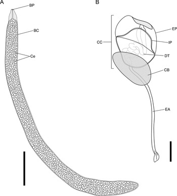
Sporocyst (fig. 3A)
Intramolluscan stages of the didymozoid infection from Thylacodes sp.: (A) sporocyst, scale: 500 μm; and (B) immature cercaria inside the sporocyst, scale: 20 μm. Abbreviations: BC, brood chamber; BP, birth pore; CC, caudal chamber; CB, cercarial body; Ce, cercariae; DT, delivery tube; EA, excretory appendage; EP, external part of the caudal chamber; and IP, internal part of the caudal chamber.

Measurements and description based on nine specimens. Sporocysts interpreted as the second intramolluscan generation contained abundant cystophorous cercariae.
Sporocysts in fleshy tissue at base of host gills. Body contractile, cylindrical to irregular elongate, tapering towards pointed conical anterior extremity and rounded posterior extremity, 1449–3356 (2385) × 183–268 (232). Tegument with fine striations most visible at anterior extremity. Birth pore at anterior extremity. Birth canal straight, sometimes scarcely visible, sometimes invaginates inside anterior extremity anteriorly to brood chamber. Brood chamber filled with active, developing cercariae.
Cercaria (fig. 3B)
Cercariae observed only inside sporocysts. None with cercarial body retracted into caudal chamber. No natural emergence observed. Measurements and descriptions based on ten cercarial bodies, 11 internal caudal cysts, six external caudal cysts and four excretory appendages.
Cystophorous cercaria. Body variable in shape, but typically short cylindrical with bluntly rounded extremities, frequently detached from tail cyst, 42–94 (65) × 14–26 (21). Suckers, mouth and digestive tract not recognizable. Excretory bladder and pore not seen. Caudal chamber rounded, composed of internal and external parts, bearing excretory appendage; internal part thick-walled, with ridge separating concave sides from convex middle, with rounded conical anterior extremity, 33–41 (36) × 31–41 (36); external part thin-walled, irregularly shaped, covering internal part, thickening and darkening at apex at junction with conical extremity of internal part, becoming indifferentiable from internal part at posterior extremity of chamber, 42–46 (43) × 42–47 (45). Delivery tube coiled tightly inside internal part of caudal chamber. Excretory appendage long, widening at posterior extremity into small paddle, 76–180 (116) × 2–4 (3.5).
Discussion
The Didymozoidae are among the most speciose hemiuroid families with 262 species currently recognized (World Register of Marine Species, 2022a). Yet, this study is only the third report of a didymozoid asexual infection host, and just the second for a gastropod. Given the rarity of hemiuroid infections in bivalves relative to other trematode groups that use bivalves as main first intermediate hosts, Louvard et al. (Reference Louvard, Cutmore, Yong, Dang and Cribb2022) hypothesized that the majority of didymozoid species use gastropod hosts as do most other hemiuroid families. The present findings, together with those of Lozano-Cobo et al. (Reference Lozano-Cobo, Oceguera-Figueroa, Silva-Segundo, Robinson and Gómez-Gutiérrez2022), confirm that didymozoids do infect both mollusc lineages. Whether gastropods are the dominant host group remains to be determined.
The paucity of records of didymozoid first intermediate hosts, the chaotic state of the phylogeny of the Didymozoidae (see Louvard et al., Reference Louvard, Cutmore, Yong, Dang and Cribb2022), and the early divergent position of the clade containing both S. magnacetabula and the new vermetid infection relative to all other didymozoids, preclude definitive conclusions about the evolution of host usage within the Didymozoidae. Still, it is noteworthy that sequences of the vermetid infection from Thylacodes sp. are phylogenetically closer to those of S. magnacetabula than to those of any other didymozoid taxon except the unidentified species from L. miniatus, despite the distant relationship of their respective intermediate hosts. Unfortunately, the cytochrome c oxidase I sequences of the two species from F. desmarestia produced by Lozano-Cobo et al. (Reference Lozano-Cobo, Oceguera-Figueroa, Silva-Segundo, Robinson and Gómez-Gutiérrez2022) are not from the same locus as those produced for S. magnacetabula and the unidentified taxa from L. miniatus and Elops hawaiensis from Louvard et al. (Reference Louvard, Cutmore, Yong, Dang and Cribb2022); thus, the phylogenetic position of the infection from Thylacodes sp. relative to that of those species is unknown.
Digenean families rarely infect more than one class of first intermediate hosts. Exceptions to this rule are understood to have arisen via host-switching and subsequent speciation. Convincing exceptions include two species of Hurleytrematoides Yamaguti, 1954 (Monorchioidea: Monorchiidae) in vermetid gastropods instead of the usual bivalves (Wee et al., Reference Wee, Cribb, Corner, Ward and Cutmore2021); Diploproctodaeum arothroni Bray and Nahhas, 1998 (Lepocreadioidea: Lepocreadiidae) in an ostreid bivalve instead of the usual gastropods (Hassanine, Reference Hassanine2006); Allocreadium handiai Pande, 1937 (Allocreadioidea: Allocreadiidae) in a bithyniid gastropod instead of the usual bivalves (Madhavi, Reference Madhavi1980); and, within the Schistosomatoidea, the use of both gastropods and polychaetes by the Spirorchiidae (Pinto et al., Reference Pinto, de Melo and Brant2015; Cribb et al., Reference Cribb, Crespo-Picazo, Cutmore, Stacy, Chapman and García-Párraga2017b; de Buron et al., Reference de Buron, Colon, Siegel, Oberstaller, Rivero and Kyle2018) and gastropods, polychaetes and bivalves by the Aporocotylidae (Evans & Heckmann, Reference Evans and Heckmann1973; Køie, Reference Køie1982; Cribb et al., Reference Cribb, Chick, O'Connor, O'Connor, Johnson, Sewell and Cutmore2017a). The Hemiuroidea are exceptional as they use more mollusc classes (i.e. Bivalvia, Gastropoda and Scaphopoda) than any other trematode superfamily (Cribb et al., Reference Cribb, Bray, Olson and Littlewood2003; fig. 1). The use of both bivalves and gastropods by the Didymozoidae mirrors the use of both the Scaphopoda and the Gastropoda by the hemiuroid family Lecithasteridae Odhner, 1905, with Lecithophyllum botryophoron (Olsson, 1868) using a scaphopod (Køie et al., Reference Køie, Karlsbakk and Nylund2002) and Lecithaster gibbosus (Rudolphi, 1802) using a gastropod (Køie, Reference Køie1989) (fig. 1). However, host use within the Lecithasteridae corresponds to a clear phylogenetic distinction between Lecithophyllum Odhner and Lecithaster Lühe (Sokolov et al., Reference Sokolov, Atopkin, Urabe and Gordeev2019), whereas such distinction is absent in the Didymozoidae.
It is understood that the eggs of hemiuroids, which are notoriously small, must be eaten by the first intermediate host for infections to develop (Szidat, Reference Szidat1956; Stunkard, Reference Stunkard1973; Martorelli, Reference Martorelli1989). The peculiar ecology of vermetid gastropods may provide clues to the mechanism of their infection by didymozoid eggs. Vermetids deploy mucus nets (Hughes, Reference Hughes1978; Kappner et al., Reference Kappner, Al-Moghrabi and Richter2000) that catch a wide variety of benthoplanktonic particles, which are ingested when the nets are retracted (Kusama et al., Reference Kusama, Nakano and Asakura2021). Presumably vermetids become infected with didymozoids by ingesting eggs via net-feeding. Net-feeding specifically, and more generally filter-feeding, are strategies strikingly different from those of most other gastropod families harbouring hemiuroids for which feeding behaviour is known (see review by Louvard et al., Reference Louvard, Cutmore, Yong, Dang and Cribb2022). Wee et al. (Reference Wee, Cribb, Corner, Ward and Cutmore2021) hypothesized that a host-switching event from gastropods to bivalves inferred for monorchiids was enabled by convergence in feeding ecologies of bivalves and vermetid gastropods, allowing both groups to ingest monorchiid eggs. It can be inferred that multiple analogous, opportunistic host-switching events have occurred within the Didymozoidae. Specifically, it can be hypothesized that the similar feeding behaviours of vermetids and bivalves (i.e. particle feeding) allowed the phylogenetically closely related S. magnacetabula and the species from Thylacodes sp. to infect hosts from different mollusc classes. In contrast, how F. desmarestia becomes infected by didymozoids is mysterious. Gabe (Reference Gabe1966) observed ‘entire animals, perfectly identifiable’ (p. 886) in the gut of some specimens, validating the interpretation of the radular and buccal structure of F. desmarestia as a clear indicator of carnivory (Buchmann, Reference Buchmann1924; Gabe, Reference Gabe1966). Indeed, pterotracheoid gastropods become exclusively visual predators (Okutani, Reference Okutani1961; Seapy, Reference Seapy1980) after larval metamorphosis (Thiriot-Quiévreux, Reference Thiriot-Quiévreux1969), catching and swallowing prey whole with their radula (Thiriot-Quiévreux, Reference Thiriot-Quiévreux1973). Species of the closely related genus Pterotrachea Forsskål (Pterotracheoidea: Pterotracheidae) eat crustaceans, siphonophores and salps (Hirsch, Reference Hirsch1915); species of Carinaria Lamarck (Pterotracheoidea: Carinariidae) eat crustaceans, chaetognaths (Okutani, Reference Okutani1961; Seapy, Reference Seapy1980), fish larvae, siphonophores, salps and other tunicates, polychaetes, pteropods and other pterotracheoids (Seapy, Reference Seapy1980); and species of Atlanta Lesueur (Pterotracheoidea: Atlantidae) eat pteropods, gastropod larvae (Thiriot-Quiévreux, Reference Thiriot-Quiévreux1969) and other atlantids (Richter, Reference Richter1968). Other than pteropods (Bonnevie, Reference Bonnevie1916) and pterotracheoids, such prey is not known to harbour first-stage trematode infections. Moreover, pterotracheoid radulas would not be able to grasp tiny hemiuroid eggs the way they seize other prey (Stunkard, Reference Stunkard1973). In the absence of a definitive explanation, the route of infection of F. desmarestia as first intermediate host for hemiuroids remains difficult to explain. However, there are at least two possible explanations: (1) Firoloida ingests eggs before larval metamorphosis; and (2) Firoloida ingests prey that have already ingested eggs, as Okutani (Reference Okutani1961) said is most probably the case for Carinaria japonica which may have diatoms in its gut from eating salps.
Prévôt (Reference Prévôt1969) reported a hemiuroid infection from a vermetid, Vermetus triquetrus Bivona-Bernardi, from Marseille, France. This infection has not been attributed to any hemiuroid family. The cercariae from Thylacodes sp. resemble those of Prévôt (Reference Prévôt1969) in the division of the caudal vesicle between posterior internal and anterior external parts and in the thickening of the external part at its anterior extremity, but differ in the absence of concentric striae and two short posterior appendages, in having a single excretory appendage, and in the presence of a paddle at the appendage's end. The significance of these distinctions is not understood, and whether Prévôt's infection relates to the Didymozoidae or another hemiuroid family remains an open question.
Acknowledgements
We thank Associate Professor Timothy Rawlings of Cape Brenton University (Canada) and Dr Rüdiger Bieler of the Field Museum of Natural History (USA) for their assistance in the identification of our vermetid specimens; the members of the Marine Parasitology Laboratory (University of Queensland, Australia) for their help during fieldwork; and the Moreton Bay Research Station (Australia) for their continued support in the field.
Financial support
This work was funded by University of Queensland Research and Training Program PhD Scholarships awarded to CL and RDC; Australia & Pacific Science Foundation and Australian Biological Resources Study (ABRS) grants (ABRS National Taxonomy Research Grant RG19-37) awarded to THC and SCC; and a Holsworth Wildlife Research Endowment administered by the Ecological Society of Australia, a Goodman Foundation (Australia) grant, a grant from the Lerner Gray Memorial Fund for Marine Research administered by the American Museum of Natural History, and a Heron Island Research Scholarship administered by Heron Island Research Station (the University of Queensland) awarded to RDC.
Conflicts of interest
None.
Ethical standards
The authors assert that all procedures contributing to this work comply with the ethical standards of the relevant national and institutional guides on the care and use of laboratory animals.