Introduction
Cryptosporidium is considered to be one of the indicators of environmental contamination and water quality (Garcia Reference Garcia and Garcia2001, Fayer Reference Fayer2004, Ramirez & Sreevatsan Reference Ramirez and Sreevatsan2006). At the present time the presence of this parasite has been recorded on all the continents, including Antarctica (Fredes et al. Reference Fredes, Raffo and Muñoz2007a).
Since the first description of Cryptosporidium by Tyzzer in 1912, 14 new species of this parasite have been described (Ryan et al. Reference Ryan, Monis, Enemark, Sulaiman, Samarasinghe, Read, Buddle, Robertson, Zhou, Thompson and Xiao2004). The interest in this parasite as a pathogenic agent began in 1971 (Panciera et al. Reference Panciera, Thomassen and Garner1971) when it was associated with diarrhoea. Today the presence of this parasite in certain biogeographical areas is often related both to the contact between animals or humans with the disease but also with perfectly healthy carriers (Soulsby Reference Soulsby1987, Cordero et al. Reference Cordero, Vasquez, Fernandez, Acedo, Rodriguez, Lopez-Cozar, Banos, Romero and Varela1999).
Cryptosporidium spp. has been found in fish, reptiles, birds, and mammals (Jellison et al. Reference Jellison, Hemond and Schauer2002, Fayer Reference Fayer2004, Xiao et al. Reference Xiao, Fayer, Ryan and Upton2004), and recently this parasite was identified in Adélie penguins (P. adeliae) by acid-fast stain (Fredes et al. Reference Fredes, Raffo and Muñoz2007a).
However, in another study during 1999 and 2000, samples of stool from this species of penguin at Peninsula Munita (64°49′S, 62°51′E) Paradise Bay, proved negative for Cryptosporidium spp. (Fredes et al. Reference Fredes, Madariaga, Raffo, Valencia, Herrera, Godoy and Alcaíno2007b).
This paper provides new data of the presence of Cryptosporidium oocysts in gentoo penguins (Pygoscelis papua) from Antarctica.
Materials and methods
Sixty-four stool samples were obtained directly from the cloacae of gentoo penguins in January–February 2005 on Ardley Island (62°13′S, 58°54′W), King George Island, South Shetland Islands from ASPA no. 150. The samples were fixed in 10% formaldehyde, stored on individual plastic bags at 4°C and sent to the Parasitology Laboratory, of the College of Veterinary Medicine of the Universidad de Chile, for analysis. The samples were centrifuged at 900 g for 15 min then a small aliquot was smeared on a glass slide. The slide was stained with fuchsine, washed with water and acid alcohol before staining with methylene blue solution. Microscopic observation under oil immersion objective (100x) of the total surface of the smear (Atías Reference Atías1998, García 2001) allowed the identification of the Cryptosporidium oocysts thanks to their acid-fast properties which makes them practically unmistakable to identify because there are very few parasitic elements that have this characteristic.
Results
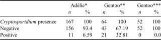
From the 64 samples analysed only 21 (32.8%) were positive. In all of these samples, spherical acid-fast structures of 5 µm diameter were found, which were compatible with Cryptosporidium spp. A comparison of the presence of Cryptosporidium oocyst finding on Antarctic penguins can be seen in Table I.
Comparison of the results obtained for the presence of Cryptosporidium spp. using acid-fast stain in faeces of pygoscelid penguins.
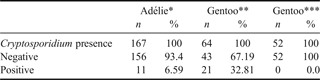
* Ardley Island (62°13′S, 58°54′W) Fredes et al. 2007a.
** Ardley Island (62°13′S, 58°54′W) present study.
*** Peninsula Munita (64°49′S, 62°51′W) Fredes et al. 2007b.
Discussion
In several locations the routine detection of Cryptosporidium oocysts in faeces is generally carried out using direct microscopic identification of oocysts by staining techniques (acid-fast) (Ramirez & Sreevatsan Reference Ramirez and Sreevatsan2006). This methodology is very useful but has some limitations. One is that it is not possible to identify species, but another important one is the possibility of false negative results, because this technique requires the presence of a significant amount of oocysts in the stool sample to give a positive result. This method is certainly less effective compared to molecular methodologies like PCR because PCR can detect around 500 oocysts per gram of stool which is around 100 to 1000 times better than traditional methodologies (Atías Reference Atías1998). Abundance values should therefore be taken with caution.
Using the same methodology, with samples of gentoo penguins taken at Munita Peninsula, (64°49′S, 62°51′E) Paradise Bay, we did not obtain positive results (Fredes et al. Reference Fredes, Madariaga, Raffo, Valencia, Herrera, Godoy and Alcaíno2007b). Therefore the finding of Cryptosporidium oocysts on the stool samples of gentoo penguins from Antarctica even though it was made under basic techniques like the Ziehl Neelsen stain makes this the first report of Cryptosporidium oocysts on this species of penguins from the Antarctic continent.
Some possible explanations can be suggested to account for Cryptosporidium spp. findings in penguins already published. More extreme climate conditions that may prevent the life cycle of the parasite, for example, when the soil temperature reaches -10°C for around 50 days (Kato et al. Reference Kato, Jenkins, Fogarty and Bowman2002), a condition that is likely at the Munita Peninsula but probably not on Ardley Island. On the other hand, the presence of human activities in the sample areas certainly increase the risk of human contamination (antropozoonosis). The first survey area (1999) had only a base camp used for short periods in summer, but the present survey area is located near two permanent bases, a small airfield and a temporary camp.
Currently we are working on genotype and subtype analyses of Cryptosporidium isolates from different animal species, including penguins. Finally the use of molecular tools to identify morphologically indistinguishable species would enable researchers to define relationships between parasite species, potential hosts and pathways of transmission (Fayer Reference Fayer2004, Ramirez & Sreevatsan Reference Ramirez and Sreevatsan2006).
Acknowledgements
We would like to thank Marcela Herrera and Claudia Godoy for the recollection of the samples used on this survey. This study was financed by the FIV 2005 grant, Favet, U. de Chile.