

Statement of research significance
Research question:
To explore the interoceptive profile after a frontal brain acquired injury.
Main Findings:
Few studies have addressed this topic; existing research indicates deficits in cardiac interoceptive accuracy and interoceptive sensitivity following frontal damage. However, results for interoceptive metacognition remain inconsistent.
Study Contributions:
This is the first systematic review on interoception following frontal brain injury, highlighting a significant lack of research and methodological differences across existing studies. It underscores the importance of investigating interoception in this population, given its critical role in emotional processes, and the prevalence of persistent emotional disorders after acquired frontal injury.
Introduction
The ‘interoceptive neural network’ contains insula as well as other brain regions including the frontal cortex and, more specifically, the prefrontal cortex (PFC) (Berntson & Khalsa, Reference Berntson and Khalsa2021). Interoception refers to the top-down and bottom-up bodily awareness processes by which an organism senses, interprets, and combines signals from within itself and below the skin. These processes involve both conscious and nonconscious levels (Desmedt et al., Reference Desmedt, Luminet, Maurage and Corneille2023). At the conscious level, Critchley and Garfinkel (Reference Critchley and Garfinkel2017) suggest three dimensions of interoception: accuracy, sensibility, and awareness. Interoceptive accuracy is the ability to accurately perceive physiological signals, generally measured with a heartbeat counting (HBC) (Schandry, Reference Schandry1981) or heartbeat discrimination (HBD) task (Whitehead et al., Reference Whitehead, Drescher, Heiman and Blackwell1977). For studies using HBC tasks, participants were typically instructed to silently count their heartbeats at rest at different time intervals. Their answers were then compared with real heartbeats recorded at intervals (Whitehead et al., Reference Whitehead, Drescher, Heiman and Blackwell1977). For studies using HBD tasks, a series of tones synchronised or asynchronized with the participants’ heartbeats were generally presented. The participants were usually then asked to indicate whether the tones were faster or slower than their heartbeats. Some studies included additional conditions beyond this accuracy condition such as a motor control condition with auditory feedback via a stethoscope held by the participants themselves or via audio recording of a sampled heartbeat, as well as a learning condition similar to the accuracy condition (Melloni et al., Reference Melloni, Sedeño, Couto, Reynoso, Gelormini, Favaloro, Canales-Johnson, Sigman, Manes and Ibanez2013; Yoris et al., Reference Yoris, García, Salamone, Sedeno, García-Cordero and Ibanez2018). The motor condition is a control of motoric performance and a measure of potential slowing, motor impairments, or attentional deficits (Canales-Johnson et al., Reference Canales-Johnson, Silva, Huepe, Rivera-Rei, Noreika, Garcia, Silva, Ciraolo, Vaucheret, Sedeño, Couto, Kargieman, Baglivo, Sigman, Chennu, Ibáñez, Rodríguez and Bekinschtein2015). The learning condition evaluated performance enhancement after interoceptive feedback. Interoceptive sensibility is the subjective perception of the ability to detect or discriminate body signals and is measured using self-report questionnaires. The most commonly used questionnaire seems to be the Multidimensional Assessment of Interoceptive Awareness Questionnaire (MAIA (Mehling, Acree, Stewart, Silas, & Jones, Reference Mehling, Acree, Stewart, Silas and Jones2018)), but many other questionnaires exist. Finally, metacognitive interoception is an awareness measure that can be measured by relating interoceptive scores on the HBC or HBD to the participants’ degree of confidence in their task performance. Although the theoretical dimensions of interoception are clearly defined, their measurement is currently limited because of the lack of validity of measurement tools (Desmedt et al., Reference Desmedt, Luminet, Maurage and Corneille2023). Although the development of more reliable tools is challenging (Garfinkel et al., Reference Garfinkel, Schulz and Tsakiris2022), this does not limit the study of interoception, which could be a potential psychiatric biomarker (Khalsa & Lapidus, Reference Khalsa and Lapidus2016; Khalsa et al., Reference Khalsa, Adolphs, Cameron, Critchley, Davenport, Feinstein and Zucker2018).
The insular cortex (IC) is a neuronal centre that underlies multimodal interoceptive integration (Herbert & Pollatos, Reference Herbert and Pollatos2012). It is situated within the lateral sulcus of the Sylvian fissure, between the frontal and temporal lobes. The insula is part of the ‘interoceptive neural network’, which includes the somatosensory and somatomotor cortices, the cingulate cortex, and both orbitofrontal and medial prefrontal cortices (Berntson & Khalsa, Reference Berntson and Khalsa2021). Berntson & Khalsa (Reference Berntson and Khalsa2021) suggested that orbitofrontal (OFC) and medial prefrontal cortices received ascendent visceral signals that contribute to interoceptive awareness. According to Craig’s (Reference Craig2009) homeostatic model, the integration of interoceptive signals progresses from the posterior insular cortex, which receives the sense of the physiological condition of the body, to the anterior IC, which integrates emotional, motivational, social, and cognitive factors form the OFC, ventromedial prefrontal cortex (VMPFC) and dorsolateral prefrontal cortex (DLPFC). The insular cortex IC has bidirectional connections with the OFC that enable conscious visceral perception (Gasquoine, Reference Gasquoine2014). Moreover, neuroimaging research has found that both the VMPFC and anterior insula (AI) are typically activated during the evaluation of emotional and bodily states (Gu et al., Reference Gu, Hof, Friston and Fan2013; Terasawa et al., Reference Terasawa, Fukushima and Umeda2013), suggesting that insular connectivity with the VMPFC is involved in emotional awareness. The fronto-insulotemporal network is also involved in social and emotional recognition tasks (Ibáñez & García, Reference Ibáñez, García, Ibáñez and García2018). Multiple connections between the insular cortex and regions involved in emotional and social processes, including the OFC and DLPFC, have been identified through functional activation and lesion studies (Omar et al., Reference Omar, Henley, Bartlett, Hailstone, Gordon, Sauter, Frost, Scott and Warren2011; Viskontas et al., Reference Viskontas, Possin and Miller2007). Recently, Sugawara et al. (Reference Sugawara, Katsunuma, Terasawa and Sekiguchi2024) found that interoceptive training enhances functional connectivity between anterior insular cortex and DLPFC cortices in particular. Literature therefore suggests interconnections between prefrontal regions such as the VMPFC, OFC and DLPFC and the insular cortex IC that contribute to the interoception. Understanding these interactions provides valuable insights into emotional disorders following injury in these prefrontal regions. Prefrontal injuries are often observed after traumatic brain injury (TBI). TBI is defined as an acute brain injury caused by an external mechanical force to the head, such as from falls, assaults, motor accidents or sport-related concussions (Gardner & Zafonte, Reference Gardner, Zafonte, Aminoff, Boller and Swaab2016). Moderate to severe TBI may cause focal injuries at the impact site or on the opposite side due to brain movement within the skull. Rapides brain acceleration and deceleration can also lead to diffuse injuries, like widespread axonal damage, vascular injury, hypoxic-ischemic injury, and swelling (Andriessen et al., Reference Andriessen, Jacobs and Vos2010), often affecting the ventral frontal and temporal cortices (Stuss, Reference Stuss2011). Focal frontal injuries may also occur after a stroke in the territory of the middle or anterior cerebral artery (Eslinger & Reichwein, Reference Eslinger, Reichwein, Bogousslavsky and Caplan2001) or following neurosurgical resection of a tumour in the frontal lobe (Fang et al., Reference Fang, Wang and Jiang2016).
Historically, the role of the PFC, in emotional processing has been identified since the case of Phineas Gage in 1848. Gage, a construction site foreman, was a victim of severe TBI resulting in prefrontal lesions. Subsequently to his TBI, Phineas Gage developed emotional and behavioural disorders similar to an ‘acquired sociopathy’. This historical case has given rise to several emotion theories, such as Damasio’s somatic marker hypothesis (Damasio et al., Reference Damasio, Everitt, Bishop, Roberts, Robbins and Weiskrantz1996). According to this hypothesis, the PFC, especially the VMPFC, records the somatic states encountered during each emotional episode as internal representations called somatic markers (Damasio et al., Reference Damasio, Everitt, Bishop, Roberts, Robbins and Weiskrantz1996) or interoceptive states (Nauta, Reference Nauta, Brady and Nauta1972). These markers are reactivated upon subsequent encounters with similar situations or stimuli in order to adapt to predictable consequences. From this perspective, interoceptive signals are integrated into the PFC, allowing for an emotional response that is appropriate to the situation. In the somatic marker hypothesis, recent updates highlight that decision-making is not solely mediated by the VMPFC but rather by a large-scale system, including several structures such as insular cortex IC (Poppa & Bechara, Reference Poppa and Bechara2018).
As interoceptive information contributes to the construction of one’s emotions (Seth, Reference Seth2013), emotional awareness is therefore linked to interoception. On the opposite, alexithymia, which refers to difficulties with emotional awareness or the ability to recognise one’s feelings, has been linking to interoceptive disorders (Murphy et al., Reference Murphy, Brewer, Hobson, Catmur and Bird2018; Shah et al., Reference Shah, Hall, Catmur and Bird2016). Moreover, emotional awareness is indispensable for social cognition which encompasses the ability to recognise emotions and interpret interpersonal cues that enable one to understand and predict others’ behaviour (Rushby et al., Reference Rushby, Honan, Kelly and Lindsey Byom2013). According to the simulation theory, the capacity to identify the emotions of others is based on the ability to replicate the emotion in one’s own mind and body (Goldman & Sripada, Reference Goldman and Sripada2005). Thus, social cognition relies on emotional awareness, itself partly based on interoceptive abilities (Gao et al., Reference Gao, Ping and Chen2019; Terasawa et al., Reference Terasawa, Moriguchi, Tochizawa and Umeda2014). This highlights the importance of considering the implications of interoceptive brain regions in alexithymia (Ricciardi et al., Reference Ricciardi, Demartini, Fotopoulou and Edwards2015; Williams et al., Reference Williams, Wood and Howe2019) and social cognition disorders frequently reported after frontal acquired brain injury (Hogeveen, Bird, Chau, Krueger, & Grafman, Reference Hogeveen, Bird, Chau, Krueger and Grafman2016).
Alexithymia has been associated with lesions in the right hemisphere after stroke (Bossu et al., Reference Bossu, Salani, Cacciari, Picchetto, Cao, Bizzoni, Rasura, Caltagirone, Robinson, Orzi and Spalletta2009; Spalletta et al., Reference Spalletta, Pasini, Costa, De Angelis, Ramundo, Paolucci and Caltagirone2001; Leszczyński et al., Reference Leszczyński, Pietras and Mokros2021) and with lesions due to tumour removal in the medial right prefrontal lobe (Campanella et al., Reference Campanella, Shallice, Ius, Fabbro and Skrap2014). For TBI, studies have found that higher alexithymia is associated with damage to the anterior insula AI compared to TBI without insular injury (Hogeveen et al., Reference Hogeveen, Bird, Chau, Krueger and Grafman2016), reinforcing the role of anterior insula AI in emotional awareness. Increased alexithymia one year after TBI has been associated with poor emotional functioning and life satisfaction, as well as poor emotional health two years after TBI (Reference Neumann, Hammond, Sander, Bogner, Bushnik, Finn, Chung, Klyce, Sevigny and Ketchum2024b, Neumann et al., Reference Neumann, Hammond, Sander, Bogner, Bushnik, Finn, Chung, Klyce, Sevigny and Ketchum2024a). Difficulties identifying negative feelings, a core feature of alexithymia, have been identified as a significant predictor of depression, anxiety, stress and adjustment issues following acquired brain injury (Fynn et al., Reference Fynn, Preece, Gignac, Pestell, Weinborn and Becerra2023). Additionally, alexithymia has been associated with poststroke depression (Hung et al., Reference Hung, Chou and Su2015). Emotional recognition has been primarily associated with occipital-temporal regions such as the fusiform gyrus, the inferior occipital gyrus, and posterior superior temporal sulcus, particularly in the right hemisphere (Zhen et al., Reference Zhen, Fang and Liu2013). However, the frontal region also seems to be involved in emotional recognition, as meta-analysis studies on patients with TBI (Murphy et al., Reference Murphy, Bennett, de la Piedad Garcia and Willis2022) and stroke (Adams et al., Reference Adams, Schweitzer, Molenberghs and Henry2019) highlight impairments in recognizing emotional displays in all modalities (e.g., face, voice, multimodal) compared to control groups. Specifically, impairment in facial emotional recognition has been associated with lesions in the frontal lobe in patients with TBI (Martins et al., Reference Martins, Faísca, Esteves, Muresan, Justo, Simão and Reis2011), and lesions in the VMPFC and OFC cortex in patients who have undergone tumour resection, stroke or TBI (Pertz et al., Reference Pertz, Okoniewski, Schlegel and Thoma2020; Willis et al., Reference Willis, Palermo, McGrillen and Miller2014). Heberlein, Padon, Gillihan, Farah, and Fellows (2008) found that patients with VMPFC damage due to stroke performed lower in facial emotional recognition tasks compared to patients with damage in other regions of the PFC and control groups. In the group with VMPFC damage, lower emotional recognition was associated with lower subjective experience of emotion during mood induction, supporting the simulation theory (see below). In line with this theory, the authors suggested that damage to the insula, primarily implicated in emotional and bodily awareness, could also be involved in facial emotion recognition (Boucher et al., Reference Boucher, Rouleau, Lassonde, Lepore, Bouthillier and Nguyen2015; Dal Monte et al., Reference Dal Monte, Krueger, Solomon, Schintu, Knutson, Strenziok, Pardini, Leopold, Raymont and Grafman2013). In summary, acquired frontal injuries are associated with emotional disorders at various levels, and when combined with anterior insula AI injuries, they can impact emotional awareness and recognition. Despite well-documented emotional disorders following acquired frontal injuries, insufficient attention has been paid to their potential relationship with interoceptive disorders. Exploring potential interoceptive deficits in patients with frontal injuries could be the next crucial step towards enhancing our understanding of emotional disorders. This is particularly relevant considering the contribution of bodily signals to emotional awareness and recognition, coupled with the bidirectional connection between the insula and the PFC.
Given the connection between the insular cortex IC and PFC, this systematic review attempts to answer the following question: Do disturbances in interoception occur systematically in patients with frontal lesions due to TBI, stroke or tumour, compared with healthy controls? To answer this question, we systematically searched for studies investigating interoceptive accuracy, sensibility, and awareness using tasks and questionnaires in participants with frontal injuries caused by TBI, stroke or tumour and compared them to healthy controls.
Methods
Protocol and registration
Prior to commencing the research procedures, the protocol for this review was submitted to the PROSPERO International Prospective Register of Systematic Reviews in March 2023, under the registration number CRD42023382648. The protocol encompasses the complete methodology of this review, and no changes were made.
Search procedure
We performed a systematic review of the literature in accordance with the PRISMA guidelines. The search was conducted using the PubMed, ProQuest, and Scopus databases. The search was limited to peer-reviewed articles published in English and French between the years 2000 and 2024. Keywords in the title or abstract were ‘traumatic brain injury’ OR ‘focal brain damaged’ OR ‘brain injury’ OR ‘stroke’ OR ‘tumour’ OR ‘cerebrovascular accident’ OR ‘CVA’ in combination with the following keywords: ‘interoception’ OR ‘interoceptive’.
Inclusion and exclusion criterion
We included studies that (1) had at least one participant with frontal brain injury after TBI, stroke, or tumours; (2) compared data of those with frontal damage to a group of healthy participants; (3) measured interoceptive accuracy, metacognition, or sensibility with tasks or questionnaires and (4) excluded studies if they (1) were animal studies; (2) included patients under 18 or over 80; (3) included patients in a persistent vegetative state; (5) targeted pathologies other than acquired frontal brain damage (TBI without brain lesion, neurodevelopmental frontal damage); (6) had no control group; (7) were case studies or (8) were conference papers, preliminary data for future publications, posters, abstracts, reviews, or meta-analyses.
The search returned 67 articles from the 3 databases. We excluded 7 duplicates and screened 60 articles. Figure 1 summarises the selection process and the reasons for article exclusion. The remaining articles were fully reviewed to assess the inclusion and exclusion criteria that led to the exclusion of 53 articles. Finally, seven were included in this review. Study selection was performed independently by two reviewers and cross-checked by two reviewers; all disagreements among the reviewers were unanimously resolved. For data extraction, two independent researchers used a standardised data collection form.
Flow diagram presenting the inclusion/exclusion of studies identified during the database search process.

Quality assessment
We used Farrington’s recommendations for the assessment of methodological quality standards (Farrington, Reference Farrington2003) to develop our own criteria for the quality of the studies included in our review (see also the systematic reviews by Lydon et al., Reference Lydon, Healy, Reed, Mulhern, Hughes and Goodwin2016 and by Bodart et al., Reference Bodart, Invernizzi, Lefebvre and Rossignol2023). We established nine criteria (Table 1), all of which were fulfilled by the seven selected studies.
Quality criteria (Bodart et al., Reference Bodart, Invernizzi, Lefebvre and Rossignol2023)

Results
First, we present the characteristics of the selected articles. Second, we address the research questions in two separate sections handling cardiac interoceptive tasks and interoceptive sensitivity questionnaires. To explore the possibility of distinct deficit profile between isolated frontal lesions and those involving the insula, studies involving patient with frontal damage without insular injury and those with insular injury are presented under different heading.
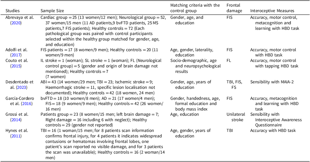
Table 2 presents qualitative information about the selected articles, including the characteristics of the sample, type of frontal lesion in our group of interest, types of interoceptive measures used.
Summary of studies

Notes: AD = Alzheimer’’s disease, bvFTD = Behavioural variant frontotemporal dementia, MS = Multiple sclerosis, FIS = Fronto-insular stroke, IL = Insular lesion, SL = Subcortical lesion, FS = Frontal stoke, FL = Frontal lesions, TMT = Trail Making Test, MAIA-2 = Multidimensional Assessment of Interoceptive Awareness questionnaire.
Sample characteristics
The sample size ranged from 14 to 149 participants, with 233 healthy controls and 129 patients with acquired frontal damage, including 91 patients with stroke and 39 patients with moderate-to-severe TBI. The mean age was 53.8 for control participants 53.9 for patients. According to the information available in articles, the frontal lobe patient sample included 77 men and 45 women, whereas the control sample included 63 men and 64 women. Abrevaya et al. (Reference Abrevaya, Fittipaldi, García, Dottori, Santamaria-Garcia, Birba and Ibáñez2020) did not specify gender distribution in group with the frontal stroke, but it was matched for gender with the control group. Grossi et al. (Reference Grossi, di Vita, Palermo, Sabatini, Trojano and Guariglia2014) did not report gender information for the control group and Couto et al. (Reference Couto, Adolfi, Sedeño, Salles, Canales-Johnson, Alvarez-Abut, Garcia-Cordero, Pietto, Bekinschtein, Sigman, Manes and Ibanez2015) for the group with frontal lesions (FL). The shortest post-injury period was 14 days (Grossi et al., Reference Grossi, di Vita, Palermo, Sabatini, Trojano and Guariglia2014) and the longest mean period was 6.2 years (SD = 2.8) (Hynes et al., Reference Hynes, Stone and Kelso2011). Moreover, the reviewed studies included groups of patients with pathologies other than frontal damage, such as Alzheimer’s disease (AD), behavioural variant frontotemporal dementia (bvFTD), cardiac disease, multiple sclerosis (MS), or insular stroke. These patients were excluded from the sample description.
Interoceptive disturbance after frontal damage
Our question addresses the systematicity of interoceptive disturbance after frontal injuries such as TBI, frontal stroke (FS) or tumours, compared to healthy controls.
Cardiac interoceptive task
Five studies examined the performance in the HBD task of patients with isolated frontal lesions (FL) (Couto et al., Reference Couto, Adolfi, Sedeño, Salles, Canales-Johnson, Alvarez-Abut, Garcia-Cordero, Pietto, Bekinschtein, Sigman, Manes and Ibanez2015) and patients with fronto-insular lesions (FIL) (Abrevaya et al., Reference Abrevaya, Fittipaldi, García, Dottori, Santamaria-Garcia, Birba and Ibáñez2020; Adolfi et al., Reference Adolfi, Couto, Richter, Decety, Lopez, Sigman, Manes and Ibáñez2017; Couto et al., Reference Couto, Adolfi, Sedeño, Salles, Canales-Johnson, Alvarez-Abut, Garcia-Cordero, Pietto, Bekinschtein, Sigman, Manes and Ibanez2015; García-Cordero et al., Reference García-Cordero, Sedeño, de la Fuente, Slachevsky, Forno, Klein, Lillo, Ferrari, Rodriguez, Bustin, Torralva, Baez, Yoris, Esteves, Melloni, Salamone, Huepe, Manes, García and Ibañez2016; Hynes et al., Reference Hynes, Stone and Kelso2011) due to stroke and TBI. All studies reported lower interoceptive accuracy in patients compared to healthy participants and the cardiac group in Abrevaya et al.’s (Reference Abrevaya, Fittipaldi, García, Dottori, Santamaria-Garcia, Birba and Ibáñez2020) study, which included patients presenting hypertensive diseases.
According to Couto et al. (Reference Couto, Adolfi, Sedeño, Salles, Canales-Johnson, Alvarez-Abut, Garcia-Cordero, Pietto, Bekinschtein, Sigman, Manes and Ibanez2015), interoceptive deficits following frontal lobe damage may be secondary to cognitive deficits. Interestingly, Couto et al. (Reference Couto, Adolfi, Sedeño, Salles, Canales-Johnson, Alvarez-Abut, Garcia-Cordero, Pietto, Bekinschtein, Sigman, Manes and Ibanez2015) compared patients with frontal lesions (FL) to a patient with a right insular lesions IL and a patient with subcortical lesion (SL) of the insular tract connecting it to the frontotemporal networks. They found that patients with frontal lesions (FL) performed better than patient with insular lesion IL but lower than patient with subcortical lesion (SL) on the HBD task. The authors suggested that the right insular cortex is involved in cardiac interoception as opposed to the subcortical frontotemporal tract or frontal lobes. They explained that the superior performance of the patient with subcortical lesions (SL) compared to the group with frontal damage was due to larger frontal lesions leading to executive deficits. However, the authors did not mention the gender of the patients with frontal lesions (FL) while the other participants (controls, IL and subcortical lesions (SL)) were women. This potential gender influence is addressed in the discussion.
The HBD task in Abrevaya et al. (Reference Abrevaya, Fittipaldi, García, Dottori, Santamaria-Garcia, Birba and Ibáñez2020) and García-Cordero et al. (Reference García-Cordero, Sedeño, de la Fuente, Slachevsky, Forno, Klein, Lillo, Ferrari, Rodriguez, Bustin, Torralva, Baez, Yoris, Esteves, Melloni, Salamone, Huepe, Manes, García and Ibañez2016) studies contained also learning and metacognition conditions. The group with fronto-insular lesions FIL in García-Cordero et al.’s (Reference García-Cordero, Sedeño, de la Fuente, Slachevsky, Forno, Klein, Lillo, Ferrari, Rodriguez, Bustin, Torralva, Baez, Yoris, Esteves, Melloni, Salamone, Huepe, Manes, García and Ibañez2016) study did not differ from the control group in the learning and metacognition conditions. Therefore, interoceptive accuracy could decrease after fronto-insular lesions FIL, but not under the conditions of learning and metacognition. However, Abrevaya et al. (Reference Abrevaya, Fittipaldi, García, Dottori, Santamaria-Garcia, Birba and Ibáñez2020) reported lower in all interoceptive conditions for the neurological group (including patients with fronto-insular lesions FIL) compared to the cardiac control group.
Moreover, Abrevaya et al. (Reference Abrevaya, Fittipaldi, García, Dottori, Santamaria-Garcia, Birba and Ibáñez2020) and García-Cordero et al. (Reference García-Cordero, Sedeño, de la Fuente, Slachevsky, Forno, Klein, Lillo, Ferrari, Rodriguez, Bustin, Torralva, Baez, Yoris, Esteves, Melloni, Salamone, Huepe, Manes, García and Ibañez2016) also compared patients presenting fronto-insular lesions FIL with those with neurodegenerative diseases such as bvFTD and Alzheimer’s disease (AD). Abrevaya et al. (Reference Abrevaya, Fittipaldi, García, Dottori, Santamaria-Garcia, Birba and Ibáñez2020) did not find any differences between neurological conditions across all dimensions of interoception, including accuracy, metacognition, and learning, suggesting that the pattern of cardiac interoceptive deficits is nonspecific and similar across neurological conditions. García-Cordero et al. (Reference García-Cordero, Sedeño, de la Fuente, Slachevsky, Forno, Klein, Lillo, Ferrari, Rodriguez, Bustin, Torralva, Baez, Yoris, Esteves, Melloni, Salamone, Huepe, Manes, García and Ibañez2016) observed a similar deficit in the accuracy condition for the three neurological conditions, but a higher performance of the group with FIS for metacognition compared to the groups presenting Alzheimer’s disease (AD) and bvFTD, and for learning compared to the group with Alzheimer’s disease (AD). This contradicts the view of Abrevaya et al. (Reference Abrevaya, Fittipaldi, García, Dottori, Santamaria-Garcia, Birba and Ibáñez2020), who suggested nonspecific interoceptive disorders in neurological conditions. Specifically, according to García-Cordero et al. (Reference García-Cordero, Sedeño, de la Fuente, Slachevsky, Forno, Klein, Lillo, Ferrari, Rodriguez, Bustin, Torralva, Baez, Yoris, Esteves, Melloni, Salamone, Huepe, Manes, García and Ibañez2016), the fronto-insular regions may not be crucial for learning, which seems to be supported by temporal regions. Furthermore, accuracy impairment in the group with fronto-insular lesions FIL was associated with fronto-insular damage and in the Alzheimer’s disease (AD) group with hippocampal and temporal atrophy, suggesting that cardiac interoception also relies on the brain memory circuit. Moreover, interoceptive accuracy scores were associated with functional connectivity in the frontotemporal-insular cortex, wherein abnormal connections were observed in each patient group. This suggests that interoceptive accuracy depends on interactions across the anterior and posterior regions and that interoceptive impairments result not only from focused brain damage but also from disruptions in this interactive network. Concerning interoceptive metacognition, performance in all groups was linked to the grey matter volume in the frontal and temporal lobes. Impairment in the groups with Alzheimer’s disease (AD) and bvFTD was associated with an abnormal OFC and prefrontal cortex and disrupted connectivity between the OFC and the hippocampus. According to the authors, the PFC plays a major role in metacognition, especially in the constant monitoring and feedback updating of internal predictions. They explained that preserved metacognition in the group with fronto-insular lesions FIL was due to sparse bilateral long-range connections. Metacognitive impairment resulted from bilateral damage, as in the groups presenting Alzheimer’s disease (AD) and bvFTD, but not from unilateral stroke. After a stroke, functional reorganization and plasticity allow metacognition to be maintained. In summary, García-Cordero et al. (Reference García-Cordero, Sedeño, de la Fuente, Slachevsky, Forno, Klein, Lillo, Ferrari, Rodriguez, Bustin, Torralva, Baez, Yoris, Esteves, Melloni, Salamone, Huepe, Manes, García and Ibañez2016) reported that interoceptive dimensions depend on a fronto-temporo-insular network in which impairment in a specific dimension could result from local damage as well as connectivity abnormalities in this network.
Taken together, these studies consistently reported lower performance in interoceptive accuracy after frontal injury, with or without insular damage, when compared to healthy participants (Adolfi et al., Reference Adolfi, Couto, Richter, Decety, Lopez, Sigman, Manes and Ibáñez2017; García-Cordero et al., Reference García-Cordero, Sedeño, de la Fuente, Slachevsky, Forno, Klein, Lillo, Ferrari, Rodriguez, Bustin, Torralva, Baez, Yoris, Esteves, Melloni, Salamone, Huepe, Manes, García and Ibañez2016; Hynes et al., Reference Hynes, Stone and Kelso2011), patient with subcortical lesion (SL) (Couto et al., Reference Couto, Adolfi, Sedeño, Salles, Canales-Johnson, Alvarez-Abut, Garcia-Cordero, Pietto, Bekinschtein, Sigman, Manes and Ibanez2015) and a cardiac group (Abrevaya et al., Reference Abrevaya, Fittipaldi, García, Dottori, Santamaria-Garcia, Birba and Ibáñez2020). However, divergences were observed in metacognitive and learning interoception. Abrevaya et al. (Reference Abrevaya, Fittipaldi, García, Dottori, Santamaria-Garcia, Birba and Ibáñez2020) found a similar deficit in the group with fronto-insular lesions FIL compared to the groups presenting Alzheimer’s disease (AD) and bvFTD, suggesting that fronto-insular damage affected these two interoceptive dimensions. However, according to García-Cordero et al. (Reference García-Cordero, Sedeño, de la Fuente, Slachevsky, Forno, Klein, Lillo, Ferrari, Rodriguez, Bustin, Torralva, Baez, Yoris, Esteves, Melloni, Salamone, Huepe, Manes, García and Ibañez2016), metacognition and learning interoception can be preserved after frontal insula damage.
Interoceptive sensibility questionnaires
Two studies investigated interoceptive sensitivity in patients with stroke and TBI (Desdentado et al., Reference Desdentado, Miragall, Llorens, Navarro and Baños2023; Grossi et al., Reference Grossi, di Vita, Palermo, Sabatini, Trojano and Guariglia2014). Both studies found lower interoceptive sensibility in the patient group. However, some methodological divergences appear between these two studies. First, the patient group of Desdentado et al. (Reference Desdentado, Miragall, Llorens, Navarro and Baños2023) was composed of patients with TBI and stroke. In contrast, the patient sample used in the Grossi et al. (Reference Grossi, di Vita, Palermo, Sabatini, Trojano and Guariglia2014) study was exclusively composed of patients with stroke presenting diffuse lesions within the vascular territories of the middle cerebral artery, including the frontal lobes and insula. Second, the authors did not use the same self-reported interoceptive questionnaire. On one hand, Desdentado et al. (Reference Desdentado, Miragall, Llorens, Navarro and Baños2023) used the well-known and validated MAIA-2 scale (Mehling et al., Reference Mehling, Acree, Stewart, Silas and Jones2018). On the other hand, Grossi et al. (Reference Grossi, di Vita, Palermo, Sabatini, Trojano and Guariglia2014) did not use a validated measure of interoception, such as the MAIA; instead, they developed a questionnaire including seven questions about interoceptive feelings (InQ) and seven control questions about emotional feelings (CEQ), with higher scores indicating higher self-reported abilities to feel these sensations. The control group had significantly higher InQ and CEQ scores than the patient group, whereas no differences were observed between patients with left or right lesions. Brain lesion analysis indicated that low InQ scores were not associated with frontal damage but rather with damage to the putamen in the left hemisphere and peri-insular white matter, insula, amygdala, and ventral portion of the external/extreme capsule in the right hemisphere. The authors suggested that the role of the insula in interoception depends on peri-insular white matter, allowing for connections in the insula-amygdala network that integrate interoceptive information. In comparison with the interoceptive accuracy dimension, the sensibility dimension is more cognizant, as it relies on self-perception, which may be associated with a broader neural network, including regions such as the amygdala (Garfinkel et al., Reference Garfinkel, Seth, Barrett, Suzuki and Critchley2015). This distinction between these two dimensions could explain the divergent results obtained by Couto et al. (Reference Couto, Adolfi, Sedeño, Salles, Canales-Johnson, Alvarez-Abut, Garcia-Cordero, Pietto, Bekinschtein, Sigman, Manes and Ibanez2015), who observed preserved cardiac interoceptive accuracy in patient with subcortical tract damage leading to the disconnection of frontotemporal networks. However, note that Grossi et al. (Reference Grossi, di Vita, Palermo, Sabatini, Trojano and Guariglia2014) did not mention the gender of the control group. This lack of gender information is addressed in the discussion regarding gender influence across interoceptive dimensions.
In summary, both Desdentado et al. (Reference Desdentado, Miragall, Llorens, Navarro and Baños2023) and Grossi et al. (Reference Grossi, di Vita, Palermo, Sabatini, Trojano and Guariglia2014) showed lower interoceptive sensitivity after acquired brain injury, which is not directly linked to frontal damage, but instead appears to be associated with damage to the insula and other limbic system structures.
To answer our research question, studies suggest that interoceptive accuracy tends to decrease after frontal injuries, with or without the involvement of the insula. However, discrepancies remain regarding the preservation of interoceptive metacognition and learning after fronto-insular lesions FIL. Finally, it should be noted that fronto-insular damages are associated with interoceptive sensitivity disorders.
Discussion
While most studies have examined interoceptive abilities after insular lesions, we aimed to summarise interoceptive alterations after acquired frontal damage in this review. According to Adolfi et al. (Reference Adolfi, Couto, Richter, Decety, Lopez, Sigman, Manes and Ibáñez2017), the insular cortex IC interacts with frontotemporal networks, allowing conscious access to interoceptive signals and their integration with more complex emotional and social cognitive processes. Interoception could be the key to a better understanding of emotional processes after frontal damage. Despite these facts, few studies have investigated interoception deficits after frontal lesions. In this review, which focused on articles published from 2000 to 2024, the seven studies included patients with frontal damage resulting from TBI (Desdentado et al., Reference Desdentado, Miragall, Llorens, Navarro and Baños2023, Hynes et al., Reference Hynes, Stone and Kelso2011) or stroke (Abrevaya et al., Reference Abrevaya, Fittipaldi, García, Dottori, Santamaria-Garcia, Birba and Ibáñez2020; Adolfi et al., Reference Adolfi, Couto, Richter, Decety, Lopez, Sigman, Manes and Ibáñez2017; García-Cordero et al., Reference García-Cordero, Sedeño, de la Fuente, Slachevsky, Forno, Klein, Lillo, Ferrari, Rodriguez, Bustin, Torralva, Baez, Yoris, Esteves, Melloni, Salamone, Huepe, Manes, García and Ibañez2016; Grossi et al., Reference Grossi, di Vita, Palermo, Sabatini, Trojano and Guariglia2014), with lesion were either limited to the frontal lobe (Couto et al., Reference Couto, Adolfi, Sedeño, Salles, Canales-Johnson, Alvarez-Abut, Garcia-Cordero, Pietto, Bekinschtein, Sigman, Manes and Ibanez2015) or extended from the frontal lobe to the insula and temporal lobe (Abrevaya et al., Reference Abrevaya, Fittipaldi, García, Dottori, Santamaria-Garcia, Birba and Ibáñez2020; Adolfi et al., Reference Adolfi, Couto, Richter, Decety, Lopez, Sigman, Manes and Ibáñez2017; García-Cordero et al., Reference García-Cordero, Sedeño, de la Fuente, Slachevsky, Forno, Klein, Lillo, Ferrari, Rodriguez, Bustin, Torralva, Baez, Yoris, Esteves, Melloni, Salamone, Huepe, Manes, García and Ibañez2016; Grossi et al., Reference Grossi, di Vita, Palermo, Sabatini, Trojano and Guariglia2014). Therefore, distinguishing the specific effect of frontal damage from that of insular lesions is challenging. However, this review attempts to provide insights into the heterogeneity of the study samples.
The five cardiac interoceptive accuracy studies reported lower performances after frontal damage, with or without damage to the insula, compared to the control groups (Abrevaya et al., Reference Abrevaya, Fittipaldi, García, Dottori, Santamaria-Garcia, Birba and Ibáñez2020; Adolfi et al., Reference Adolfi, Couto, Richter, Decety, Lopez, Sigman, Manes and Ibáñez2017; Couto et al., Reference Couto, Adolfi, Sedeño, Salles, Canales-Johnson, Alvarez-Abut, Garcia-Cordero, Pietto, Bekinschtein, Sigman, Manes and Ibanez2015; García-Cordero et al., Reference García-Cordero, Sedeño, de la Fuente, Slachevsky, Forno, Klein, Lillo, Ferrari, Rodriguez, Bustin, Torralva, Baez, Yoris, Esteves, Melloni, Salamone, Huepe, Manes, García and Ibañez2016; Hynes et al., Reference Hynes, Stone and Kelso2011). This is consistent with studies in healthy participants showing increased frontal connectivity during the HBC task (Candia-Rivera et al., Reference Candia-Rivera, Sappia, Horschig, Colier and Valenza2022) and activation of the inferior frontal operculum and middle frontal gyrus during heartbeat sounds (Kleint et al., Reference Kleint, Wittchen, Lueken and Bodurka2015). However, interoceptive accuracy tasks are cognitively demanding, and the cognitive disorders frequently reported after frontal damage may play a role in lower interoceptive accuracy. Therefore, understanding the involvement of cognitive function in interoception could be useful in determining whether interoceptive disorders after frontal damage can be attributed to cognitive disorders. However, none of the studies carried out analyses to confirm or deny the influence of this cognitive factor. Future research is needed to clarify this point.
Cognitive involvement in interoception requires distinguishing between cognitive resources needed for interoception and those independent of interoceptive processes but necessary to complete interoceptive tasks. HBD tasks involve attention to internal signals, which may explain why cardiac awareness has been positively associated with selective and divided attention abilities (Matthias et al., Reference Matthias, Schandry, Duschek and Pollatos2009). These internal signals, detected by the salience network (which comprises the anterior insula, ventrolateral prefrontal cortex, and anterior cingulate cortex), are relayed to the executive control network in the dorsolateral frontal and parietal neocortices (Weng et al., Reference Weng, Feldman, Leggio, Napadow, Park and Price2021). Therefore, executive functions are necessary to integrate and analyse internal signals to complete interoceptive tasks. For instance, interventions, such as meditation, rely on executive networks to increase interoceptive engagement (Weng et al., Reference Weng, Feldman, Leggio, Napadow, Park and Price2021). Quadt et al. (Reference Quadt, Critchley and Garfinkel2018) described an executive dimension of interoception as the individual’s ability to attend flexibly to and utilise interoceptive and/or exteroceptive signals. As metacognitive self-awareness decreases after frontal injury and is linked to executive disorders (Bivona et al., Reference Bivona, Ciurli, Barba, Onder, Azicnuda, Silvestro, Mangano, Rigon and Formisano2008), future research should investigate executive interoception to further understand the interweaving of cognitive and interoceptive disorders after frontal injury.
However, HBC and HBD tasks are cognitively demanding, and this load may affect interoceptive performance even without a primary interoceptive disorder. HBC tasks involve internal counting, which relies on working memory (Haustein et al., Reference Haustein, Thomas, Scheer and Denburg2023). HBD tasks have lower cognitive demands, as they require no internal counting, but may involve splitting attention between internal heartbeats and external sounds, as in Hynes et al. (Reference Hynes, Stone and Kelso2011), potentially causing interference. To avoid this, Abrevaya et al. (Reference Abrevaya, Fittipaldi, García, Dottori, Santamaria-Garcia, Birba and Ibáñez2020), Adolfi et al. (Reference Adolfi, Couto, Richter, Decety, Lopez, Sigman, Manes and Ibáñez2017), Couto et al. (Reference Couto, Adolfi, Sedeño, Salles, Canales-Johnson, Alvarez-Abut, Garcia-Cordero, Pietto, Bekinschtein, Sigman, Manes and Ibanez2015), and García-Cordero et al. (Reference García-Cordero, Sedeño, de la Fuente, Slachevsky, Forno, Klein, Lillo, Ferrari, Rodriguez, Bustin, Torralva, Baez, Yoris, Esteves, Melloni, Salamone, Huepe, Manes, García and Ibañez2016) used an HBD tapping paradigm comprising an interoceptive condition in which participants followed their heartbeat without external stimulation. This type of task might be adapted for patients with cognitive disorders. However, we recommend controlling for cognitive influences by assessing executive functions and working memory.
Exploring other interoceptive modalities can reduce the cognitive interference that constitutes methodological limitations in cardiac tasks. Respiratory interoception can be assessed through tasks that involve the detection of change in breathing resistance, such as load detection tasks (Zhao et al., Reference Zhao, Martin and Davenport2002), or through discrimination tasks that require participants to discern the strongest respiratory load among multiple loaded breaths (Webster & Colrain, Reference Webster and Colrain2000). These tasks reduce cognitive load, as they do not involve internal counting or splitting attention between internal and external signals. Respiratory signals are more consciously detectable than cardiac signals and can be easily modified noninvasively by altering the respiratory rate or holding the breath. Moreover, improving respiratory awareness through practices such as meditation or slow-paced respiration (cardiac-coherence methods) has been shown positive effects on disorders such as depression, anxiety (Fournié et al., Reference Fournié, Chouchou, Dalleau, Caderby, Cabrera and Verkindt2021; Payne & Crane-Godreau, Reference Payne and Crane-Godreau2013) or panic disorder (Meuret, Rosenfield, Hofmann, Suvak, & Roth, 2009). According to the neurovisceral perspective, the interaction between the respiratory rhythm and cardiac responses influences emotional regulation through heart rate variability (HRV) (Mather & Thayer, Reference Mather and Thayer2018). HRV, a variation in the heartbeat time interval, indexes the influence of the autonomic nervous system (Laborde et al., Reference Laborde, Mosley and Thayer2017). Higher HRV is associated with better emotional regulation, which can be achieved through paced breathing exercises that increase HRV and strengthen the prefrontal network dynamics (Mather & Thayer, Reference Mather and Thayer2018). HRV is decreased in individuals with acquired brain injury and correlated with poorer outcomes (Lee et al., Reference Lee, Walsh, Fong, Sykora, Doering and Wong2021) but paced breathing biofeedback can improve emotional functioning post-TBI (Talbert et al., Reference Talbert, Kaelberer, Gleave, Driggs, Driggs, Steffen, Baldwin and Larson2023). Investigating respiratory interoception post-frontal damage is therefore more relevant than cardiac interoception. However, existing respiratory tasks require advanced, nonportable equipment and future research should focus on developing simpler tasks for frontal brain-damaged patients (Bodart et al., Reference Bodart, Invernizzi, De Leener, Lefebvre and Rossignol2024).
Three reviewed articles suggest a relationship between interoception and emotional disorders. Hynes et al. (Reference Hynes, Stone and Kelso2011) highlighted the link between lower interoceptive accuracy and changes in emotional awareness after TBI. Adolfi et al. (Reference Adolfi, Couto, Richter, Decety, Lopez, Sigman, Manes and Ibáñez2017) reported reduced social cognition following fronto-insular damage, and Desdentado et al. (Reference Desdentado, Miragall, Llorens, Navarro and Baños2023) explored the connection between interoceptive sensibility, alexithymia, and emotional regulation after acquired brain injury. However, none of these studies included causal analysis limiting the ability to verify the link between interoception and emotional disorders. Emotional deficits after frontal damage can disturb daily functioning and socio-professional reintegration (Milders, Reference Milders2019). It is therefore crucial to understand their origins for better patient support. Acquired alexithymia, often reported after frontal damage (Hobson et al., Reference Hobson, Hogeveen, Brewer, Catmur, Gordon, Krueger, Chau, Bird and Grafman2018; Ricciardi et al., Reference Ricciardi, Demartini, Fotopoulou and Edwards2015), is associated with altered interoceptive accuracy on HBC tasks (Murphy, Brewer, Hobson, Catmur, & Bird, 2018) and on noncardiac tasks (Nord & Garfinkel, Reference Nord and Garfinkel2022). Some authors consider alexithymia to be a general deficit of interoception, as it has been associated with difficulties in perceiving nonaffective interoceptive signals (Brewer et al., Reference Brewer, Cook and Bird2016). Desdentado et al. (Reference Desdentado, Miragall, Llorens, Navarro and Baños2023) suggested that interoceptive sensibility predicted the alexithymia level in participants with or without an acquired brain injury ABI. Since emotions arise from both physiological arousal and cognitive appraisal (Schachter & Singer, Reference Schachter and Singer1962), interoceptive deficits can impair emotional awareness. Accordingly, emotional impairment after frontal injury could be partly caused by interoceptive alterations. Indeed, Hynes et al. (Reference Hynes, Stone and Kelso2011) proposed using HBD tasks to measure emotional awareness changes after TBI. Several studies have shown that interventions targeting interoception such as mindfulness meditation, or yoga decreased alexithymia and improved mental health after TBI (Acabchuk et al., Reference Acabchuk, Brisson, Park, Babbott-Bryan, Parmelee and Johnson2021, Neumann et al., Reference Neumann, Malec and Hammond2017). Further studies should clarify the relationship between emotional changes and interoceptive after frontal injury, integrating interoception accuracy assessment into clinical practice to improve patient rehabilitation.
Limitations
Our review presented some limitations. First, it included only seven studies, limiting our ability to draw significant conclusions. However, it highlighted an important gap in the literature, stressing the need for future studies to fill this gap. Second, differences in age, gender and the type of pathology (TBI or stroke) across studies samples should be considered, as these factors may influence interoception.
First, age variations should be taken into account, as HBD performance declines with age (Khalsa et al., Reference Khalsa, Rudrauf and Tranel2009). This variation can be due to the inclusion of studies involving both TBI and stroke patients. TBI is more common in young men due to risk-taking behaviours or accidents in male-dominated professions (Colantonio, Reference Colantonio2016; Iverson et al., Reference Iverson, Lange, Brooks and Rennison2010), while age is the strongest risk factor for stroke (Gibson, Reference Gibson2013). In our review, the sample of TBI participants in Hynes et al. (Reference Hynes, Stone and Kelso2011) was much younger than the other samples, which mainly comprised stroke participants. Second, gender influences interoception, with variations across dimensions. For self-report, women report paying more attention to bodily sensations compared to men (Grabauskaitė et al., Reference Grabauskaitė, Baranauskas and Griškova-Bulanova2017), but men generally outperform women on cardiac interoceptive tasks (Reference Ma-Kellams, Prentice, Spooner, Murphy, Murphy and BrewerMa- Kellams et al., Reference Ma-Kellams, Prentice, Spooner, Murphy, Murphy and Brewer2024; Prentice et al., Reference Prentice, Hobson, Spooner and Murphy2022). Several factors contribute to men’s higher cardiac interoception accuracy, including larger heart volume (Legato & Leghe, 2010), stronger heart muscle contraction (Shephard & Miller, Reference Shephard and Miller1998), and difference in body fat composition, as higher body fat, which is more common in women, can reduce the perception of heartbeats (Jackson et al., Reference Jackson, Stanforth, Gagnon, Rankinen, Leon, Rao, Skinner, Bouchard and Wilmore2002). In our case, the patient group counted more men than women, while the control group had more women. Since the patient group showed lower interoceptive accuracy, and men generally performed better on such tasks, these differences cannot be attributed to gender influence. In contrast, among the two studies using self-reported measures, Desdentado et al. (Reference Desdentado, Miragall, Llorens, Navarro and Baños2023) controlled for gender influence by matching groups, while Grossi et al. (Reference Grossi, di Vita, Palermo, Sabatini, Trojano and Guariglia2014) did not specify the gender composition of the control group. Since the patient group in Grossi et al. (Reference Grossi, di Vita, Palermo, Sabatini, Trojano and Guariglia2014) contained more men, if the control group had a higher proportion of women, the difference in interoceptive sensibility could be attributed to gender rather than brain injuries. The result of this study should therefore be nuanced by a plausible gender influence. Third, the studies included both stroke and TBI patients, and the exact location of frontal damage was not systematically documented. Moderate to severe TBI could lead to both focal and diffuse damage, whereas strokes typically cause more focal damage (Bramlett & Dietrich, Reference Bramlett and Dietrich2004; Kunz et al., Reference Kunz, Dirnagl and Mergenthaler2010). However, interoception appears to be a potential transdiagnostic and dimensional biomarker not only for neurological disorders (Couto et al., Reference Couto, Adolfi, Sedeño, Salles, Canales-Johnson, Alvarez-Abut, Garcia-Cordero, Pietto, Bekinschtein, Sigman, Manes and Ibanez2015; García-Cordero et al., Reference García-Cordero, Sedeño, de la Fuente, Slachevsky, Forno, Klein, Lillo, Ferrari, Rodriguez, Bustin, Torralva, Baez, Yoris, Esteves, Melloni, Salamone, Huepe, Manes, García and Ibañez2016; Yoris et al., Reference Yoris, García, Salamone, Sedeno, García-Cordero and Ibanez2018) but also across a spectrum of psychiatric disorders (e.g., schizophrenia), neurodevelopmental disorders (e.g., autism), and peripheral conditions (e.g., functional motor disorder) (Bonaz et al., Reference Bonaz, Lane, Oshinsky, Kenny, Sinha, Mayer and Critchley2021). Regardless of the type of pathology or brain damage, the identification of interoceptive markers could allow for better treatment.
Conclusion
In conclusion, this review is the first to systematically study interoceptive disorders following acquired frontal brain injury. The findings suggest deficits in cardiac interoceptive accuracy after frontal damage, as well as reduced interoceptive sensitivity. Inconsistent results were found for interoceptive metacognition. Methodological variations, particularly cognitive load in cardiac tasks may bias performances of brain-damaged patients with cognitive disorders. Additionally, the use of self-reported questionnaires to measure interoceptive sensibility in patients with impaired insight also raises concern. Overall, we believe interoceptive deficits contribute to emotional disorders after frontal injury and are key to better understanding and managing these disorders. However, studies using objective tasks adapted to this population are needed to confirm interoceptive deficits and clarify their relationship with emotional changes after frontal brain damage.
Funding statement
AB and SI were funded by a doctoral award from the University of Mons. MR received support from the Research Foundations FWO and F.R.S.-FNRS under the Excellence of Science (EOS) program (EOS 40007528).
Competing interests
The authors report no financial interests or other potential conflicts of interest.