1. INTRODUCTION
Laser-induced forward transfer or laser-induced film transfer (LIFT) is a micro/nano printing technique. Usually pulsed laser is focused onto the back side of thin film, which is deposited on the transparent substrate (also called as donor substrate). Interface of the thin film gets heated up due to laser pulse which ultimately melts and vaporizes. The pressure induced by the vapor at the interface ejects the film towards the accepter substrate which is kept sufficiently close to donor substrate.
This method was first demonstrated by Bohandy et al. (Reference Bohandy, Kim and Adrian1986), for depositing copper metal patterns under vacuum using nanosecond excimer laser (λ = 193 nm) pulses. LIFT was demonstrated in atmospheric condition by the same group using Ag thin films (Bohandy et al., Reference Bohandy, Kim, Adrian and Jette1988). Later on LIFT was used for a variety of metallic films such as Al (Schultze & Wagner, Reference Schultze and Wagner1991), W (Tóth et al., Reference Tóth, Szorenyi and Tóth1993; Kántor et al., Reference Kántor, Toth and Szorenyi1994a), and Cr (Zergioti et al., Reference Zergioti, Mailis, Vainos, Papakonstantinou, Kalpouzos, Grigoropoulos and Fotakis1998). All these references cited so far shows that superheating of the donor metal thin film is necessary to realize transfer. Hence LIFT was not seen as a practical option for sensitive and soft materials.
To overcome this, number of modifications in the LIFT techniques have been suggested and implemented which results into multiple variants of the technique. These variants are Matrix-Assisted Pulsed Laser Evaporation-Direct Write (MAPLE-DW) technique (Pique et al., Reference Pique, Chrisey, Auyeung, Fitz-Gerald, Wu, McGill, Lakeou, Wu, Nguyen and Duignan1999), Dynamic Release Layer-LIFT (Tolbert et al., Reference Tolbert, Lee, Doxtader, Ellis and Dlott1993), Hydrogen Assisted LIFT (Toet et al., Reference Toet, Thompson, Smith and Sigmon1999), Laser-Induced Thermal Imaging (LITI) technique (Blanchet et al., Reference Blanchet, Loo, Rogers, Gao and Fincher2003), Long-Pulsed LIFT (Kántor et al., Reference Kántor, Tóth, Szorenyi and Tóth1994b; Kántor & Szorenyi, Reference Kántor and Szorenyi1995). Variants of the LIFT technique had been tested on diverse varieties of materials, from a gamut of hazardous material (Veiko et al., Reference Veiko, Shankhno, Smirnov, Miaskovski and Nikishin2006) to soft biological matter (Colina et al., Reference Colina, Serra, Fernández-Pradas, Sevilla and Morenza2005). Recently, LIFT has been used for fabrication of multilayer metamaterials (Tseng et al., Reference Tseng, Wu, Sun, Chang, Chen, Chu, Chen, Zhou, Huang, Yen and Tsai2012), three-dimensional plasmonic cavities (Chen et al., Reference Chen, Tseng, Liao, Wu, Sun, Huang, Chang, Lu, Zhou, Huang, Liu and Tsai2013), miniature sensors and microbattery systems (Piqué et al., Reference Piqué, Arnold, Wartena, Weir, Pratap, Swider-Lyons, Kant and Chrisey2002), bio-printing (Mézel et al., Reference Mézel, Souquet, Hallo and Guillemot2010), and many more.
During their work on the dynamic release method Tolbert et al. (Reference Tolbert, Lee, Doxtader, Ellis and Dlott1993) showed that laser pulse width has an effect on laser threshold needed for transfer. Laser fluence threshold was found to be order of magnitude smaller than for picoseconds laser pulses compared with nanosecond laser pulses. These findings were verified by other researches using femtosecond laser pulses. Use of femtosecond laser pulses for the LIFT process allows greater precision of energy deposition compared with that of nanosecond pulses (Banks, Reference Banks2008). Femtosecond-LIFT has successfully been applied recently to transfer bio-materials (Karaiskou et al., Reference Karaiskou, Zergioti, Fotakis, Kapsetaki and Kafetzopoulos2003).
Recently, Thomas et al. (Reference Thomas, Bernard, Thomas, Alti, Santhosh, Kumari, Khare and Mathur2014) demonstrated utilization of only a femtosecond laser oscillator with MHz repetition rate for implementation of LIFT. With low pulse energy (~nJ) but with high pulse frequency (MHz), heat accumulation takes place which ultimately melts the thin film and subsequently ejects it out. This paper explored the effects of laser energy and scanning speed on the efficacy of LIFT and showed that that one can control the LIFT pattern by controlling the relative motion between the acceptor and donor substrates. However, from an applications perspective, it would be more appropriate to combine various experimental parameters such as laser energy, translation speed, laser repetition rate, and focusing conditions into one single parameter to compare results obtained using LIFT instead of, as is currently the vogue in existing literature, merely mentioning a set of single parameters, such as laser energy, speed, repetition rate or focusing conditions. In this paper, the concept of deposition dose is used which slightly differs from energy dose concept used by Thomas et al. (Reference Thomas, Bernard, Alti, Dharmadhikari, Dharmadhikari, Bhatnagar, Santhosh and Mathur2013). This single parameter will be representation of all the experimental conditions used.
Deposition dose can be defined as
 $$D_{\rm dose} = \displaystyle{{E_{\rm pulse} R} \over {hdv}} \, {\rm \mu J}\,{\rm \mu m}^{-3}$$
where E pulse is the input pulse energy (in μJ), R is the pulse repetition rate (in Hz), v is the speed (in μm s−1) with which donor substrate moves and h is the donor film thickness and d is the laser spot diameter (both in μm). The highest deposition dose reported for LIFT of Indium thin films was ~89.5 μJ μm−3 (Thomas et al., Reference Thomas, Bernard, Thomas, Alti, Santhosh, Kumari, Khare and Mathur2014). This paper reports further experimental work that seeks to probe the following outstanding questions:
• Is surface micro-patterning possible with higher deposition dose values like the sub-surface pattern formation reported by Thomas et al. (Reference Thomas, Bernard, Alti, Dharmadhikari, Dharmadhikari, Bhatnagar, Santhosh and Mathur2013)?
• Will deposition on the accepter substrate also be patterned?
• What will be the effect of movement of accepter substrate along with donor substrate on the re-deposition?
The lowest deposition dose used in the present study is 192.5 μJ μm−3, which is much more than that of reported by Thomas et al. (Reference Thomas, Bernard, Thomas, Alti, Santhosh, Kumari, Khare and Mathur2014).
2. EXPERIMENTAL SET-UP
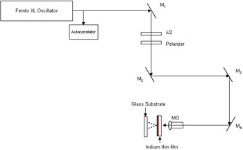
The experimental set-up used for this work is essentially similar to that described in our earlier report (Thomas et al., Reference Thomas, Bernard, Thomas, Alti, Santhosh, Kumari, Khare and Mathur2014) with only relatively small modifications. In brief, a high energy Ti: Sapphire femtosecond laser oscillator (800 nm wavelength, 200 nJ output energy, 50 fs pulse duration, 5.2 MHz repetition rate) was used for our LIFT measurements (Fig. 1). A single-shot autocorrelator was used to monitor the laser pulse width. The pulse energy was attenuated with the help of a half wave plate and polarizer assembly. The laser beam was directed towards the sample using suitable optics, as shown in Figure 1, and finally focused on the rear side of the donor Indium thin film using a 10X (NA = 0.25) microscopic objective. The focal spot size after the microscopic objective was measured using a beam profiler and it was found to be 2.7 μm. An integrating sphere was used to measure the transmission of the microscope objective; it was found to be 93%.
Schematic depiction of our LIFT experimental set up using a Ti:Sapphire femtosecond laser oscillator.

The Indium thin film (donor) was mounted on a one-dimensional (1D) motorized translation stage. The accepter substrate was kept on another, similar, 1D motorized translation stage. Accepter substrate was kept stationary in one set of experiments while in another, it moved with the same velocity as that of the donor substrate. Measurements were made at different values of laser energy. The distance between the accepter substrate and the donor thin film was kept constant to within 400 μm.
The Indium thin film used in this work was deposited on cleaned microscope glass slides (size 2.5 × 7.5 cm) via a thermal evaporation coating unit kept at a base pressure of 10−6 Torr. Indium metal used for the deposition was of 99.995% purity (purchased from Nuclear Fuel Complex, Hyderabad). Thickness of the thin films was found to be 2 μm. Machined donor substrates and LIFT patterns on acceptor substrates were characterized using optical microscopy, scanning electron microscopy (SEM), and energy-dispersive X-ray diffraction (EDAX).
3. RESULTS AND DISCUSSIONS
Often, femtosecond micromaching at MHz repetition rate is compared with KHz repetition rate. In KHz machining individual pulses are responsible for inducing change in the refractive index of the material, whereas in MHz regime, cumulative effect of many pulses is responsible for index change of the material. Due to dominant cumulative effects, heating of the material has been observed which leads to melting and subsequent re-solidification (Eaton et al., Reference Eaton, Zhang, Herman, Yoshino, Shah, Bovatsek and Arai2005).
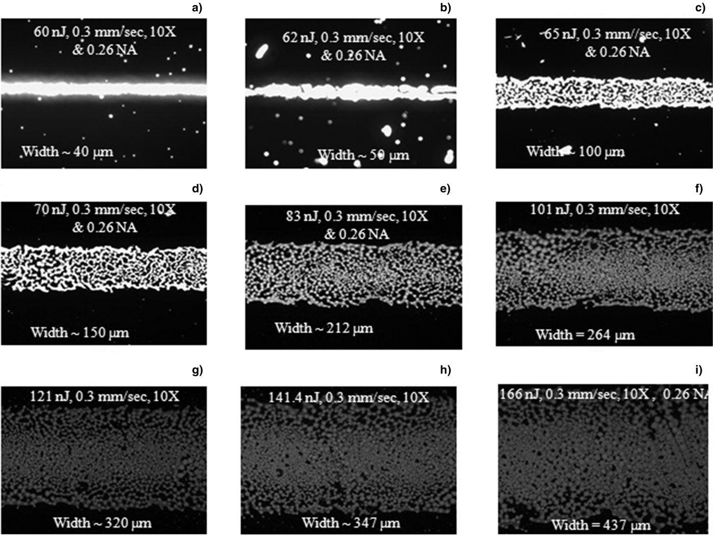
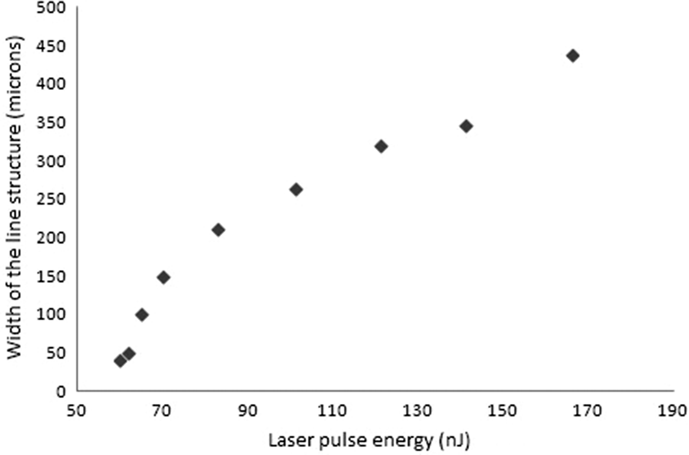
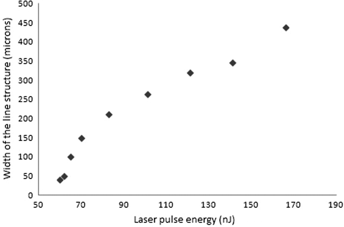
In this work, micron sized line structure was written on the donor Indium thin film by focusing the femtosecond laser pulses via a 10X microscopic objective from the rear side of the Indium thin film, as shown in Figure 1. Line structures were written at different values of incident laser energy, with values which varied over the range 60–166 nJ. During the course of each experiment, the donor thin film was moved at a constant speed of 0.3 mm s−1, a value that is significantly lower than previously used by us (Thomas et al., Reference Thomas, Bernard, Thomas, Alti, Santhosh, Kumari, Khare and Mathur2014). Figure 2 shows typical optical micrographs of the machined donor thin films with 10X magnification for different laser energies. The dark region in each micrograph indicates presence of Indium while the bright region corresponds to absence of it. Laser pulse energy was increased from 60 to 166 nJ as one go from Figures 2a to 2i. With just 5 nJ increase in the laser pulse energy, the width of the machined region was found to increase from 40 to 100 μm, as seen in images shown in Figures 2a and 2c. More interestingly, grain/mesh like structure is seen in Figure 2c whereas Figure 2a shows a clean, machined region with no visible leftovers at 10X magnification. Thereafter, all the subsequent images show similar grain/mesh like structure. Width of the machined region was found to increase with increase in laser pulse energy as shown in Figure 3. In a report by Thomas et al. (Reference Thomas, Bernard, Alti, Dharmadhikari, Dharmadhikari, Bhatnagar, Santhosh and Mathur2013), sub-surface patterning of glass has been reported at higher energies. In this article, varieties of sub-surface patterns were reported in different transparent materials. As reported, such patterns were obtained only if energy dose is above a certain fixed value. Below which, the change in the refractive index was found to be smooth and continuous and as it goes above certain threshold value, it becomes discontinuous pearl/rod like structures. However, these results were reported for bulk transparent material. Figure 2 reports similar transition but for a thin film.
Micrograph showing modified region of Indium thin film when femtosecond laser pulses of energy; (a). 60 nJ, (b). 62 nJ, (c) 65 nJ, (d) 70 nJ, (e) 83 nJ, (f) 101 nJ, (g) 121 nJ, (h) 141 nJ, and (i) 166 nJ were used for machining.

Graph showing increase in the width of line structure written on donor substrate with increase in the laser pulse energy.
From Figure 2, it is quite clear that the micro-pattering of the donor substrate only becomes possible when the deposition dose is equal to or above 210 μJ μm−3 (corresponds to Fig. 2c–i). The highest deposition dose used in this experiment (Fig. 2i) corresponds to 532 μJ μm−3.
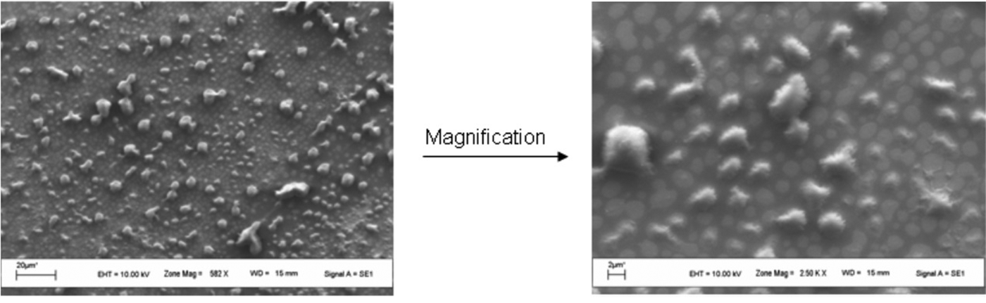
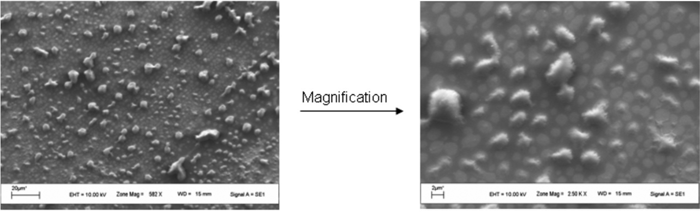
Next the accepter substrate, which was kept 400 μm away from the donor thin film, was analyzed. The accepter substrate was kept stationary during this experiment. SEM images of the accepter substrate (corresponding to the donor substrate shown in Fig. 2i) were acquired and are shown in Figure 4. These images confirm deposition of Indium particles on the accepter substrate. Spattered deposition of Indium particles on the accepter substrate correlates to micro-patterned donor substrate (Fig. 2i). Micron and few submicron size particles are clearly visible in the SEM images. EDAX measurement confirms presence of Indium on the accepter substrate.
SEM images of accepter substrate showing deposition of micro and some submicron particles at two different magnifications.
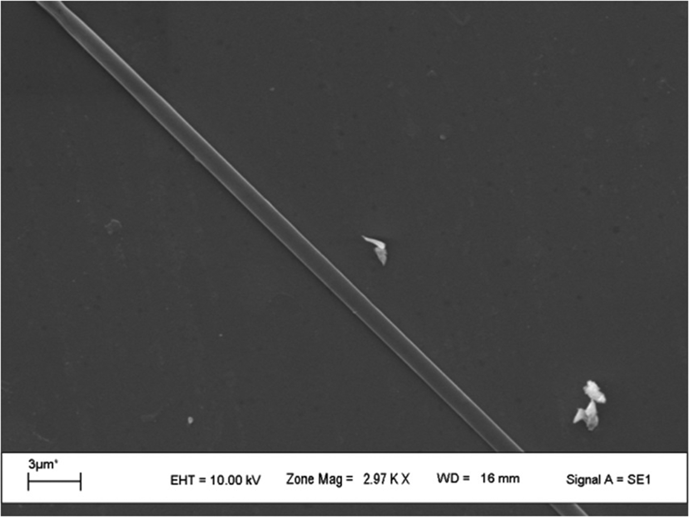
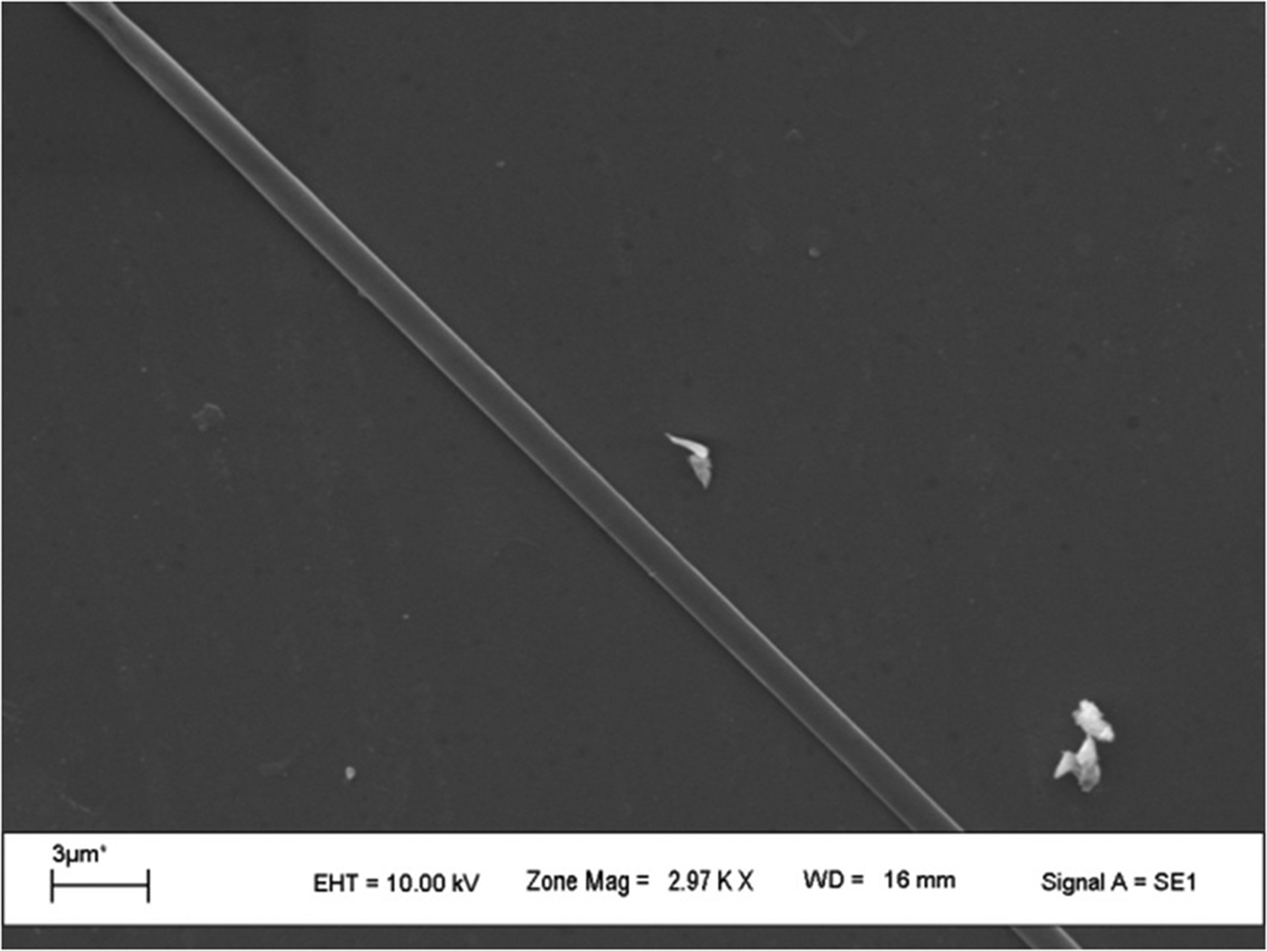
In a different experimental set, accepter substrate was moved with the same velocity as that of the donor substrate, namely 0.3 mm s−1, during transfer. Forty linear structures were written on the donor substrate using a deposition dose of 192.5 μJ μm−3, which is below the micro-pattering threshold. At this deposition dose, machined donor substrate looks similar to as shown in Fig. 2a. Each time, to write a new linear structure, the donor substrate was laterally displaced so as to get the next unexposed thin film region for transfer: The accepter substrate was not moved laterally during the entire experiment. This implies that the ejected material from all the 40 line structures written on the donor substrate might have got deposited linearly on the accepter substrate at one location. SEM images were recorded for the accepter substrate and a typical image is shown in Figure 5. It shows linear deposition of material, in conformity with our expectation. The width of this structure is nearly 1 μm. It should be noted again that at 192.5 μJ μm−3 deposition dose value, there was clean ejection of material from the donor substrate, as seen in Figure 2a. It seems that the ejected material from all the written 40 lines coalesced and compressed to a single line structure with a width of just 1 μm. A similar experiment was done before by Tan et al. (Reference Tan, Venkatakrishnan and Tok2003), in which they have used maximum deposition dose of ~44.5 μJ μm−3 to deposit micro lines of gold on the accepter substrate. Deposition dose used in this paper to produce micro lines of Indium is much more than that used by Tan et al. (Reference Tan, Venkatakrishnan and Tok2003). However micro line structure in our case is much cleaner, compress and coalesce compared with work presented in Tan et al. (Reference Tan, Venkatakrishnan and Tok2003).
SEM image of accepter substrate showing linear deposition.
4. CONCLUSION
The experimental work reported here suggests that as in the case of sub-surface pattern formation using a femtosecond laser, surface machining also requires a threshold deposition dose. Micro-patterning is possible at or above this threshold deposition dose. In the case of Indium deposition in our experiments, the threshold deposition dose was found to be 210 μJ μm−3. This value may vary for different thickness of thin film or for different thin film material. Spattered micron and submicron particles of Indium were observed on the acceptor substrate, which correlated with the micro-patterned donor substrate. Interesting, clean line-like deposition having width of ~1 μm was seen on the acceptor substrate when it was moved at the same velocity as that of the donor substrate during the experiment. Therefore this technique has potential application in fabrication of optoelectronic and photonic devices.
ACKNOWLEDGEMENT
This work constituted part of the activities of I-CUP, the Indian Cluster for Ultrafast Photonics, which was supported by the Office of the Principal Scientific Adviser to the Government of India.