Implications
Nile tilapia is one of the most produced fish species worldwide. The adaptability to a wide environmental and dietary conditions justifies their success. The expected population growth requires the development of more efficient and productive food production systems. Disease outbreaks are major constraints to aquaculture, as the aquatic environment provides a close contact with opportunistic pathogens. Thus, the improvement of resistance to diseases and the enhancement of zootechnical performances are desired production goals. Probiotics constitute a nutritional strategy that may accomplish both. This biological approach, instead of pharmaceuticals, may contribute to a more environmentally friendly and sustainable aquaculture activity.
Introduction
Tilapias are among the most produced farmed fish in the world constituting the most widespread type of aquaculture in the world (Food and Agriculture Organization, 2016). Their success lies on their resistance towards diseases, the support of large aquatic conditions, adaptability to several production systems and omnivorous diet, feeding on a low trophic level (Watanabe et al., Reference Watanabe, Losordo, Fitzsimmons and Hanley2002). It is not surprising that the scientific investigation on tilapia species raised significantly in the last years and a good demonstration of their global importance are the international symposiums dedicated exclusively to tilapia aquaculture, currently in its 11th edition.
Although tilapia presents the good supra cited attributes for aquaculture, the intensification of the production can be associated to poorest growth performances (Telli et al., Reference Telli, Ranzani-Paiva, Dias, Sussel, Ishikawa and Tachibana2014) and to increased risk of diseases, requiring the application of more intense prophylactic methodologies (Watanabe et al., Reference Watanabe, Losordo, Fitzsimmons and Hanley2002). Current strategies can include diet supplementation with probiotics or ‘live organisms that, when administered in adequate amounts, confer a health benefit on the host’ (Food and Agriculture Organization/WHO, 2002). Probiotics act in complex multifactorial ways, depending on species, strain and host (Lazado and Caipang, Reference Lazado and Caipang2014), but mostly by gut microbiota modulation (Balcazar et al., Reference Balcazar, De Blas, Ruiz-Zarzuela, Vendrell, Calvo, Marquez, Girones and Muzquiz2007; Merrifield and Carnevali, Reference Merrifield and Carnevali2014) and their consequent influence on gut mucosal immunity. Probiotics can alter the intestinal immunology and morphology towards a more tolerant and less inflammatory mucosa (Ringø et al., Reference Ringø, Salinas, Olsen, Nyhaug, Myklebust and Mayhew2007; Lazado and Caipang, Reference Lazado and Caipang2014), ameliorating the epithelial barrier function (Madsen et al., Reference Madsen, Cornish, Soper, McKaigney, Jijon, Yachimec, Doyle, Jewell and De Simone2001). Probiotics can also provide nutrients, digestive enzymes (Ray et al., Reference Ray, Ghosh and Ringø2012) and enhance the absorptive surface area through longer villi (Pirarat et al., Reference Pirarat, Pinpimai, Endo, Katagiri, Ponpornpisit, Chansue and Maita2011) and longer (Merrifield et al., Reference Merrifield, Harper, Dimitroglou, Ringø and Davies2010) and denser microvilli (Standen et al., Reference Standen, Rodiles, Peggs, Davies, Santos and Merrifield2015). Some experiments report the stimulation of systemic innate defences as plasma lysozyme, complement or respiratory burst by probiotic microorganisms (Balcazar et al., Reference Balcazar, De Blas, Ruiz-Zarzuela, Vendrell, Calvo, Marquez, Girones and Muzquiz2007) and their implication in the oxidative stress response (Castex et al., Reference Castex, Lemaire, Wabete and Chim2009; Ozório et al., Reference Ozório, Kopecka-Pilarczyk, Peixoto, Lochmann, Santos, Santos, Weber, Calheiros, Ferraz-Arruda, Vaz-Pires and Gonçalves2016). As result, animals become more resistant to infectious diseases (Castex et al., Reference Castex, Lemaire, Wabete and Chim2009) and/or present improved feed and growth performances (Pirarat et al., Reference Pirarat, Pinpimai, Endo, Katagiri, Ponpornpisit, Chansue and Maita2011; Ramos et al., Reference Ramos, Gonçalves, Batista, Costas, Pires, Rema and Ozório2015). The properties and effects described are not all displayed by a single microorganism, explaining why microbial consortia can be more effective (Verschuere et al., Reference Verschuere, Rombaut, Sorgeloos and Verstraete2000).
Probiotic are very promising in animal production but particularly in aquaculture, given the abundance of pathogen and other antigenic stimulus in aquatic environment and their intimate contact with the animals (Merrifield and Ringø, Reference Merrifield and Ringø2014). The proofs of their interest are accumulating, as reviewed in several specialized papers and books (Merrifield and Ringø, Reference Merrifield and Ringø2014) and Nile tilapia is no exception (Pirarat et al., Reference Pirarat, Pinpimai, Endo, Katagiri, Ponpornpisit, Chansue and Maita2011; Telli et al., Reference Telli, Ranzani-Paiva, Dias, Sussel, Ishikawa and Tachibana2014). Even so, those putative beneficial microorganisms must be tested at every situation. The probiotic efficacy is host, strain, dosage and timing dependent (Merrifield and Ringø, Reference Merrifield and Ringø2014). In that way, the present work aimed to study two dosages (3 and 6 g/kg) of a commercial multi-strain probiotic formulation (AquaStar® Growout, Biomin, Getzersdorf, Austria: Bacillus sp., Pediococcus sp., Enterococcus sp., Lactobacillus sp.) on growth performance, immune and oxidative stress status and intestinal morphology of juvenile Nile tilapia (Oreochromis niloticus). Recent studies evaluated the current product in this fish species at three dosages (1.5, 3 and 5 g/kg) with distinct outcomes (Standen et al., Reference Standen, Rodiles, Peggs, Davies, Santos and Merrifield2015 and Reference Standen, Peggs, Rawling, Foey, Davies, Santos and Merrifield2016). Nevertheless the current histological investigation performed at different gut sections and the immune and stress parameters were not evaluated previously.
Material and methods
The current study was performed according to the guidelines on the protection of animals used for scientific purposes from the European directive 2010/63/UE, under the supervision of an accredited expert in laboratory animal science by the Portuguese Veterinary Authority (1005/92, Direção-Geral de Alimentação e Veterinária-Portugal).
Experimental diets
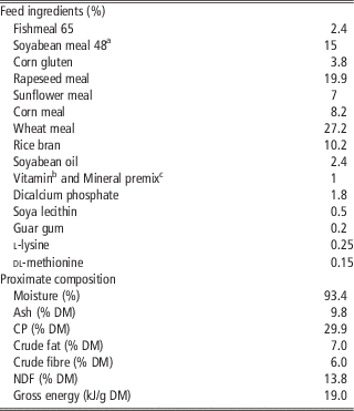
One commercial multi-species probiotic formulation (AquaStar® Growout: Bacillus sp., Pediococcus sp., Enterococcus sp., Lactobacillus sp.; 1×10¹² colony forming unit (CFU)/kg) was incorporated at 3 g/kg (A1, 1.0×10⁶ CFU/g of diet) and 6 g/kg (A2, 2.3×10⁶ CFU/g of diet). A basal diet (composition presented on Table 1) without probiotic was used as a control diet (A0). The defined quantity of lyophilized probiotic preparation was dissolved in tap water (100 ml/kg of diet) for 1 h and coated the basal extruded diet, by slowly mixing in a kitchen mixer (Kenwood, Havant, UK). The diets were prepared, 1 kg at the time, started with the control diet (A0) without probiotic addition, then A1 and A2 at the end. After homogenization, diets were dried in a ventilation oven (48 h at 35°C) and stored at 4°C until use. During growth trial, fresh diets were prepared every month to prevent the decrease of the probiotic viability. The procedure of probiotic incorporation for the complementary digestibility trial was similar to the formerly described but, previous to probiotic incorporation, the basal diet was milled and 1% of chromic oxide (Cr2O3) was added. The homogenized mixture was pelleted (2.4 mm) using a laboratory pellet press (CPM, C-300 model, San Francisco, CA, USA).
Feed ingredients and proximate composition of the experimental basal diet given to juvenile Nile tilapia

DM=dry matter.
a Soyabean meal 48: solvent extracted dehulled soyabean meal: 47% CP, 2.6% crude fat, SORGAL SA, Ovar, Portugal.
b Vitamins (mg or IU/kg diet): vitamin A (retinyl acetate), 20.000 IU; vitamin D3 (dl-cholecalciferol), 2000 IU; vitamin E (Lutavit E50), 100 mg; vitamin K3 (menadione sodium bisulfite), 25 mg; vitamin B1 (thiamine hydrochloride), 30 mg; vitamin B2 (riboflavin), 30 mg; calcium pantothenate, 100 mg; nicotinic acid, 200 mg; vitamin B6 (pyridoxine hydrochloride), 20 mg; vitamin B9 (folic acid), 15 mg; vitamin B12 (cyanocobalamin), 100 mg; vitamin H (biotin), 3000 mg; vitamin C (Lutavit C35), 1000 mg; inositol, 500 mg; colin chloride, 1000 mg; betaine (Betafin S1), 500 mg.
c Minerals (mg or %/kg diet): Co (cobalt carbonate), 0.65 mg; Cu (cupric sulphate), 9 mg; Fe (iron sulphate), 6 mg; I (potassium iodide), 0.5 mg; Mn (manganese oxide), 9.6 mg; Se (sodium selenite), 0.01 mg; Zn (zinc sulphate) 7.5 mg; Ca (calcium carbonate), 18.6%; KCl, 2.41%; NaCl, 4.0%.
Animals and experimental conditions
Nine homogeneous groups of 20 male tilapia (initial BW (IBW): 12.82±0.02 g) were stocked at an initial density of 2.6 kg/m3 in square glass tanks (100 l), reared in a closed recirculating freshwater system at 24.5±1.1°C and photoperiod 12 : 12 h (light : dark), at Universidade de Trás-os-Montes e Alto Douro experimental research station. Fish were kept under quarantine conditions for 2 weeks, during which their health conditions were monitored. There were no visible signs of external injuries or infections, and the behaviour of the animals was normal. After the acclimation period, each experimental diet was randomly attributed to triplicate tanks. Diets were given for 8 weeks by hand three times a day (0900, 1230 and 1800 h), 6 days/week, until apparent visual satiety.
The digestibility trial was performed after 20 weeks of feeding with the experimental diets (15 fish/treatment; 98.94±25.81 g of BW) in duplicate cylindrical fibreglass tanks (75 l) adapted with a ‘Choubert system’ of faeces collection. The water temperature and photoperiod were the same as the growth trial. Faeces were collected for 7 days, after a 2-week period adaptation to diets with Cr2O3. At the end of the digestibility trial faeces were freeze-dried for chemical analyses.
Sampling procedures
The growth trial lasted for 8 weeks during which fish weighting and tissue collection were done at 2, 4 and 8 weeks of feeding. Before samplings, fish were fasted for 24 h and then sacrificed with an overdose of ethylene glycol monophenyl ether (MS 222; 250 mg/l). At each sampling time, two fish per tank were sacrificed and blood collected from the caudal vein, using heparinized syringes. Plasma was obtained after centrifugation (3000×g for 10 min at 4°C) and maintained at −80°C for further analysis. Liver was collected and stored at −80°C for the analyses of the oxidative stress indicators. Head-kidney (HK) was aseptically collected and immediately processed as will be described hereinafter. Additional two fish per tank were sacrificed to collect blood for haematocrit and cell counting, and plasma. In addition, intestine were sampled and fixed in 4% formalin buffered solution for histological assessment. Fish were individually weighed at the end of the trial for the calculation of growth performance parameters. In all, 10 fish from the initial stock and three fish from each tank at the end of trial (nine fish per treatment) were sampled and stored at −20 °C for body composition analyses. At the end of the digestibility trial, eight animals per treatment were sacrificed for plasma analyses and intestine for digestive enzymes determination.
Probiotic detection in diets and chemical analysis of diets, faeces and body composition
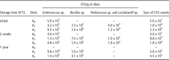
A probiotic count of the diets was made at the beginning of the growth trial. Diets were homogenized with phosphate buffered saline, then serial dilutions were spread in de Man, Rogosa and Sharpe (MRS), Tryptone Soya Agar (TSA) and Bile Aesculin Azide Agar – TSA medium plates, respectively, for Pediococcus and Lactobacillus, Bacillus and Enterococcus and incubated at 37°C for 24 to 48 h. To selectively grow only bacillus on TSA medium, before serial dilutions the sample was subjected to a 10-min bath at 80°C. Pediococcus and Lactobacillus incubation was done anaerobically. Samples of the first diets were maintained at 4°C, and new counts were made after 2 weeks of storage and after a year of storage (Table 2). Fish from the same tank were pooled and homogenized. Dry matter (DM) content was determined in fresh samples of diets, faeces and carcass, whereas the proximate composition analyses was carried out in freeze-dried material using standard procedures as described in Ramos et al. (Reference Ramos, Gonçalves, Batista, Costas, Pires, Rema and Ozório2015).
Probiotic counting of experimental diets (colony forming units (CFU)/g) given to Nile tilapia juveniles

Growth performance and somatic indices
Specific growth rate (SGR; % BW gain/day) was calculated as ((ln final BW)−(ln initial BW)/days)×100. Feed conversion ratio (FCR) was determined as dry feed intake/weight gain. Voluntary feed intake (VFI) was calculated as (dry feed intake/average BW/days)×100, where average BW is (final BW+initial BW)/2. Protein efficiency ratio (PER) was determined as weight gain/protein intake.
Condition factor or K=(weight (g)×100)/length3 (cm), hepatosomatic index (%)=100×(liver weight (g)/BW (g)), intestine somatic index (ISI, %)=100×(intestine weight (g)/BW (g)), spleen somatic index (%)=100×(spleen weight (g)/BW (g)). Apparent digestibility coefficients (ADC) were calculated with the following formulae:
 $$\eqalign{ \hskip -2.2pc{\rm ADC}_{{{\rm nutrient \,\,or \,\,energy}}} (\,\%\,)\,{\equals}\, 100{\times}\Bigg ( {1{\minus}{{(\,\%\,)\,{\rm marker}_{{{\rm diet}}} } \over {(\,\%\,)\,{\rm marker}_{{{\rm faeces}}} }}} \cr {{\times}} {{{(\,\%\,)\,{\rm nutrient \,\,or \,\,energy}_{{{\rm faces}}} } \over {(\,\%\,)\,{\rm nutrient \,\,or \,\,energy}_{{{\rm diet}}} }}} \Bigg).}}$$
ADC of DM were calculated as:
 $${\rm ADC}_{{{\rm DM}}} (\,\%\,)\,{\equals}\,100{\times}\left( {1{\minus}{{(\,\%\,)\,{\rm marker}_{{{\rm diet}}} } \over {(\,\%\,)\,{\rm marker}_{{{\rm faeces}}} }}} \right).$$
Haematological and innate immune parameters
Immediately after blood sampling (n=6/treatment), aliquots of blood were introduced into capillary tubes, centrifuged (microhaematocrit centrifuge, Hermle Z320, Wehingen, Germany; 10 000×g, 5 min, room temperature) and the haematocrit values were measured and read as % packed cell volume. Another aliquot was diluted in Natt–Herricks stain solution for total erythrocytes and leucocytes counts using a haemocytometer. The proceedings were done in duplicate per fish. The alternative complement pathway (ACH50), lysozyme and peroxidase activities were determined in plasma (n=12/treatment) by references methodologies as described in Ramos et al. (Reference Ramos, Gonçalves, Batista, Costas, Pires, Rema and Ozório2015). The respiratory burst activity and the production of nitric oxide (NO) was evaluated on HK leucocytes. The isolation and culture of HK leucocytes were done aseptically. The cells were isolated, adhered to plates and maintained to conduct respiratory burst and NO assays, as described in Ramos et al. (Reference Ramos, Gonçalves, Batista, Costas, Pires, Rema and Ozório2015), after 24 and 72 h of incubation at 25°C, respectively. Respiratory burst activity of HK leucocytes was based on the reduction of ferricytochrome C method for the detection of extracellular O2-production.
Oxidative stress indicators
Liver samples (n= 6/treatment) were individually diluted and homogenized with ultra-pure water (1 : 10) and an ultrasonic processor (<20 s/sample) on ice. Part of the liver homogenate was used to evaluate lipid peroxidation (LPO) measuring the presence of thiobarbituric acid reactive substances as described by Gravato et al. (Reference Gravato, Teles, Oliveira and Santos2006). The remaining liver homogenate was centrifuged (10 000×g, 20 min, 4°C) to isolate de post-mitochondrial supernatant where catalase (CAT), glutathione peroxidase (GPx), glutathione reductase (GR) enzymes and total glutathione content (GSSG+GSH), in its reduced (GSH) or oxidized (GSSG; glutathione disulfide) forms were determined, as described by Gravato et al. (Reference Gravato, Teles, Oliveira and Santos2006).
Intestine histological examination
After 2, 4 and 8 weeks of experimental feeding, intestinal samples (n=6/treatment) were collected for histological examination by light microscopy. From each animal, three samples, with ~2 cm, were taken from the proximal, medial and distal intestine and fixed in 4% buffered formalin solution. Fixed tissues were processed according to standard histological techniques. Sections of 3 μm were cut and stained for histological purposes with haematoxylin–eosin and periodic acid–Schiff reagent (PAS). Histological examinations were made using light microscopy (Olympus BX51, cell^B software, GmbH, Hamburg, Germany).
From each intestine section, two segments were analysed, measuring the following parameters: perimeter and area; tunica muscularis thickness, mucosal fold (or ‘villi’) height and goblet cell presence. The t. muscularis thickness was measured in eight points of each analysed transverse section and an average was computed. The mucosal folds length or height was measured at 10 highest folds. The measures were done from the fold tip to the bottom, following the curves of the fold. Goblet cells stained with PAS were counted on the previous selected highest villi, then the average number of goblet cell per villus was determined.
Digestive enzymes
Intestine sample preparation and digestive enzymes analyses were performed as described by Thongprajukaew et al. (Reference Thongprajukaew, Kovitvadhi, Kovitvadhi, Somsueb and Rungruangsak-Torrissen2011). Intestine samples (n=6/treatment) were diluted, homogenized, centrifuged and the supernatant used for analyse. The amylase, lipase, trypsin and chymotrypsin activities were evaluated measuring the product formation after sample incubation with the adequate substrate (starch, p-nitrophenyl palmitate, benzoyl-l-arginine-p-nitroanilide and N-succinyl-ala-ala-pro-phe-p-nitroanilide, respectively). The specific activity of the enzymes, as the activity of an enzyme per milligram of total protein (expressed in μmol/min per mg) was calculated dividing the sample enzyme activity by their protein content.
Statistical analysis
Statistical analyses were carried out using STATISTICA software (2008, version 8; StatSoft, Inc.). Data were submitted to Kolmogorov–Smirnov and Levene’s tests, to verify normal data distribution and homogeneity of variances, respectively. Data were analysed by one-way or two-way ANOVA. Post hoc Dunnett’s or Tukey’s test was used when significant differences between means were detected. Non-parametric test (Kruskal–Wallis) was used in non-normally distributed data. Significance differences were considered when P<0.05. Results are presented as mean±SD.
Results
Growth performance and carcass composition
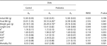
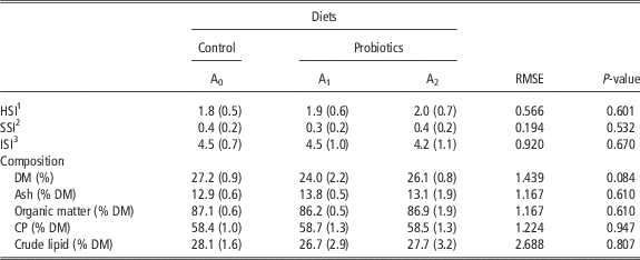
The growth, feed intake and feed conversion performances after the 8 weeks of trial are displayed in Table 3. After 2 weeks of feeding trial, the VFI of A1 group was higher (A0 2.64±0.04; A1 3.07± 0.14; A2 2.86±0.14; P=0.01) regarding the control group and no other parameter differed between treatments. After 4 weeks none of the followed parameters were significantly different. However, after 8 weeks of probiotic supplementation, the animals receiving A1 diet presented an increased (P<0.05) weight and SGR. The length of A1 animals was higher but not significant (P>0.05) and, therefore, the condition factor was similar between the all the experimental groups. Although the FCR and PER of A1 group were numerically improved, towards the control group values, the difference had no statistical significance (P>0.05). The carcass proximate composition showed no significant differences (P>0.05), as liver, spleen or intestine somatic indices (Table 4).
Growth performance of Nile tilapia fed the experimental diets for 8 weeks

SGR=specific growth rate; FCR=feed conversion ratio; PER=protein efficiency ratio; VFI=voluntary feed intake.
Values are means (SD), n=3; except length and k, n=12.
*In each row indicates significant differences regarding control diet (ANOVA and Dunnett’s test P-value<0.05).
1 Condition factor=(weight (g)×100)/length3 (cm).
2 SGR (% BW gain/day)=((ln final BW)−(ln initial BW)/days)×100.
3 FCR=dry feed intake/weight gain.
4 PER=weight gain/protein intake.
5 VFI=(dry feed intake/average BW/days)×100.
Organosomatic indices and carcass proximate composition after 8 weeks of probiotic administration to Nile tilapia juveniles

HSI=hepatosomatic index; SSI=spleen somatic index; ISI=intestine somatic index; DM=dry matter.
Values are means (SD), n=12 for organ indices and n=3 for carcass composition.
In each row absence of distinct letters or symbols indicate no significant difference between means (ANOVA P-value>0.05).
1 HSI (%)=100×(liver weight (g)/BW (g)).
2 SSI (%)=100×(spleen weight (g)/BW (g)).
3 ISI (%)=100×(intestine weight (g)/BW (g)).
Haematological and innate immune status
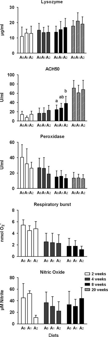
Haematological results (data not shown) were not affected by dietary treatment. No significant impact of probiotic feeding was detected in haematocrit, leucocytes or erythrocytes count over the feeding period (P>0.05). Data from the innate immune parameters are presented in Figure 1. Respiratory burst activity and NO production of HK leucocytes were not significantly (P>0.05) affected by probiotic treatment at any sampling moment. Plasma lysozyme and peroxidase presented the same trend. After 8 weeks of trial, alternative complement activity was significantly higher (P<0.05) in fish-fed A2 diet (38.3±15.3 U/ml) when compared with fish-fed A0 diet (24.7±9.7 U/ml); the remaining values did not differ from control ones.
Lysozyme, alternative complement (ACH50) and peroxidase activities in plasma and respiratory burst and nitric oxide in head-kidney (HK) leucocytes measured in Nile tilapia specimens receiving control (A0) or probiotic supplemented diets (A1 and A2) for 2, 4, 8 and 20 weeks. Results are expressed as means±SD (n=12, except HK parameters where n=6, and 20 weeks where n=16). a,bAt each sampling time, values with different superscript letters indicate significant differences between treatments (one-way ANOVA, Tukey’s test, P<0.05).

Oxidative stress indicators
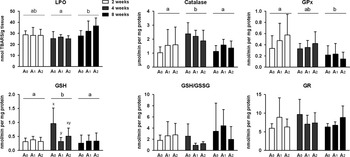
Along the growth trial, the LPO level and some enzymatic and non-enzymatic antioxidant defences were followed at the liver (Figure 2). A large variation was observed among individuals fed the same diets in the majority of the parameters analysed. The LPO increased significantly (P<0.05) over time, as global values at 8 weeks were higher than at 4 weeks. Although probiotic impact had no statistical significance, at the end of the trial, the peroxidation increased proportionally to the probiotic dosage. The antioxidant enzymes studied were not significantly (P>0.05) affected by probiotic supplementation at any sampling period, but changed significantly over time: GPx decreased, whereas CAT increased and then decreased globally. GR presented no specific tendency over the trial and was very variable among specimens. GSH levels after 4 weeks of administration were significantly increased and very variable in control group (A0) comparatively to A1 values. After 2 or 8 weeks of supplementation, GSH levels were not different (P>0.05) between experimental treatments. GSH/GSSG ratio, although highly variable, did not differ between diets or with time (P>0.05).
Liver oxidative stress parameters of Nile tilapia along the growth trial: lipid peroxidation (LPO), catalase, glutathione peroxidase (GPx) and glutathione reductase (GR) activities; glutathione (GSH) and the ratio between reduced and oxidized glutathione levels (GSH/GSSG). Results are expressed as means±SD (n=6) measured in Nile tilapia specimens receiving control (A0) or probiotic supplemented diets (A1 and A2) for 2, 4 and 8 weeks. x,yAt each sampling time, values with different superscript letters indicate significant differences between treatments (one-way ANOVA, Tukey’s test, P<0.05). a,bDistinct superscript letters between sampling times, indicate overall significant differences regardless of the diets. TBARS=thiobarbituric acid reactive substances.

Intestinal histomorphology
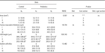
Table 5 displays the data regarding intestine morphometric evaluation at the end of the growth trial. The area of proximal (I), medial (II) or distal (III) intestine was not affected by diet treatment at any sampling moment (P>0.05) and decreased significantly along the gut, as expected in this fish species. The t. muscularis did not differ significantly among experimental groups or intestine sections (P>0.05) at any sampling time. The villi height was not affected by probiotic treatment after 2 or 4 weeks of supplementation. However, after 8 weeks of feeding a significant difference (P<0.05) was observed among control (A0) or probiotic diets (A1 and A2), being the control globally shorter than probiotics, and markedly distinct at proximal intestine. The presence of positive PAS goblet cells was similar among experimental groups after 2 and 4 weeks of trial (data not shown). Yet, after 8 weeks of probiotic feeding, the presence of goblet cells were significantly higher on fish supplemented with both probiotic diets independently of intestine section, comparatively with the control. Regarding the distribution along the gut, the goblet cells presence decreased in number.
Intestinal histological parameters of Nile tilapia after 8 weeks of feeding the experimental diets, by gut part (I-proximal, II-medial, III-distal)

ns=no statistical significance (ANOVA P-value>0.05).
Results are expressed as means (SD), n=6.
Different symbols (¥, §) indicate significant differences between diets, independently of the gut section; different letters (a, b, c) indicate significant differences between gut sections, independently of the diet; x,y,zdifferent superscript letters indicate significant differences between diets and gut section.
ANOVA P-value: *P<0.05, **P<0.01, ***P<0.001.
Apparent digestibility coefficients and digestive enzymes
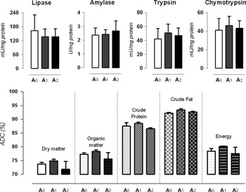
The ADC of nutrients and energy of experimental diets after 20 weeks of experimental feeding were not significantly different (P>0.05; Figure 3, bottom), even if the coefficients presented by A1 diets were numerically higher towards the others. At that time, the specific activity of amylase, lipase, trypsin and chymotrypsin (Figure 3, top) were measured in the intestine, and no significant difference was observed between the specimens-fed probiotic or control diets (P>0.05).
Top: digestive enzymes specific activity after 20 weeks of experimental feeding in Nile tilapia (control, A0 or probiotic supplemented diets, A1 and A2). Values are means±SD (n=8). 1 U=amount of enzyme that catalyses the formation of 1 µmol of product per min at specific conditions (by the respective order 35°C, 30°C, 25°C and 25°C and 7.4, 9, 7.5 and 7.5 pH). Bottom: apparent digestibility coefficients (ADC) of nutrients and energy of experimental diets (A0, A1 and A2) after 20 weeks of probiotic supplementation to Nile tilapia. Values are means±SD (n=2). No significant differences were observed among the diet treatments in any of the parameters (ANOVA P-value>0.05). Average fish weight (g): A0 96.10±28.12; A1 108.87±17.81; A2 92.03±28.37.

Discussion
The improvement of growth performance after probiotic feeding has been observed previously in tilapias receiving several probiont species (Lara-Flores et al., Reference Lara-Flores, Olvera-Novoa, Guzmán-Méndez and López-Madrid2003; Pirarat et al., Reference Pirarat, Pinpimai, Endo, Katagiri, Ponpornpisit, Chansue and Maita2011; Standen et al., Reference Standen, Peggs, Rawling, Foey, Davies, Santos and Merrifield2016). The current study is in agreement with Standen et al. (Reference Standen, Peggs, Rawling, Foey, Davies, Santos and Merrifield2016) supporting the use of the tested commercial product as growth promoter, when supplemented at 3 g/kg (A1). Current results points to the hypothesis that possibly the dosage for best growth performance is not the same as the best dosage for the prevention of infection breaks. Higher dosages may enhance changes in the microbiota and/or immune responses (Li et al., Reference Li, Zhu, Zhang, Yue, Cai, Lu, Zhang, Weng, Zhang, Zhou, Yang and Wang2012), consequently increasing the energy expenditure, at expenses of growth. Other recent studies on bacterial consortia in rainbow trout juveniles observed an improvement on growth rate and FCR at 1.5 g/kg dietary probiotic supplementation, but not at 3 g/kg (Ramos et al., Reference Ramos, Gonçalves, Batista, Costas, Pires, Rema and Ozório2015) and at 1 g/kg but not at and 5 g/kg (Giannenas et al., Reference Giannenas, Karamaligas, Margaroni, Pappas, Mayer, Encarnação and Karagouni2015).
Similarly to previous findings (Standen et al., Reference Standen, Peggs, Rawling, Foey, Davies, Santos and Merrifield2016), we have not noticed influence on FCR. Several management aspects may explain the lack of effects on FCR, such as lower water temperature and dietary protein level, the feeding frequency and the incorporation method of the probiotic. However, different hosts and rearing conditions, with their particular water and gut microbiota (Giatsis et al., Reference Giatsis, Sipkema, Smidt, Heilig, Benvenuti, Verreth and Verdegem2015), may be sufficient for some unpredictability of the results. In a previous study in rainbow trout, the authors observed a decrease in VFI in fish-fed probiotics when compared with the control group (Ramos et al., Reference Ramos, Gonçalves, Batista, Costas, Pires, Rema and Ozório2015). Some reports indicated the possibility of the enhancement of appetite by probiotics (Robertson et al., Reference Robertson, O’Dowd, Burrells, Williams and Austin2000), especially at the beginning of the feeding trial as reported by Irianto and Austin (Reference Irianto and Austin2002) and in the current study. Those studies used coating as the incorporation method, which may explain the findings. Nevertheless, current trial applied water as suspension carrier, whereas Irianto and Austin (Reference Irianto and Austin2002) used saline solution and Robertson et al. (Reference Robertson, O’Dowd, Burrells, Williams and Austin2000) used fish oil. The possible influence of the incorporation method of probiotic on feed intake may deserve further attention. Lara-Flores et al. (Reference Lara-Flores, Olvera-Novoa, Guzmán-Méndez and López-Madrid2003) observed an influence of probiotics on carcass composition in fingerlings. However, as the fish grow, the effect on body composition seems to attenuate (Telli et al., Reference Telli, Ranzani-Paiva, Dias, Sussel, Ishikawa and Tachibana2014; Ramos et al., Reference Ramos, Gonçalves, Batista, Costas, Pires, Rema and Ozório2015; Standen et al., Reference Standen, Peggs, Rawling, Foey, Davies, Santos and Merrifield2016) as in the current study.
Probiotics administered as dietary supplement, pass across the gut and are able to have an impact on the microbiota, even without colonization of the intestine (Merrifield and Carnevali, Reference Merrifield and Carnevali2014). The microbiota influences the mucosal and systemic immune system of fish (Lazado and Caipang, Reference Lazado and Caipang2014). Recent study with the commercial probiotic used in the current study reported changes in intestine microbiota in tilapia, as well as up-regulation of intestine genes involved in immune responses (Standen et al., Reference Standen, Peggs, Rawling, Foey, Davies, Santos and Merrifield2016). In the current study, the alternative complement pathway, a non-specific mechanism of protection against putative pathogens as bacteria, fungi, viruses or parasites (Merrifield and Ringø, Reference Merrifield and Ringø2014), was significantly higher in fish-fed diets supplemented with 2.3×10⁶ CFU/g diet (A2, 6 g/kg) but the not with the lower dosage (A1, 3 g/kg, 1×10⁶ CFU/g). Nevertheless the lower dosage is able to stimulate the tilapia intestinal expression of several immune genes as observed by Standen et al. (Reference Standen, Peggs, Rawling, Foey, Davies, Santos and Merrifield2016), although the authors did not evaluated alternative complement protein genes. It is plausible to think that locally (at the gut) the immune response would be more notorious at lower dosages of probiotics than systemically, as the response studied by us. Our data indicates that alternative complement stimulation requires (at least at circumstances not very challenging for fish) higher dosages of probiotics. It is also necessary to attend to the distinct temperatures of the experiments (28°C v. 24.5°C) that can also influence enormously the fish responsiveness. Previous studies on rainbow trout using the same probiotic blend reported an increase on the alternative (Ozório et al., Reference Ozório, Kopecka-Pilarczyk, Peixoto, Lochmann, Santos, Santos, Weber, Calheiros, Ferraz-Arruda, Vaz-Pires and Gonçalves2016) or total (Giannenas et al., Reference Giannenas, Karamaligas, Margaroni, Pappas, Mayer, Encarnação and Karagouni2015) complement and no significant effect on lysozyme activity. Conversely Ramos et al. (Reference Ramos, Gonçalves, Batista, Costas, Pires, Rema and Ozório2015) observed the significant enhancement of lysozyme while higher but not significantly alternative complement activity. Interestingly, on tilapia, Telli et al. (Reference Telli, Ranzani-Paiva, Dias, Sussel, Ishikawa and Tachibana2014) observed no probiotic influence on lysozyme level of fish maintained at low densities, whereas the protective effect was noticed at higher stocking densities.
The immune system is closely linked to antioxidant system. Recent findings of antioxidant activities of some lactic acid bacteria strains brings to attention the potential role of probiotic treatments on reactive oxygen species (ROS) clearance (Castex et al., Reference Castex, Lemaire, Wabete and Chim2009). Oxidative stress results from an imbalance between oxidative processes and antioxidant mechanisms of defence, causing damage to cellular lipids, protein and DNA (Lushchak, Reference Lushchak2011). Liver is highly adaptive to oxidative damage and main organ used for the studies on the effects of pollutants in fish (Gravato et al., Reference Gravato, Teles, Oliveira and Santos2006). Conversely to the findings of Ozório et al. (Reference Ozório, Kopecka-Pilarczyk, Peixoto, Lochmann, Santos, Santos, Weber, Calheiros, Ferraz-Arruda, Vaz-Pires and Gonçalves2016) in rainbow trout, the hepatic LPO was not reduced in the present study. Similarly, no probiotic influence was observed on the antioxidant enzymes GR, CAT and GPx. After 4 weeks of feeding, fish-fed control diet showed higher GSH levels when compared with fish-fed probiotic diets, especially in fish-fed A1 diet. The reduction of GSH levels can hamper the ROS clearance, causing damage to cellular components (Lushchak, Reference Lushchak2011). Several lactic acid bacteria were able to induce the biosynthesis of GSH in rat ileal mucosa or pancreas (Lutgendorff et al., Reference Lutgendorff, Nijmeijer, Sandström, Trulsson, Magnusson, Timmerman, Minnen, Rijkers, Gooszen, Akkermans and Söderholm2009). Clear positive effects of probiotics have been often observed on immune or oxidative stress status when fish are subjected to stress situations. Telli et al. (Reference Telli, Ranzani-Paiva, Dias, Sussel, Ishikawa and Tachibana2014) reported no differences regarding haematological parameters and plasma lysozyme levels between tilapia-fed Bacillus subtilis or the control diet, when stocked at lower densities. However, after stocking the animals at higher densities, the effect of probiotic administration was noticed, as probiotic-treated fish showed increased lysozyme level, phagocytic activity and a decrease in the number of erythrocytes and in the haematocrit level, but increased levels of mean corpuscular haemoglobin. Inasmuch, Castex et al. (Reference Castex, Lemaire, Wabete and Chim2009) observed a lower oxidative stress level in the digestive gland of shrimps receiving Pediococcus acidilactici, after a Vibrio nigripulchritudo challenge. Previous to that infection, the positive impact of the probiotic was much less noted. Moreover, Reyes-Becerril et al. (Reference Reyes-Becerril, Tovar-Ramírez, Ascencio-Valle, Civera-Cerecedo, Gracia-López, Barbosa-Solomieu and Esteban2011) observed similar findings in leopard grouper-fed yeast after Aeromonas hydrophila infection.
The evidences of probiotics implication in the maintenance of intestinal barrier homoeostasis are accumulating in humans and animals (Madsen et al., Reference Madsen, Cornish, Soper, McKaigney, Jijon, Yachimec, Doyle, Jewell and De Simone2001). In fish, recent findings reported their role on intestinal morphology modulation, feature that may contribute to the improvement of zootechnical parameters. In the current study, the villi length were increased in the anterior intestine of fish supplemented with probiotics for 8 weeks. Longer villi enable an increase in absorption surface area, improving the dietary nutrient utilization, and ultimately improving the growth performance. A recent work in Nile tilapia observed no significant effect on the mid-intestine villi length using a distinct level (5 g/kg) of the same probiotic formula (Standen et al., Reference Standen, Rodiles, Peggs, Davies, Santos and Merrifield2015), consistent with our findings at that gut section.
Pirarat et al. (Reference Pirarat, Pinpimai, Endo, Katagiri, Ponpornpisit, Chansue and Maita2011) working with tilapia and Merrifield et al. (Reference Merrifield, Harper, Dimitroglou, Ringø and Davies2010) working with trout observed that intestinal morphology might be significantly affected by probiotic treatment, especially the proximal intestine. McGuckin et al. (Reference McGuckin, Lindén, Sutton and Florin2011) observed, in mammals, a less thicker mucus layer in small intestine relative to large intestine, correlating it with the facilitation of nutrient absorption. That feature, together with a lower microorganism content comparatively with more distal segments (Ringø et al., Reference Ringø, Olsen, Mayhew and Myklebust2003), might explain a higher impact of microbial feed additives in proximal intestine morphology, towards distal segments.
In the current study, goblet cells were more abundant in fish treated for 8 weeks with both diets supplemented with probiotic. Goblet cells produce mucus that prevents dehydration, lubricates and protects the gastrointestinal tract against physical damage and injurious agents. The mucus, predominantly mucins (gel-forming glycoproteins), play an important role in innate immune defence mechanism (McGuckin et al., Reference McGuckin, Lindén, Sutton and Florin2011). In fish, the increase in goblet cell count as an effect of probiotics treatment has been previously observed (Pirarat et al., Reference Pirarat, Pinpimai, Endo, Katagiri, Ponpornpisit, Chansue and Maita2011; Standen et al., Reference Standen, Peggs, Rawling, Foey, Davies, Santos and Merrifield2016).
Fish gastrointestinal microbiota produces several enzymes like amylase, cellulase, chitinase, lipase, phytase and/or proteases as extensively reviewed by Ray et al. (Reference Ray, Ghosh and Ringø2012). That supplementary enzymes may help to digest substrates, especially those for which the host has no endogenous enzymatic equipment. In that sense, gut microbiota can influence the digestibility of nutrients and/or provide additional nutrients to the host (Merrifield and Ringø, Reference Merrifield and Ringø2014). In fish starting the exogenous feeding, lactic acid bacteria were able to stimulate an early maturation of digestive system, resulting in an increase of intestinal and pancreatic digestive enzyme activity (Suzer et al., Reference Suzer, Çoban, Kamaci, Saka, Firat, Otgucuoğlu and Küçüksari2008). Essa et al. (Reference Essa, Serafy, El-Ezabi, Daboor, Esmael and Lall2010) observed an increase in amylase, lipase and protease-specific activity in tilapia juveniles supplemented with B. subtilis or Lactobacillus plantarum. However, the findings of Essa et al. (Reference Essa, Serafy, El-Ezabi, Daboor, Esmael and Lall2010) were not consistent with the observations in the current study and of Gisbert et al. (Reference Gisbert, Castillo, Skalli, Andree and Badiola2013). The specific activities of amylase, lipase, trypsin and chymotrypsin, and diet digestibility were not significantly affected by long-term probiotic supplementation.
In summary, the dietary supplementation of multi-species probiotic at 3 g/kg (1.0×10⁶ CFU) improved the growth performance, whereas 6 g/kg (2.3×10⁶ CFU) stimulated the innate immune response in tilapia. Both dietary probiotic levels had a positive impact on intestinal morphology. The dietary supplementation of mixed species probiotic may constitute a valuable nutritional approach towards a sustainable tilapia aquaculture. Additional probiotic studies are necessary to study microbiota modulation and the effective protective abilities face to challenges with abiotic and biotic stressful factors, investigating antioxidant and immune parameters in tilapia. Future studies should also investigate digestive enzymes activity and apparent digestibility using internal markers.
Acknowledgements
M.A.R. is very grateful to Cátia Ramos, António J. Pinto and Carla Tomás for their precious collaboration along the experiment, to Carla Dias and Susana Gonçalves for their help with the microbiology work, to Augusto Queiroz for the analytical help, to Mrs Ligia Lourenço (UTAD) for histology technical assistance and to Fundação para a Ciência e a Tecnologia (FCT) by the CECAV funding (ref. UID/CVT/00772/2013).