Introduction
Signal transducers and activators of transcription (STATs) are transcription factors implicated in cellular proliferation, differentiation and metabolism. After ligand binding-induced activation of cognate receptors, STAT proteins become phosphorylated, hetero- or homodimerize, and enter the nucleus (Wakao et al., Reference Wakao, Gouilleux and Groner1994). STAT5A mediates, through the STAT/JAK pathway, signals of hormones with important functions in bovine reproduction – growth hormone (GH), leptin and prolactin (Darnell et al., Reference Darnell, Kerr and Stark1994; Duncan et al., Reference Duncan, Zhong, Wen and Darnell1997). This signalling cascade is extraordinarily versatile and leads both to general phenomena, such as cell proliferation, and to highly specialized signalling, such as tissue-specific expression of individual genes. It is also known that GH exerts distinct effects on the development, differentiation and metabolism of preimplantation embryos. The development of bovine embryos cultured in vitro can be improved by supplementing the medium with GH (Kolle et al., Reference Kolle, Stojkovic, Prelle, Waters, Wolf and Sinowatz2001). GH and its receptor (GHR) mRNA are detectable from the second stage (2-cell stage) of bovine embryogenesis onwards (Kolle et al., Reference Kolle, Sinowatz, Boie, Lincoln, Palma, Stojkovic and Wolf1998, Reference Kolle, Stojkovic, Boie, Wolf and Sinowatz2002; Joudrey et al., Reference Joudrey, Lechniak, Petrik and King2003). Leptin, another possible ligand for the STAT5 gene family, is known to regulate diverse reproductive functions, and recent studies suggest its involvement in early embryo development (Levy et al., Reference Levy, Cordonier, Czyba and Guerin2001; Hansis & Edwards, Reference Hansis and Edwards2003; Madeja et al., Reference Madeja, Warzych, Peippo, Lechniak and Switonski2009). In the study by Madeja et al. (Reference Madeja, Warzych, Peippo, Lechniak and Switonski2009), the expression of leptin and its functional receptor was shown in mouse oocytes and preimplantation embryos.
The aim of the present study was to evaluate the level of STAT5A mRNA expression by quantitative real-time reverse transcription-polymerase chain reaction (RT-PCR) in bovine preimplantation embryos at different stages. The results obtained enhance understanding of the functioning of the GH/STAT5A pathways during embryo development.
Materials and methods
In vitro production of bovine embryos
Bovine ovaries were collected at a local slaughterhouse (University of Munich) and washed in pre-heated phosphate-buffered saline (PBS) (30°C). Cumulus–oocyte complexes (COCs) were obtained by aspirating follicles (2–8 mm diameter) with a 20-gauge needle and a vacuum of approximately 13 kPa. Only COCs with a complete dense cumulus and dark, evenly granulated cytoplasm were selected for in vitro maturation. COCs were collected in a 50 ml centrifuge tube and washed twice with pre-incubated (39°C, 5% CO2 in air) tissue culture media 199 (Invitrogen) supplemented with 10% heat-inactivated serum from cows at estrus (OCS). They were washed again in medium 199 supplemented with 10% OCS and 10 μg/ml bovine follicle stimulating hormone (FSH) and leutenizing hormone (LH) and matured in this medium for 22–24 h at 39 °C in an atmosphere of 5% CO2 in air and maximum humidity. After maturation, COCs were maintained in Tyrode's–albumin–lactate–pyruvate (TALP) medium that contains 6 mg/ml BSA, 100 μg/ml heparin (Sigma). Motile spermatozoa were obtained by the swim-up procedure and by centrifugation of semen from the upper phase for 10 min at 700 g at room temperature. Viable spermatozoa (final concentration 106 spermatozoa/ml) were added to matured COCs for 22 h under the same conditions as used for in vitro maturation. Semen from the same bulls was used for all experiments.
For in vitro culture, cumulus cells were removed from presumptive zygotes by vortexing for 3 min. Groups of 30–40 presumptive zygotes were washed three times and cultured for 8 days in 400 μl synthetic oviduct fluid (SOF) supplemented with 10% OCS and covered with pre-equilibrated mineral oil (Sigma). Embryos were cultured in incubator chambers at 38.5 °C in an atmosphere of 5% CO2, 10% O2, 85% N2. The cleavage rate was evaluated 72 h post-insemination (hpi) and the number of blastocysts scored at 186 hpi. Three replicates of each experiment were performed. Overall, high rates of maturation (82 ± 4%), fertilization (68 ± 2%), cleavage (52 ± 3%) were obtained, and a moderate rate of blastocyst formation (32 ± 3%), in the in vitro produced (IVP) experiments described.
Oocyte and embryo collection
Oocytes were collected 22–24 h after maturation and cumulus cells removed by vortexing. Zygotes were collected 18–20 hpi, 2-cells 26–30 hpi, 4-cells 37–39 hpi, 8-cells at day 3 (d3), 16-cells d4, morula d5, expanded d7 and hatched blastocysts d8. Embryos were stored in groups of four in 10 μl RNAlater (Qiagen) at –80 °C until extraction of RNA. Three replicates of pools of five in vitro matured oocytes and pools of five in vitro embryos were used for each developmental stage.
RNA isolation and reverse transcription
Total RNA was isolated with TriZol reagents (Invitrogen), essentially according to the method of Chomczynski & Sacchi (Reference Chomczynski and Sacchi1987). RNA preparations were treated with 0.25 μl DNase (10 IU/μl; Invitrogen) by incubation at 25 °C for 15 min. The enzyme was inactivated by adding 1 μl 25 mM EDTA and heating at 65 °C for 10 min. Samples were chilled rapidly on ice and reverse transcription directly followed. Reverse transcription was carried out in a total volume of 40 μl according to the Superscript II reverse transcriptase protocol (Invitrogen). Samples were stored at –80 °C until quantitative PCR (qPCR).
Gene cloning
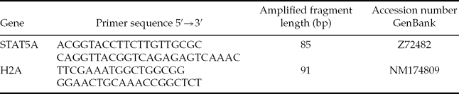
Amplification primers (Table 1) were designed according to the sequences found in GenBank and using the software Primer Express 3 (Applied Biosystems).
Primers used for RT-qPCR determination of mRNA abundance and their localization in respective genes

For qPCR, plasmid standards were established for each gene of interest with the TOPO cloning kit (Invitrogen). Prior to the cloning step PCR was performed in a total volume of 20 μl with 0.25 μl HotStarTaq (Qiagen), 1 μl of each primer listed in Table 1 (final concentration 300 nM), 1 μl cDNA, 2 μl 1 mM dNTPs (Fermentas), 1.25 μl MgCl2 (0.25 mM) and 11.5 μl water. Thermal cycling conditions were as follows: 15 min at 95 °C, then 35 cycles of 95 °C for 15 s, 60 °C for 30 s and 72 °C for 30 s, terminating with 10 min at 72 °C. Samples comprising 10% of the PCR reaction were analysed by 2.5% agarose gel electrophoresis and ethidium bromide fluorescence.
For the cloning step, a PCR aliquot of 2 μl was mixed with 1 μl salt solution, 1 μl TOPO vector and 2 μl water (Invitrogen). Reactions were incubated for 10 min at room temperature and 2 μl ligation mix added to the bacteria. Plasmids were isolated using the QIAprep® spin Mini kit (Qiagen) following the manufacturer's instructions. Sequence integration was tested by digestion of 2 μl plasmid isolate with EcoRI (Fermentas) and gel electrophoresis.
Quantitative PCR (qPCR)
Quantification of mRNA abundance was performed by SybrGreen-based real-time PCR detection using an ABI PRISM 7000 apparatus (Applied Biosystems). Amplification mixes (25 μl) contained 2 μl cDNA, 12.5 μl SybrGreen PCR Mix, 0.25 IU AmpErase uracil N–glycosylase (Applied Biosystems), 300 nM each primer and 7.25 μl water.
Standard curves were obtained using plasmids prepared as described above, and seven serial dilutions (1:5) used for all selected genes, providing a range of 1 million to 64 copies. The abundance of STAT5A mRNA was calculated using the standard curve method, with determination of PCR amplification efficiency and normalization for histone 2A mRNA as the internal reference. qPCR was started with 2 min at 50°C for AmpErase activation and 10 min at 95 °C for denaturation. The programme continued with 45 cycles of 15 s at 95°C, 30 s at 60°C and 30 s at 72°C. Each assay included triplicates of cDNA primed separately for the two genes of interest. The significance of the differences in the expression levels of STAT5A was estimated using Duncan's new multiple range test using data from different embryo stages (mean ± standard deviation (SD)).
Results and Discussion
Real-time RT–PCR was performed to detect and quantify STAT5A mRNA expression. Real-time RT-PCR with an external calibration curve is a fully quantitative method, and thus allows an absolute comparison of the abundance of individual transcripts in RNA preparations at each embryonic stage. RT-PCR products showed single bands of the lengths predicted: 85 bp for STAT5A and 91 bp for H2A (data not shown). The abundance of mRNA encoding STAT5A was normalized relative to H2A mRNA. The CT values obtained for H2A mRNA were similar in all embryonic stages (data not shown), as reported earlier (Robert et al., Reference Robert, McGraw, Massicotte, Pravetoni, Gandolfi and Sirard2002).
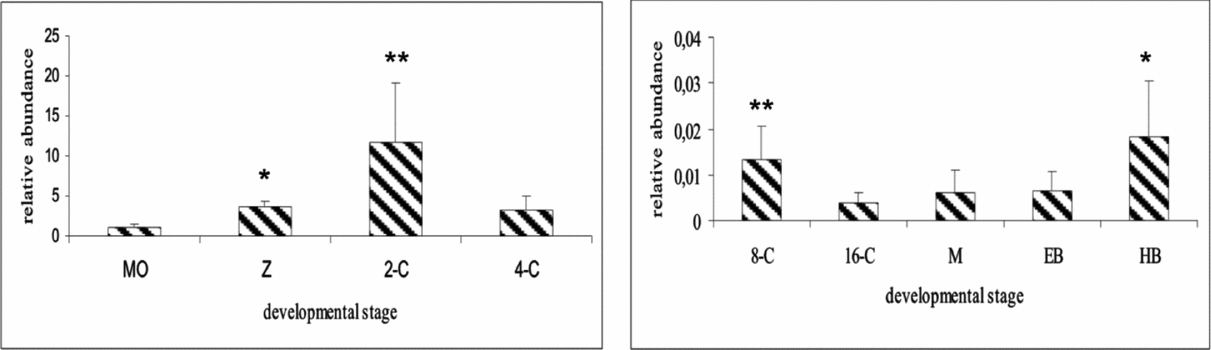
Figure 1 shows relative expression measurements for STAT5A. The expression level at the mature oocyte stage was significantly lower than at zygote (P < 0.05) or 2-cell stage (P < 0.01). STAT5A expression reached a peak at the 2–cell stage and showed a sudden drop at the 8-cell stage (P < 0.01). STAT5A expression in hatched blastocysts appeared somewhat higher than in 8-cell, morula and expanded blastocyst stages (P < 0.05), but was still approximately 100-fold lower than at the 4-cell stage. Quantification of STAT5A expression in bovine embryos revealed that the abundance of the transcript decreased markedly at the 8-cell stage.
Relative expression levels of the STAT5A gene during early embryogenesis. Expression of STAT5A was normalized to the H2A gene. Mean ± standard error (SE) for nine determinations, five embryos each. Abbreviations: 2-C, 2-cell stage; 4-C, 4-cell stage; 8-C, 8-cell stage; 16-C, 16-cell stage; EB, expanded blastocyst; HB, hatched blastocyst; M, morula; MO, matured oocyte; Z, zygote. *Differences statistically significant at P < 0.05. **Differences statistically significant at P < 0.01.
Low fertilization rate and embryonic mortality are significant factors contributing to failed pregnancies in cattle (Diskin & Morris, Reference Diskin and Morris2008). Among the genetic factors, the genes of the pituitary-specific positive transcription factor 1 (POU1F1) pathway play a crucial role in embryonic survival rate (Khatib et al., Reference Khatib, Maltecca, Monson, Schutzkus and Rutledge2009a). Growth hormone, prolactin and their main mediator, STAT5A, belong to this pathway. Growth hormone supports mouse (Wright & Bondioli, Reference Wright and Bondioli1981) and bovine (Kolle et al., Reference Kolle, Stojkovic, Prelle, Waters, Wolf and Sinowatz2001) preimplantation embryo development in vitro. Also, in both species GH transcripts were detected at different developmental stages of early embryos. In bovine embryos the GH transcript has been detected in oocytes and all pre-attachment stages including blastocysts (Kolle et al., Reference Kolle, Sinowatz, Boie, Lincoln, Palma, Stojkovic and Wolf1998; Joudrey et al., Reference Joudrey, Lechniak, Petrik and King2003). It has also been shown that the quality of bovine blastocysts derived from non-early cleaving zygotes is improved by GH supplementation (Pers-Kamczyc et al., Reference Pers-Kamczyc, Warzych, Peippo and Lechniak2010). However, little information is known about the major mediator of the GH signal, embryonic STAT5A. Stat5a−/− double knockout mice exhibit female infertility (Teglund et al., Reference Teglund, McKay, Schuetz, van Deursen, Stravopodis, Wang, Brown, Bodner, Grosveld and Ihle1998), thus STAT5A is considered to be involved in oogenesis and early embryo development. Two studies showed upregulation of STAT5A expression in bovine degenerate embryos compared with blastocysts (Khatib et al., Reference Kolle, Sinowatz, Boie, Lincoln, Palma, Stojkovic and Wolf2009b; Laporta et al., Reference Laporta, Driver and Khatib2011).
In mice, STAT5A transcription decreases between MII and 4-cell stages, and then increases from the morula stage (Nakasato et al., Reference Nakasato, Shirakura, Ooga, Iwatsuki, Ito, Kageyama, Sakai, Nagata and Aoki2006). The present paper demonstrates developmental changes in STAT5A gene expression during early bovine preimplantation embryogenesis. STAT5A mRNAs are present throughout the early developmental stages but fall to low levels after embryonic genome activation, which is different to mice. The bovine embryonic genome undergoes a major transcriptional activation at the 8-cell stage (66–72 hpi) and in vitro cultured bovine embryos can struggle to pass the so-called ‘8–16-cell developmental block’ (Wolf et al., Reference Wolf, Arnold, Bauersachs, Beier, Blum, Einspanier, Frohlich, Herrler, Hiendleder, Kolle, Prelle, Reichenbach, Stojkovic, Wenigerkind and Sinowatz2003). The marked drop in STAT5A expression after the 4-cell stage suggests that abundant STAT5A expression observed in the early stage embryos is a consequence of activation by maternal gene products, and the subsequent low level of STAT5A expression represents regulation by the embryonic genome. The different expression pattern before and after developmental block suggests different activation mechanism for STAT5A. It is known that mammary gland STAT5A expression is induced by GH treatment (Boutinaud & Jammes, Reference Boutinaud and Jammes2004). Therefore, maternal GH or other cytokines may also stimulate expression of STAT5A before the 8-cell stage in cattle. Indeed, mice experiments showed that dynamic changes in the expression of cytokine receptors activate the STAT5 signalling pathway in preimplantation embryos (Nakasato et al., Reference Nakasato, Shirakura, Ooga, Iwatsuki, Ito, Kageyama, Sakai, Nagata and Aoki2006). The constantly low level of STAT5A expression suggests that autocrine cytokine stimulation of STAT5A pathway may be activated at later stages in bovine embryos. It should also be mentioned that the presence of GH in unknown concentrations during maturation and culture could alter STAT5A expression. In contrast, expression of the related factor STAT3 declines steadily from the oocyte to the 16-cell stage and then increases until hatched blastocyst (Leidenfrost et al., Reference Leidenfrost, Boelhauve, Reichenbach, Gungor, Reichenbach, Sinowatz, Wolf and Habermann2011). A similar expression pattern to STAT3 has also been observed for the related LEP gene in in vitro produced bovine embryos (Madeja et al., Reference Madeja, Warzych, Peippo, Lechniak and Switonski2009). In the same experiment LEPR mRNA expression was detected in all but 4-cell bovine embryos (Madeja et al., Reference Madeja, Warzych, Peippo, Lechniak and Switonski2009). These data illustrate the complexity and dynamic nature of mRNA expression changes in preimplantation embryos.
These results provide new data regarding the embryonic STAT5A expression and new insight for the GH pathways regulation in bovine early embryonic development.