Implications
In farm animals, implantation problems contribute significantly toward embryo mortality. It has been suggested that leptin plays an important role in the control of implantation and placentation. In this study, we demonstrated that the levels of leptin and OB-Rb gene expression in porcine ovarian cells differ in the luteal phase of the cycle and early pregnancy. Our results indicate that the hormonal milieu affects the leptin system in the porcine ovary. Detailed knowledge of the regulatory mechanisms during pregnancy and a better understanding of leptin's role in the porcine ovary could contribute toward improving reproductive effectiveness.
Introduction
Leptin is an adipocyte-derived hormone that consists of 146 amino acids. It influences satiety, adiposity, metabolism and reproduction (Zhang et al., Reference Zhang, Proenca, Maffei, Barone, Leopold and Friedman1994; Barash et al., Reference Barash, Cheung, Weigle, Ren, Kabigting, Kuijper, Clifton and Steiner1996). Leptin exerts its effects via a specific leptin receptor. This receptor is a member of the cytokine receptor superfamily, and it can take on several forms. The long form (OB-Rb) is capable of full signal transduction, the short form (OB-Ra, -Rc, -Rd, -Rf) transports leptin across physiological barriers, whereas the soluble receptor (OB-Re) lacks intracellular and transmembrane domains and acts as a binding protein for circulating leptin (Tartaglia et al., Reference Tartaglia, Dembski, Weng, Deng, Culpepper, Devos, Richards, Campfield, Clark, Deeds, Muir, Sanker, Moriarty, Moore, Smutko, Mays, Wool, Monroe and Tepper1995; Bjorbaek et al., Reference Bjorbaek, Uotani, da Silva and Flier1997).
Leptin has been found to exert endocrine and/or paracrine effects on the ovary. The expression of leptin receptors was observed in human and rat granulosa (GC) and theca cells (Cioffi et al., Reference Cioffi, Van Blerkom, Antczak, Shafer, Wittmer and Snodgrass1997; Karlsson et al., Reference Karlsson, Lindell, Svensson, Bergh, Lind, Billig, Carlsson and Carlsson1997; Agarwal et al., Reference Agarwal, Vogel, Weitsman and Magoffin1999; Ryan et al., Reference Ryan, Van der Hoek, Robertson and Norman2003), in luteinized GC and theca cells harvested from prepubertal gilts (in a single in vitro study) and in the porcine corpus luteum (CL) and ovarian stroma (OS; Ruiz-Cortes et al., Reference Ruiz-Cortes, Men, Palin, Downey, Lacroix and Murphy2000; Smolinska et al., Reference Smolinska, Kaminski, Siawrys and Przala2007a). The ovary is also believed to be a site of leptin synthesis. The presence of leptin mRNA and protein was observed in human GC and theca cells, follicular fluid, oocytes and CL (Antczak and Van Blerkom, Reference Antczak and Van Blerkom1997; Cioffi et al., Reference Cioffi, Van Blerkom, Antczak, Shafer, Wittmer and Snodgrass1997; Loffler et al., Reference Loffler, Aust, Kohler and Spanel-Borowski2001; Welt et al., Reference Welt, Schneyer, Heist and Mantzoros2003), in murine GC and theca cells, oocytes, CL and OS (Antczak and Van Blerkom, Reference Antczak and Van Blerkom1997; Ryan et al., Reference Ryan, Woodhouse, Van Der Hoeck, Gilchrist, Armstrong and Norman2002 and Reference Ryan, Van der Hoek, Robertson and Norman2003; Archanco et al., Reference Archanco, Muruzabal, Llopiz, Garayoa, Gómez-Ambrosi, Frühbeck and Burrell2003) and in porcine CL and OS (Smolinska et al., Reference Smolinska, Kaminski, Siawrys and Przala2010). The expression of leptin and OB-Rb in porcine ovarian follicles during the cycle and in early pregnancy (implantation) has scarcely been investigated to date. The objective of this study was to identify the cellular distribution of leptin and OB-Rb genes in porcine ovarian follicles and to compare the expression levels of leptin and OB-Rb mRNAs by in situ hybridization in porcine GC and theca cells during the mid- and late-luteal phases of the estrous cycle and at early stages of pregnancy (days 14 to 16 and 30 to 32).
Material and methods
Experimental animals
The studies were carried out in accordance with the principles and procedures of the Animal Ethics Committee at the University of Warmia and Mazury in Olsztyn. Mature gilts (Large White × Polish Landrace), 7 to 8 months old, weighing 120 to 130 kg, were obtained from private breeders. The pigs were last fed the afternoon of the day before their slaughter. A total of sixteen gilts were assigned to one of four experimental groups (n = 4 per group) as follows: the mid-luteal group (days 10 to 12 of the cycle coinciding with a period of fully active corpora lutea corresponding to the activity of corpora lutea in the period of pregnancy), the late-luteal group (days 14 to 16 of the cycle coinciding with a period of luteal regression and development of a new cohort of follicles), the early-implantation group (days 14 to 16 of gestation) and the post-implantation (placentation) group (days 30 to 32 of gestation). Females were monitored daily for estrus behavior in the presence of an intact boar. The day of onset of the second estrus was designated as day 0 of the estrous cycle. The phase of the estrous cycle was also confirmed on the basis of the morphology of the ovaries. Insemination was performed on days 1 to 2 of the estrous cycle. Pregnancy was detected by ultrasound scanning before killing the gilts (days 30 to 32). Additionally, pregnancy was confirmed by the presence of embryos/fetuses after flushing the uterine horns with 20 ml of sterile saline (days 14 to 16 and 30 to 32). Within a few minutes after slaughter, ovaries with healthy follicles without signs of atresia, subcutaneous adipose tissue and medial basal hypothalamus samples were collected. All the samples were frozen in liquid nitrogen and maintained at −80°C until RNA analyses were performed.
In situ hybridization
Hybridization was carried out according to Smolinska et al. (Reference Smolinska, Kaminski, Siawrys and Przala2007a). Briefly, paraformaldehyde-fixed sections (6 μm) through porcine ovary, subcutaneous adipose tissue (positive control for leptin) and medial basal hypothalamus (positive control for OB-Rb) samples were acetylated for 10 min in 0.25% acetic anhydride (Fluka, St. Louis, MO, USA) in 0.1 M triethanolamine/0.9% NaCl and then dehydrated with ethanol series. The antisense oligonucleotide probes (5′-TAG AGG GAG GCT TCC AGG AC-3′ for leptin, 5′-TTG GGA TGC TGA TCT GAT AA-3′ for OB-Rb) were labeled with [35S]-αdATP (Perkin Elmer, Waltham, MA, USA) at the 3′-end using terminal deoxynucleotidyl transferase (Roche, Meylan, France). The sense oligonucleotide probes (5′-ATC TCC CTC CGA AGG TCC TG-3′ for leptin, 5′-AAC CCT ACG ACT AGA CTA TT-3′ for OB-Rb) were used as a negative control for hybridization specificity. Tissue sections were air-dried and incubated for 22 h with 100 μl of hybridization solution containing 50% formamide, 10% dextran sulfate, 1× Denhardt's solution, 4× SSC (saline sodium citrate), 0.5 mg/ml salmon sperm DNA, 0.25 mg/ml transfer RNA and appropriate probes (107 cpm/ml) under parafilm coverslips at 42°C in humidified chambers. After hybridization, the slides were subjected to several washes in 1× SSC for 10 min, 2× SSC/50% formamide at 42 to 45°C for 15 min, 1× SSC for 15 min and finally in distilled water for 1 min. The slides were then serially dehydrated in ethanol and air-dried. The [35S]-αdATP-labeled sections were dipped in LM-1 emulsion (Amersham Biosciences, Little Chalfont, UK), exposed for 21 days (leptin) or 9 days (OB-Rb) at 4°C, developed in D-19 (4 min) and fixed in Fixer (5 min; Eastman Kodak, Rochester, NY, USA). In addition, the sections were stained with eosin/hematoxylin, dehydrated in a graded series of ethanol and coverslipped with Entellan (Merck, Darmstadt, Germany). All sections were photographed using the CH30/CH40 microscope (equipped for bright-field and dark-field microscopy) and a C-5060 WZ digital camera (Olympus, Tokyo, Japan), and then analyzed by densitometry using the analySIS 5 Soft Imaging System (Olympus). Data were expressed as the absolute values in optical density.
Statistical analysis
All data reported as mean ± s.e.m. were analyzed by two-way ANOVA with ovarian follicular cell type and the day of the estrous cycle or pregnancy as the independent variables, followed by a least significant difference post hoc test (n = 4). Statistical analyses were performed using the Statistica Program (Stat Soft Inc., Tulsa, OK, USA). The level of significance was set at P < 0.05 for all analyses.
Results
Two-way ANOVA indicated that, for both leptin and OB-Rb gene expression, the differences due to the phase of the estrous cycle or pregnancy and the kind of examined cells, and the interaction between the phase and the cells were significant (P < 0.05).
Leptin mRNA expression
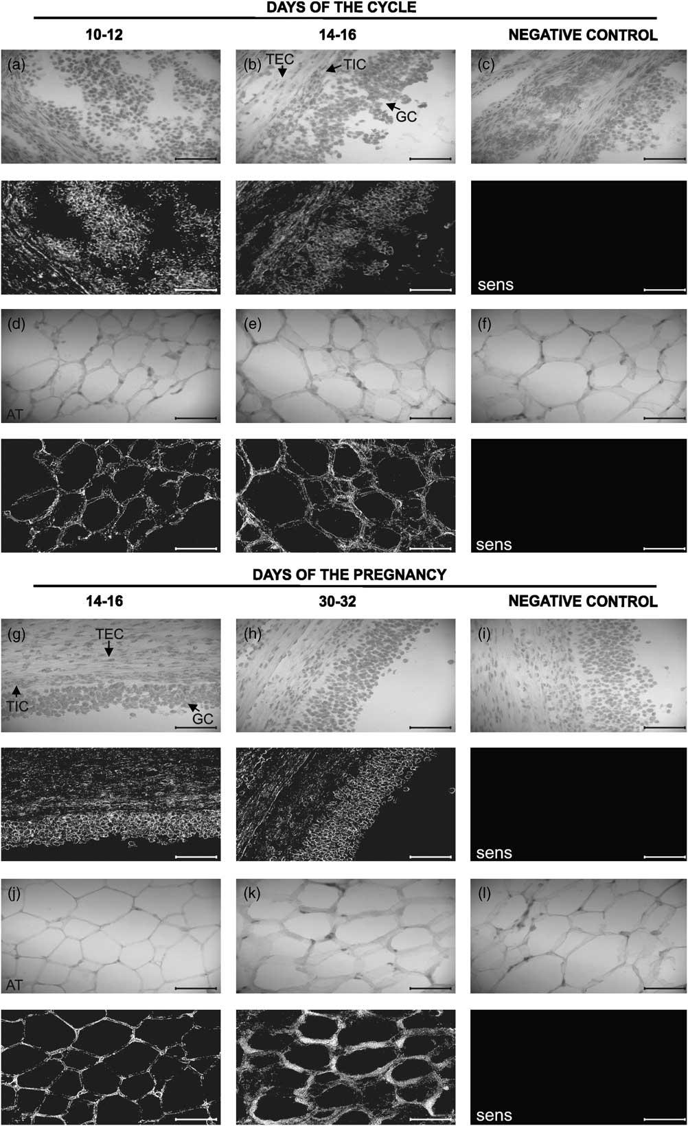
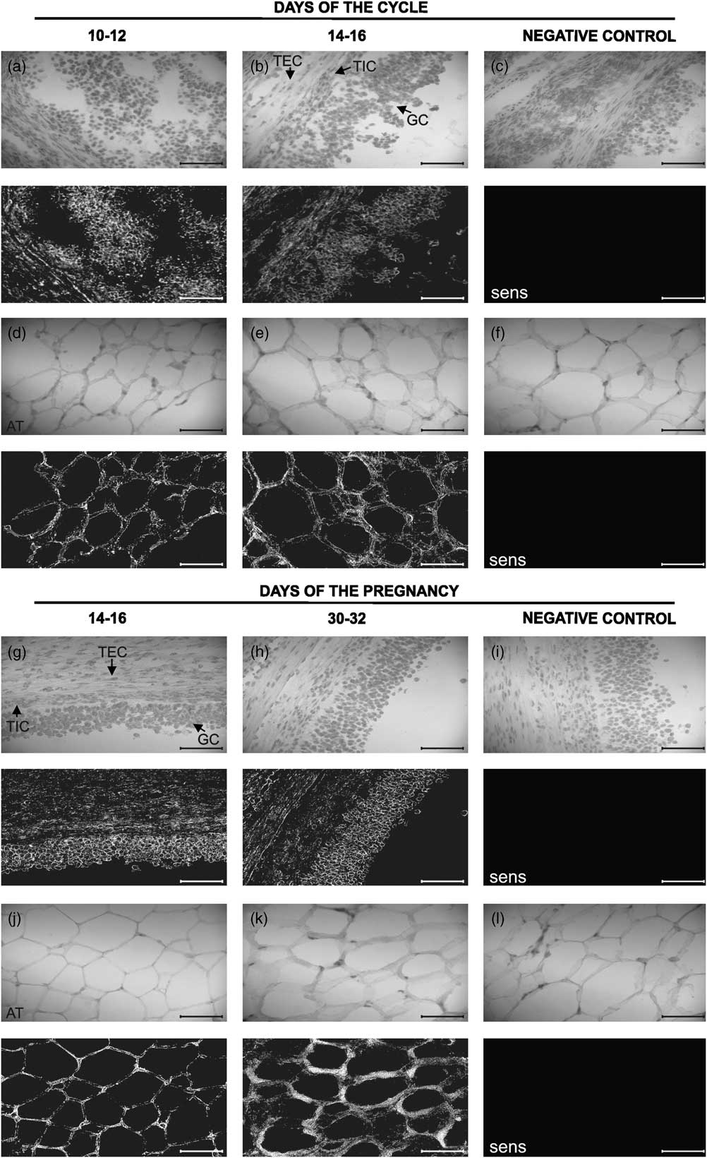
Leptin mRNA expression was detected in porcine GC, theca interna (TIC) and theca externa cells (TEC) of antral follicles on days 10 to 12 and 14 to 16 of the cycle and days 14 to 16 and 30 to 32 of pregnancy. When the sense probes were used, no stain was detected in porcine ovarian cells (Figure 1).
Expression of leptin mRNA determined by in situ hybridization (dark-field images) in porcine granulosa (GC, a to b and g to h), theca interna (TIC, a to b and g to h) and theca externa cells (TEC, a to b and g to h), and adipose tissue (AT: as a positive control, d to e and j to k) on days 10 to 12 and 14 to 16 of the cycle and days 14 to 16 and 30 to 32 of pregnancy. Corresponding bright-field images show hematoxylin–eosin staining. Negative controls with the sense sequence (c and f; i and l). Bar = 20 μm.
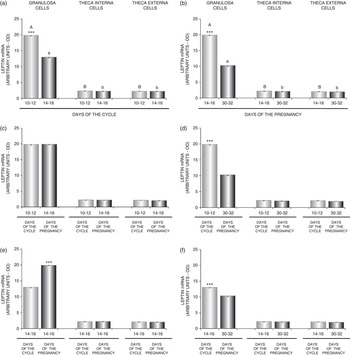
During the two periods of pregnancy and the luteal phase of the cycle, leptin gene expression was higher (P < 0.001) in GC in comparison with TIC and TEC. In GC, gene expression was higher (P < 0.001) on days 10 to 12 compared with days 14 to 16 of the cycle and on days 14 to 16 of pregnancy compared with days 30 to 32 of pregnancy. However, no significant differences were found in leptin expression in TIC and TEC within the cycle or during pregnancy (Figure 2a and b).
Comparison of leptin mRNA expression determined by in situ hybridization in porcine follicular cells between (a, b) two stages of the cycle or two periods of pregnancy and between granulosa (GC), theca interna (TIC) and theca externa (TEC); (c, d) pregnancy and the mid-luteal phase of the cycle; (e, f) pregnancy and the late-luteal phase of the cycle (n = 4). Values are expressed as means ± s.e.m. in arbitrary optical density units (OD). Bars with different superscripts are significantly different. Capital letters indicate differences between GC, TIC and TEC on days 10 to 12 of the cycle and days 14 to 16 of pregnancy (P < 0.001). Small letters indicate differences between GC, TIC and TEC on days 14 to 16 of the cycle and days 30 to 32 of pregnancy (P < 0.001). Asterisks indicate the differences between GC isolated during both examined periods of the cycle and/or pregnancy ***P < 0.001.

Significantly higher (P < 0.001) leptin gene expression was detected in GC on days 10 to 12 and 14 to 16 of the cycle in comparison with days 30 to 32 of pregnancy (Figure 2d and f). In addition, on days 14 to 16 of pregnancy, the content of leptin transcript was higher (P < 0.001) than that on days 14 to 16 of the cycle (Figure 2e). There were no significant differences in leptin mRNA expression in GC between days 10 to 12 of the cycle and 14 to 16 of pregnancy, and in TIC and TEC between the two periods of the cycle and pregnancy (Figure 2c to f).
OB-Rb gene expression
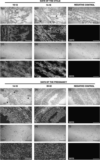
OB-Rb mRNA expression was localized in porcine GC, TIC and TEC cells of antral follicles on days 10 to 12 and 14 to 16 of the cycle and days 14 to 16 and 30 to 32 of pregnancy. When the sense probes were used, no stain was detected in porcine ovarian cells (Figure 3).
Expression of OB-Rb mRNA determined by in situ hybridization (dark-field images) in porcine granulosa (GC, a to b and g to h), theca interna (TIC, a to b and g to h) and theca externa cells (TEC, a to b and g to h), and medial basal hypothalamus (MBH: as a positive control, d to e and j to k) on days 10 to 12 and 14 to 16 of the cycle and days 14 to 16 and 30 to 32 of pregnancy. Corresponding bright-field images show hematoxylin–eosin staining. Negative controls with the sense sequence (c and f; i and l). Bar = 20 μm.

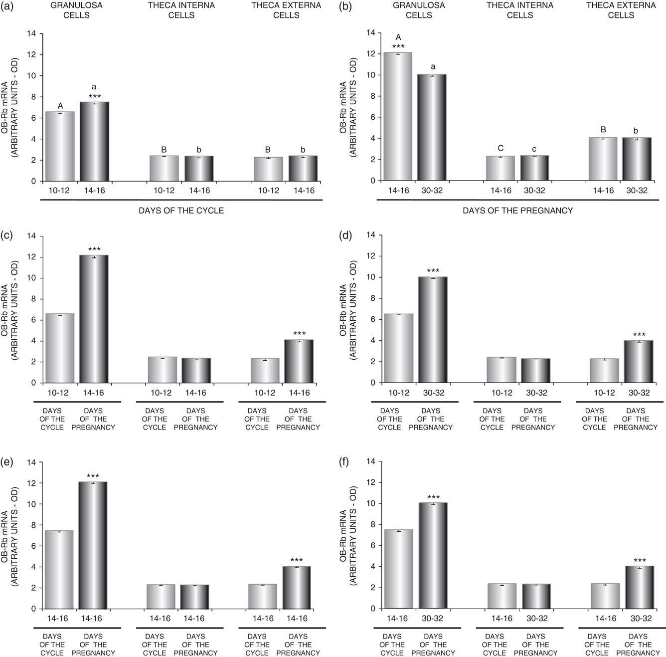
OB-Rb gene expression during both the stages of the cycle examined was higher (P < 0.001) in GC compared with TIC and TEC. During the two periods of pregnancy, the expression of OB-Rb mRNA was the greatest (P < 0.001) in GC in relation to TIC and TEC and the lowest in TIC. In GC during the cycle, a higher (P < 0.001) OB-Rb mRNA expression was observed on days 14 to 16 compared with days 10 to 12. In pregnant animals, OB-Rb gene expression decreased (P < 0.001) from days 14 to 16 to 30 to 32 after conception. Similar to leptin mRNA expression, there was no change in the gene expression within the cycle or pregnancy in TIC and TEC (Figure 4a and b).
Comparison of leptin OB-Rb expression determined by in situ hybridization in porcine GC, TIC and TEC between: (a, b) two stages of the cycle and two periods of pregnancy and between GC, TIC and TEC; (c, d) pregnancy and the mid-luteal phase of the cycle; (e, f) pregnancy and the late-luteal phase of the cycle (n = 4). Values are expressed as means ± s.e.m. in arbitrary optical density units (OD). Bars with different superscripts are significantly different. Capital letters indicate differences between GC, TIC and TEC on days 10 to 12 of the cycle and days 14 to 16 of pregnancy (P < 0.001). Small letters indicate differences between GC, TIC and TEC on days 14 to 16 of the cycle and days 30 to 32 of pregnancy (P < 0.001). Asterisks indicate differences between GC or TEC isolated during both examined periods of the cycle and/or pregnancy ***P < 0.001.
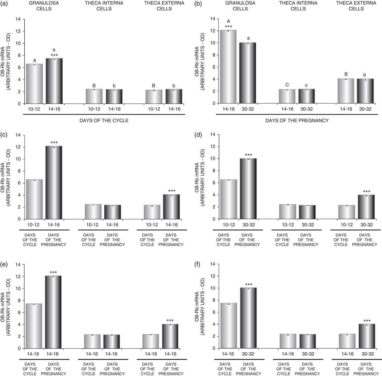
In GC, OB-Rb gene expression was higher (P < 0.001) on days 14 to 16 and 30 to 32 of pregnancy compared with the mid- and late-luteal phases of the cycle. Similarly, comparison of the OB-Rb transcript content in TEC between the two stages of the pregnancy and the cycle showed higher expression (P < 0.001) during pregnancy. However, no differences were found in TIC between days of the cycle and pregnancy (Figure 4c to f).
Discussion
In this study, we have shown that leptin and OB-Rb genes are present in porcine ovarian follicles. We compared the luteal phase of the cycle with early pregnancy to show how the expressions of leptin and OB-Rb genes differ in porcine GC and theca cells in cyclic and pregnant pigs. Leptin gene expression in GC was significantly lower on days 30 to 32 of pregnancy than that on days 10 to 12 and 14 to 16 of the luteal phase. The content of leptin transcript was higher on days 14 to 16 of pregnancy than that on days 14 to 16 of the cycle. The expression of the OB-Rb gene in GC and TEC was higher on days 14 to 16 and 30 to 32 of pregnancy than in the luteal phase of the cycle. In addition, during the luteal phase of the cycle and the two periods of pregnancy, the expression of leptin and OB-Rb genes was higher in GC than that in TIC and TEC. To our knowledge, this is the first report to show in situ leptin and OB-Rb expression in the GC and theca cells of mature pigs.
The role of ovarian follicles during pregnancy is controversial. On the one hand, a study has shown that removal of the follicles did not alter the duration of gestation and litter size in pigs (Nara et al., Reference Nara, Darmadja and First1981). On the other hand, in later studies by Duda et al. (Reference Duda, Burek, Galas, Kozioł, Koziorowski and Słomczyńska2004) and Knapczyk et al. (Reference Knapczyk, Duda, Durlej, Galas, Koziorowski and Slomczynska2008), it has been reported that estrogen receptor α and androgen receptor proteins are present in the porcine GC on days 10, 18, 32, 71 and 90 post coitum, estrogens and androgens are essential for the maintenance of a pregnancy and that ovarian follicles are important during pregnancy.
We have previously shown that, during the luteal phase and early pregnancy in the porcine CL and OS, leptin mRNA and protein (Smolinska et al., Reference Smolinska, Kaminski, Siawrys and Przala2010), and OB-Rb mRNA and protein are expressed (Smolinska et al., Reference Smolinska, Kaminski, Siawrys and Przala2007a), as is OB-Ra mRNA (Bogacka et al., Reference Bogacka, Przała, Siawrys, Kaminski and Smolinska2006). This study, our previous findings and the results obtained in other species support the conclusion that the ovary is an important source of leptin, that the hormone may act within the ovary as an autocrine factor or a paracrine factor or both and that leptin plays a direct role in the regulation of ovarian functions. Leptin mRNA and protein have been identified in humans in GC and theca cells, CL, oocytes and follicular fluid (Antczak and Van Blerkom, Reference Antczak and Van Blerkom1997; Cioffi et al., Reference Cioffi, Van Blerkom, Antczak, Shafer, Wittmer and Snodgrass1997; Loffler et al., Reference Loffler, Aust, Kohler and Spanel-Borowski2001; Welt et al., Reference Welt, Schneyer, Heist and Mantzoros2003); in mice in GC and theca cells, CL, oocytes and OS (Antczak and Van Blerkom, Reference Antczak and Van Blerkom1997; Ryan et al., Reference Ryan, Woodhouse, Van Der Hoeck, Gilchrist, Armstrong and Norman2002); and in rats in theca cells, CL and oocytes (Archanco et al., Reference Archanco, Muruzabal, Llopiz, Garayoa, Gómez-Ambrosi, Frühbeck and Burrell2003; Ryan et al., Reference Ryan, Van der Hoek, Robertson and Norman2003). OB-Rb gene and protein expression has been found in human GC and theca cells (Cioffi et al., Reference Cioffi, Van Blerkom, Antczak, Shafer, Wittmer and Snodgrass1997; Karlsson et al., Reference Karlsson, Lindell, Svensson, Bergh, Lind, Billig, Carlsson and Carlsson1997; Agarwal et al., Reference Agarwal, Vogel, Weitsman and Magoffin1999); mouse follicular cells, CL, OS and oocytes (Ryan et al., Reference Ryan, Woodhouse, Van Der Hoeck, Gilchrist, Armstrong and Norman2002); rabbit and rat follicular cells, CL and oocytes (Zamorano et al., Reference Zamorano, Mahesh, De Sevilla, Chorich, Bhat and Brann1997; Ryan et al., Reference Ryan, Van der Hoek, Robertson and Norman2003); and porcine GC and theca cells harvested from prepubertal gilts, CL and OS (Ruiz-Cortes et al., Reference Ruiz-Cortes, Men, Palin, Downey, Lacroix and Murphy2000 and Reference Ruiz-Cortes, Martel-Kennes, Gévry, Downey, Palin and Murphy2003; Smolinska et al., Reference Smolinska, Kaminski, Siawrys and Przala2007a).
Our present findings lend further weight to the observation that the expression of leptin and leptin receptor genes depends on the stage of the cycle or pregnancy. These cycle- and pregnancy-related differences might be linked to the hormonal milieu of animals; they suggest an important role for steroid hormones in the regulation of gene expression. The leptin concentration in blood increased during the luteal phase in women (Lukaszuk et al., Reference Lukaszuk, Liss, Kusiak and Wojcikowski1998). Leptin mRNA expression in GC (present paper) and in OS (Smolinska et al., Reference Smolinska, Kaminski, Siawrys and Przala2010) has been shown to be higher on days 10 to 12 than that on days 14 to 16 of the cycle. OB-Rb protein expression in OS is also higher on days 10 to 12 than days 14 to 16 (Smolinska et al., Reference Smolinska, Kaminski, Siawrys and Przala2007a). Similarly, Ruiz-Cortes et al. (Reference Ruiz-Cortes, Men, Palin, Downey, Lacroix and Murphy2000) demonstrated that both gene and protein expression of the leptin receptor peaked in porcine CL during the mid-luteal phase of the cycle, and was the lowest in regressed CL. Furthermore, leptin receptor mRNA increased as the porcine GC luteinized during 96 h in culture. Moreover, OB-Rb gene expression increased during follicular development and in CL of mature porcine ovaries compared with prepubertal ovaries (Gregoraszczuk et al., Reference Gregoraszczuk, Ptak, Wojciechowicz and Nowak2007). These results suggest that the expression of leptin and of the leptin receptor correlates with the maximal progesterone content. Consistent with these results, it has been reported that the content of the leptin receptor in rat ovaries is the highest during the stages of the estrous cycle with the maximum progesterone concentration (estrus and diestrus I) and that it decreased significantly after these stages (Duggal et al., Reference Duggal, Weitsman, Magoffin and Norman2002a). Furthermore, the number of luteal cells containing leptin protein increased as the CL matured and decreased during regression of the CL (Archanco et al., Reference Archanco, Muruzabal, Llopiz, Garayoa, Gómez-Ambrosi, Frühbeck and Burrell2003). In our study, however, OB-Rb mRNA expression in GC was higher during the late-luteal phase than on days 10 to 12 of the cycle. Our previous research has also shown that this is true for leptin gene and protein expression in the CL (Smolinska et al., Reference Smolinska, Kaminski, Siawrys and Przala2010). In addition, ovarian leptin protein levels in rats increased from estrus to metestrus and decreased in diestrus (Archanco et al., Reference Archanco, Muruzabal, Llopiz, Garayoa, Gómez-Ambrosi, Frühbeck and Burrell2003).
The expression of both leptin and the leptin receptor in uterine, trophoblast and placental tissues in humans, rodents and pigs (Hoggard et al., Reference Hoggard, Hunter, Duncan, Williams, Trayhurn and Mercer1997; Masuzaki et al., Reference Masuzaki, Ogawa, Sagawa, Hosoda, Matsumoto, Mise, Nishimura, Yoshimasa, Tanaka, Mori and Nakao1997; Zamorano et al., Reference Zamorano, Mahesh, De Sevilla, Chorich, Bhat and Brann1997; Gonzalez et al., Reference Gonzalez, Caballero-Campo, Jasper, Mercader, Devoto, Pellicer and Simon2000; Bogacka et al., Reference Bogacka, Przała, Siawrys, Kaminski and Smolinska2006; Smolinska et al., Reference Smolinska, Siawrys, Kaminski and Przala2007b and Reference Smolinska, Kaminski, Siawrys and Przala2009) and the elevation in leptin levels in pregnant humans and rats (Amico et al., Reference Amico, Thomas, Crowley and Burmeister1998; Helland et al., Reference Helland, Reseland, Saugstad and Drevon1998) suggest that leptin may play a role in pregnancy. It has been suggested that leptin may be essential in embryo development before implantation and in the apposition and adhesion phases of implantation (Castellucci et al., Reference Castellucci, De Matteis, Meisser, Cancello, Monsurro, Islami, Sarzani, Marzioni, Cinti and Bischof2000; Craig et al., Reference Craig, Zhu, Dyce, Wen and Li2005). The expression of OB-Rb mRNA in GC and TEC (present study) as well as in CL and OS (Smolinska et al., Reference Smolinska, Kaminski, Siawrys and Przala2007a) was greater during pregnancy compared with the cycle, and the expressions of leptin and OB-Rb genes were higher in GC in comparison with TIC and TEC on days 14 to 16 and 30 to 32 of pregnancy (present study). The latter may imply that estrogens increased and androgens decreased this expression. These results may also suggest that the leptin system existing in GC, TEC, CL and OS plays an important role in controlling pregnancy. The lack of differences in the expression of leptin and OB-Rb in TIC during pregnancy and between the cycle and pregnancy seems to indicate that the leptin system existing in TIC is not involved in the regulation of pregnancy. The differences in both leptin (present study, Smolinska et al., Reference Smolinska, Siawrys, Kaminski and Przala2007b and Reference Smolinska, Kaminski, Siawrys and Przala2010) and OB-Rb (present study, Smolinska et al., Reference Smolinska, Kaminski, Siawrys and Przala2007a and Reference Smolinska, Kaminski, Siawrys and Przala2009) gene and protein expression in the reproductive organs of pregnant pigs on days 14 to 32 of pregnancy, a period in which implantation and embryo development take place, may indicate that leptin plays a role in the control of these processes.
One of the ways in which leptin influences the implantation process is by participation in steroidogenesis control. However, the action of leptin on ovarian steroidogenesis is controversial. It has been reported that leptin affects the secretion of steroids in a dose-dependent manner. In cultured human GC, low doses of leptin increased both basal estradiol (1 and 10 ng/ml) and progesterone (10 ng/ml) secretion, but high doses inhibited estradiol (40 to 100 ng/ml) and progesterone (60 to 100 ng/ml) secretion (Tsai et al., Reference Tsai, Yang, Chen, Liu, Chen, Hsu and Lee2002; Karamouti et al., Reference Karamouti, Kollia, Kallitsaris, Vamvakopoulos, Kollios and Messinis2009). Similarly, in porcine GC, leptin at physiological doses (10 ng/ml) increased the activity of steroidogenic acute regulatory protein and the accumulation of progesterone, whereas a high dose (1000 ng/ml) inhibited them (Ruiz-Cortes et al., Reference Ruiz-Cortes, Martel-Kennes, Gévry, Downey, Palin and Murphy2003). On the other hand, in mice, at a high concentration (200 ng/ml), leptin significantly stimulated follicular estradiol, progesterone and testosterone production (Swain et al., Reference Swain, Dunn, McConnell, Gonzalez-Martinez and Smith2004). On the basis of these results, it seems that leptin affects steroid secretion in a species-related manner. Moreover, it has been found that leptin impaired the insulin-induced secretion of progesterone and estradiol by bovine GC (Spicer and Francisco, Reference Spicer and Francisco1997) and the insulin-stimulated release of progesterone and androstenedione by theca cells (Spicer and Francisco, Reference Spicer and Francisco1998). In rats, the hormone inhibits the augmentation of IGF-I (insulin-like growth factor I) in FSH-induced estradiol secretion by GC (Zachow and Magoffin, Reference Zachow and Magoffin1997). Consistent with these findings, it has been demonstrated that leptin suppresses LH-stimulated estradiol (Karlsson et al., Reference Karlsson, Lindell, Svensson, Bergh, Lind, Billig, Carlsson and Carlsson1997) and hCG (human chorionic gonadotropin)-induced progesterone (Brannian et al., Reference Brannian, Zhao and McElroy1999) production in human GC. Agarwal et al. (Reference Agarwal, Vogel, Weitsman and Magoffin1999) reported an inhibitory effect of this hormone on combined IGF-I and FSH-stimulated estradiol production by human GC, and IGF-I- and LH-induced androstenedione secretion by cultured theca cells. In addition, in porcine preovulatory follicles, leptin attenuated basal and IGF-I- and growth hormone (GH)-stimulated estradiol production (Gregoraszczuk et al., Reference Gregoraszczuk, Wojtowicz, Ptak and Nowak2003 and Reference Gregoraszczuk, Ptak, Wojtowicz, Gorska and Nowak2004). However, other studies showed that leptin increased estrogen secretion in cultured human GC (Kitawaki et al., Reference Kitawaki, Kusuki, Koshiba, Tsukamoto and Honjo1999), and basal and IGF-I- and GH-stimulated progesterone release by porcine preovulatory follicles (Gregoraszczuk et al., Reference Gregoraszczuk, Wojtowicz, Ptak and Nowak2003 and Reference Gregoraszczuk, Ptak, Wojtowicz, Gorska and Nowak2004). Furthermore, some results have indicated that leptin has no effect on the synthesis of estradiol and progesterone in human (Karlsson et al., Reference Karlsson, Lindell, Svensson, Bergh, Lind, Billig, Carlsson and Carlsson1997; Agarwal et al., Reference Agarwal, Vogel, Weitsman and Magoffin1999; Brannian et al., Reference Brannian, Zhao and McElroy1999), rat (Zachow and Magoffin, Reference Zachow and Magoffin1997; Duggal et al., Reference Duggal, Ryan, Van der Hoek, Ritter, Armstrong, Magoffin and Norman2002b) and bovine (Spicer and Francisco, Reference Spicer and Francisco1997) GC. Therefore, the effect of leptin on ovarian follicle steroidogenesis remains to be determined.
In summary, our paper represents the first ever presentation of gene expression of leptin and its receptor in porcine GC, TIC and TEC during the luteal phase of the estrous cycle and early pregnancy. This study lends further weight to the observation that not only CL and OS but also follicular cells are an important source of leptin and leptin may directly affect porcine follicles. These data imply that leptin affects ovarian follicle functions. In addition, our present findings confirm that the expression of leptin and OB-Rb in porcine GC, TIC and TEC depends on the stage of the cycle and pregnancy, and these differences might be linked to the hormonal milieu of animals, including the intrafollicular milieu. These results suggest the role of the leptin system existing in GC and TEC in the control of pregnancy. Moreover, these findings are in agreement with earlier suggestions indicating that locally synthesized leptin may affect the reproductive functions of pigs.
Acknowledgments
This research was supported by the State Committee for Scientific Research (Project nos. PBZ KBN-084/P06/2002JP, 0206.911 and 0206.0805) and The European Social Fund.