Introduction
Nasopharyngeal carcinoma is an epithelial neoplasm known for its marked invasive and metastatic potential. It is more common in men, with an age standardised male to female ratio of 2–3:1. External beam radiotherapy, with a dose of 60–70 Gy, is the core treatment for locoregional disease.Reference Chew, Kerr and Hibbert1–Reference Wei, Ho, Yuen, Fung, Sham and Lam4 Although nasopharyngeal carcinoma is radiosensitive, local residual or recurrent disease is not uncommon. Post-irradiation local residual disease has been observed in 8–13 per cent of patients, with a cumulative local failure rate of 21–24 per cent over a five-year surveillance period.Reference Lee, Law, Foo, Poon, Chan and O5, Reference Teo, Yu, Lee, Leung, Kwan and Yu6
Early detection of residual or recurrent disease is important in order to achieve better control and survival for nasopharyngeal carcinoma patients. Therefore, regular follow up is crucial. Follow-up measures include clinical examination, radiological imaging and histopathological studies.
Clinical examination can be performed using a postnasal mirror or a fibre-optic endoscope. Such mirror examination is technically challenging, requires the patient's cooperation and commonly misses early local failures. Thorough examination of the nasopharynx with a fibre-optic endoscope is currently considered the most efficient clinical tool.Reference Chew, Kerr and Hibbert1
Imaging techniques include computed tomography (CT), magnetic resonance imaging (MRI), positron emission tomography and single photon emission CT, with standard CT being the current first-line imaging modality of choice. Each technique has its own limitations in detecting residual and recurrent nasopharyngeal disease. Postradiotherapy changes affect the credibility of CT and MRI results. Computed tomography may not be able to differentiate between local failures and postradiotherapy changes, except in the presence of bony erosions. Magnetic resonance imaging shows an overlap of signal intensity between, on the one hand, recurrences and residuals and, on the other, oedema, inflammation and immature fibrosis after radiotherapy.Reference Gong, Zheng and Zhu7, Reference Olmi, Fallai, Colagrande and Giannardi8 The disadvantage of positron emission tomography is that it is optimally undertaken six months or more after radiotherapy.Reference Kao, ChangLai, Chieng, Yen and Yen9, Reference Peng, Yen, Liu, Tsay and Liu10 Single photon emission CT is currently under investigation, with preliminary results suggesting its superiority to standard CT in detecting local residual and recurrent nasopharyngeal carcinoma after radiotherapy.Reference Tai, Shiau, Wang, Ho, Ho and Kao11
Histopathological examination, on the other hand, is the backbone of nasopharyngeal carcinoma detection. Clinicians tend to take multiple biopsies, motivated by anxiety over inefficient clinical and radiological examination. This is expensive, invasive and time-consuming.
Therefore, we conducted a prospective study (1) to evaluate and compare the efficacies of nasopharyngeal endoscopy and CT in the diagnosis of local failures following external beam radiotherapy for nasopharyngeal carcinoma, and (2) to assess whether multiple nasopharyngeal biopsies were superior to a single, targeted biopsy, for the same purpose.
Patients and methods
Over a five-year period, all patients diagnosed with undifferentiated nasopharyngeal carcinoma and selected to receive treatment with external beam radiation therapy (with or without adjuvant chemotherapy) were prospectively invited to take part in the study.
The exclusion criteria included distant metastases, pregnancy, significant psychological problems, withholding of consent, inability to comply with study protocol, children under 18 years of age, systemic diseases affecting the nose, systemic diseases preventing participation in the study, and medical and/or surgical treatments influencing the study. Patients who declined any of the three main procedures – CT, nasopharyngeal endoscopy and nasopharyngeal biopsy – were also excluded.
Forty-six patients (35 men and 11 women), with a mean age of 52 ± 11 years (range 27–71) met the inclusion and exclusion criteria and were enrolled in the study. At presentation, before radiotherapy, eight (17.3 per cent) patients had stage I disease, 12 (26.1 per cent) had stage II disease, 17 (37 per cent) had stage III disease and nine (19.6 per cent) had stage IV disease, according to the staging criteria of the American Joint Committee on Cancer.Reference Fleming, Cooper, Henson and Hutter12 For every patient recruited, CT, nasopharyngeal endoscopy and nasopharyngeal biopsy were performed 12 weeks post-treatment. The end of treatment was considered to be the last day of radiotherapy.
Treatment regimen
All patients were treated with external beam radiotherapy. The treated volume encompassed the nasopharynx, adjacent parapharyngeal tissues with adequate margin (1–2 cm), and all of the cervical lymphatics. The treated volume was delivered through two lateral opposing fields and a lower anterior neck field with or without a supplementary anterior field with anterior extension only. The primary tumour and positive lymph nodes received a total dose of 65–75 Gy. The dose per fraction was 180 cGy, with five daily fractions given per week. Negative lymph nodes were irradiated to a minimal dose of 45–50 Gy. Doses to critical normal tissues were not exceeded. Chemotherapy was administered to some patients with stage III or stage IV disease. The chemotherapy regimen consisted of intravenous cisplatin 100 mg/m2 on day one and intravenous 5-fluorouracil 1 g/m2 from day one to day five, every four weeks for four to six cycles.
Computed tomography
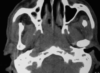
Computed tomography was performed in the axial and coronal planes with intravenous injection of 80 ml of non-ionic contrast medium. Axial sections, of a thickness of 5 mm, were taken from skull base to manubrium. The CT images were reviewed by an experienced radiologist blinded to the endoscopic and biopsy findings. The diagnostic criterion for a positive CT was the presence of a mass or an asymmetry in the nasopharynx. Changes beyond this were regarded as expected postradiotherapy changes. Figure 2 shows an axial CT with a persistent nasopharyngeal mass 12 weeks after radiotheraphy.
Nasopharyngeal endoscopy
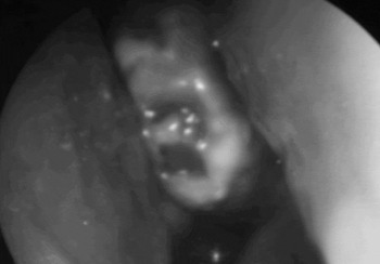
Before endoscopy, the nasal cavities were anaesthetised with 10 per cent xylocaine for 10 minutes. Endoscopy was performed using a rigid endoscope (Karl Storz, Germany; 0° and 30° if needed, 4 mm in diameter). Illumination was provided by a 150-W xenon light source. The nasopharynx was judged to be normal if it appeared to be smooth, and suspicious if masses, ulcers, submucosal bulges or irregular mucosa were seen. In all cases, the endoscopic results were interpreted in the presence of the most senior author. The otolaryngologists were blinded to the radiological findings at the time of examination. Figure 1 shows nasopharyngeal endoscopic view of a nasopharyngeal ulcer 12 weeks after radiotheraphy.
Nasopharyngeal endoscopic view 12 weeks after radiotherapy, showing a nasopharyngeal ulcer.

Axial computed tomography scan taken 12 weeks after radiotherapy, showing a persistent nasopharyngeal mass.

Nasopharyngeal biopsy
The first biopsy was taken under endoscopic control, being targeted at the most suspicious area of the nasopharynx. Further biopsies were taken from the right and left fossa of Rosenmüller, and from the roof and the posterior wall of the nasopharynx. The pathologist was blinded to the endoscopic and CT findings at the time of assessment.
Statistical analysis
Data were expressed in terms of absolute values, percentages and means ± standard deviations, as indicated. The sensitivity, specificity, positive predictive value and negative predictive value were calculated for rigid endoscopy, CT and targeted biopsy, and compared with results for histological examination of multiple biopsy specimens. The Kappa reliability test was employed to evaluate the degree of agreement between each follow-up tool and the histological examination of multiple biopsy specimens. A value of p < 0.05 was considered significant. To improve the consistency of results and to eliminate interobserver discrepancy, all the histological and radiological slides were analysed by the same pathologist and radiologist, respectively. In addition, all endoscopic examinations were performed in the presence of the most senior otolaryngologist. The interpretation of the results of each follow-up team was unknown to the others at the time of assessment.
Results
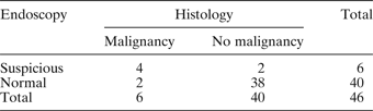
In the 12-week follow-up period after radiotherapy, six patients (13 per cent) were discovered to have persistent or recurrent nasopharyngeal malignancy on histological examination. The nasopharyngeal endoscopic appearance was normal in 40 patients (86.9 per cent), of whom two were discovered to have persistent or recurrent malignancy on examination of nasopharyngeal biopsies. Six patients had a suspicious endoscopic appearance; of these, only four were found to have evidence of malignancy. The sensitivity, specificity, positive predictive value and negative predictive value of nasopharyngeal follow-up endoscopy were 66.6, 95, 66.6 and 95 per cent, respectively. There was statistically significant agreement between the endoscopic and histological findings (Kappa reliability coefficient = 0.617, p < 0.01). Table I shows the nasopharyngeal endoscopic findings versus the histological findings.
Patients' nasopharyngeal endoscopic vs histological findings

Data represent number of patients.
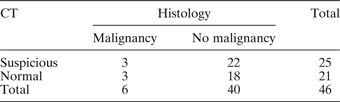
Computed tomography results were suspicious in 25 (54.3 per cent) patients, of whom three had histological evidence of disease. Of the 21 patients with normal CT scans, 18 were histologically disease-free. The sensitivity, specificity, positive predictive value and negative predictive value of CT were 50, 45, 12 and 85.7 per cent, respectively. There was no statistically significant agreement between the CT and histological findings (Kappa reliability coefficient = 0.021, p > 0.05). Table II shows the CT findings versus the histological findings.
Patients' CT vs histological findings

Data represent number of patients. CT = computed tomography
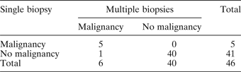
Findings for a single, targeted endoscopic biopsy failed to match those for multiple endoscopic biopsies in only one out of the 46 cases (2 per cent). The sensitivity, specificity, positive predictive value and negative predictive value of single, targeted endoscopic biopsy were 83.3, 100, 100 and 97.5 per cent, respectively. The Kappa test showed a highly statistically significant agreement between the histological findings for single and multiple endoscopic biopsies (Kappa reliability coefficient = 0.897, p < 0.001). Table III shows the findings for single, targeted endoscopic biopsy versus those for multiple biopsy specimens.
Patients' results for single, targeted biopsy vs multiple biopsies

Data represent number of patients.
Discussion
Salvage treatments are commonly used to manage local failures following primary radiotherapy for nasopharyngeal carcinoma, in the form of surgery or re-irradiation. Patients with limited local treatment failure are often salvaged by brachytherapy or nasopharyngectomy. Patients with more extensive local failures often require external re-irradiation. A significant proportion of these patients can still achieve long-term disease control (up to 87 per cent) and survival (up to 85 per cent).Reference Chua, Sham, Kwong, Wei, Au and Choy13–Reference Leung, Tung, Sze, Sze, Wong and O17 The prognosis for patients with local failure depends on the extent of disease at the time of detection.Reference Leung, Tung, Sze, Sze, Wong and O17–Reference Wei23 This highlights the importance of early detection and management of local residual and recurrent nasopharyngeal carcinoma, following irradiation.
After treatment of nasopharyngeal carcinoma, follow up includes clinical, radiological and histological monitoring. Flexible and rigid fibre-optic endoscopes are the currently accepted clinical tools for examining the nasopharynx. In our study, rigid endoscopy was chosen as our clinical tool. Its sensitivity and specificity for prediction of local failures were 66.6 and 95 per cent, respectively, and its positive and negative predictive values were 66.6 and 95 per cent, respectively. The excellent specificity and negative predictive value imply that if endoscopic examination is normal, the possibility of detecting residual or recurrent disease in histological biopsies is very low. Therefore, it would be unnecessary to pursue further radiological or histological examination. This will reduce patient discomfort, save resources and decrease associated morbidity.
Using flexible endoscopy to detect local failures after nasopharyngeal carcinoma treatment, Kwong et al. reported a low sensitivity (40.4 per cent) and a low positive predictive value (16.3 per cent), and a specificity and negative predictive value of 84.4 and 95 per cent, respectively.Reference Kwong, Nicholls, Wei, Chua, Sham and Yuen24 Our results were comparatively better, which could be explained by the following.
Firstly, Kwong and colleagues' study had all the inherent problems of retrospectivity, including very wide variation in the time period between end of radiotherapy and endoscopic examination, which ranged between four and 16 weeks. Secondly, the outcome of Kwong and colleagues' study depended on the six-week endoscopic examination, although interpretation of endoscopic findings is very difficult at this time due to radiation-induced mucosal changes; this is clearly shown by the very low sensitivity and positive predictive value, in contrast with the specificity and negative predictive value. Thirdly, we used rigid endoscopy, which gives better illumination, a wider field, excellent resolution and a sharper image compared with flexible endoscopy; these properties assist detection of subtle residual and recurrent disease.
Computed tomography results showed lower indices and efficacy than our endoscopic results in detecting local failures of nasopharyngeal carcinoma treatment. Radiotherapy induces a variety of post-treatment changes, such as oedema, loss of tissue planes, fibrosis and scarring, which may interfere with the detection of recurrent or residual nasopharyngeal carcinoma. Computed tomography cannot differentiate between inflammatory tissue, post-radiotherapy fibrosis, and recurrent or residual tumour, and thus cannot reliably indicate the presence or absence of recurrent or residual nasopharyngeal carcinoma.Reference Ahmad and Stefani25–Reference Chong and Fan29 In addition, a diagnosis of local treatment failure based on nasopharyngeal asymmetry is often difficult, since identification of the point at which asymmetry is considered to be beyond the expected postradiotherapy changes is totally subjective. Asymmetry of the nasopharyngeal mucosal outline with straight margins suggests a benign lesion, whereas a lobulated appearance suggests malignancy.Reference Chong and Fan29 On the other hand, it is not ethical to repeat CT scanning during every follow-up visit, since this exposes patients to significant radiation hazards. It should be mentioned here that although the sensitivity (64 per cent) and specificity (96 per cent) of single photon emission CT in detecting local residual and recurrent nasopharyngeal carcinoma have recently been reported as superior to corresponding values for CT,Reference Tai, Shiau, Wang, Ho, Ho and Kao11 these reported results are very comparable to the sensitivity (66.6 per cent) and specificity (95 per cent) of our endoscopic results. There is no doubt about the importance of imaging in identifying the anatomical plane(s) affected by nasopharyngeal carcinoma. However, its use as a follow-up tool should be reserved for patients with histological or symptomatic indications, rather than being routine.
• This prospective, controlled study evaluated different methods of detecting local failures following nasopharyngeal cancer treatment
• Rigid nasopharyngeal endoscopy should be considered the primary follow-up tool
• Computed tomography should be reserved for patients with histological or symptomatic indications
• A single, targeted endoscopic biopsy represents an excellent alternative to the standard multiple biopsies, and also reduces cost, time, morbidity and patient discomfort
Histological examination is the ‘gold standard’ methodology in detecting residual and recurrent nasopharyngeal carcinoma. The currently accepted practice is to obtain multiple biopsies, irrespective of whether residual tumour is observed during endoscopic examination.Reference Kwong, Nicholls, Wei, Chua, Sham and Yuen24 In our study, results for 12-week single, targeted endoscopic biopsy showed excellent reliability and efficacy when compared with those for multiple biopsies. The former approach was also cost-effective, less invasive and time-sparing.
Taking biopsies earlier than 12 weeks post-treatment has been shown to result in a high rate of false positive results. Kwong et al. reported that 66.5 per cent of patients with positive histological findings in the sixth week after radiotherapy achieved spontaneous remission over time and had negative histological findings in subsequent biopsies.Reference Kwong, Nicholls, Wei, Chua, Sham and Yuen24 These authors also found that 95.4 per cent of patients with positive histological findings who achieved spontaneous histological remission had done so by the end of the 10th week.
Conclusions
Rigid nasopharyngeal endoscopy should be considered the primary follow-up tool for monitoring nasopharyngeal carcinoma patients after radiotherapy, with CT being reserved for patients with histological or symptomatic indications. Routine postnasal biopsies are not necessary, considering the excellent specificity and negative predictive value of rigid nasopharyngeal endoscopy. A single, targeted endoscopic biopsy provides an excellent alternative to the standard multiple biopsies, and also reduces cost, time, morbidity and patient discomfort.