INTRODUCTION
With the advancement of new chemotherapy drugs and radiotherapy techniques, the long-term survival of young female cancer patients has dramatically increased.Reference Dickman and Adami 1 The major side effects of these treatments frequently include ovarian failure and infertility. Whole pelvic radiotherapy in particular can result in ovarian atrophyReference Mundt, Lujan and Rotmensch 2 and reduced follicle stores; also known as early menopause.Reference Dryden, Ussher and Perz 3 Several cancers that afflict pre-menopausal women, where pelvic radiotherapy occupies an important role in treatment, include cervical, vaginal and anorectal carcinomas, dysgerminoma, Hodgkin’s disease and central nervous system tumours.Reference Mundt, Lujan and Rotmensch 2 For younger women with pelvic cancer, the resulting infertility can cause significant psychological distress.Reference Greil, Slauson-Blevins and McQuillan 4 Doses of 5–10 Gy are commonly accepted as a sterilising dose for a woman in her thirties with a 1992 paperReference Chambers, Chambers, Kier and Peschel 5 reporting a steep dose response effect at 3 Gy for induction of early menopause. A more recently published paperReference Wallace, Thomson, Saran and Kelsley 6 produced a model which calculated that a dose of 2 Gy is sufficient to bring about ovarian failure. The evidence does, therefore, suggest that the dose limit for ovarian failure differs in each patient. There is also a range of responses that are age-dependent with lowest risks seen in the under-25 age group.
Traditionally ‘ovarian transposition’, in which one or both ovaries are surgically fixed outside the irradiated area (typically in the lateral abdomen) is used to reduce dose to the ovaries.Reference Mazonakis, Damilakis, Varveris and Gourtsoyiannis 7 The advent of intensity-modulated radiotherapy (IMRT), however, suggests the possibility of shaping dose around the ovaries in situ and therefore sparing dose without the need for surgical intervention. The current evidence does not seem to provide a clear answer as to which of these techniques will provide optimal outcomes for the patient; accordingly, this review aims to identify the most effective intervention in terms of ovarian function preservation. The review will compare and contrast the impact of ovarian transposition with the use of modern radiotherapy techniques.
METHODS
A literature search was performed to identify original and review articles related to ovarian function preservation techniques for pelvic radiotherapy. Case reports, editorials and letters were excluded. Articles were sourced from the main medical databases, comprising MedLine, the Science Citation Index, Scopus, ScienceDirect and CINAHL Plus. Search terms utilised were ‘ovary’, ‘ovarian’, ‘transposition’, ‘radiotherapy’ and ‘pelvic IMRT’. Combinations of these terms using Boolean operators enabled the search to be focussed more clearly within the desired topics. Abstract analysis of the retrieved studies was performed and articles that were irrelevant or written in languages other than English were excluded. Further evidence and additional potential search terms were sourced from retrieved papers. A quality assessment of the papers collected was performed using the well-validated Critical Appraisal Skills Programme (CASP) tool in order to ensure only high-quality evidence was utilised. Articles were to be rejected if they fell too far below the relevant CASP quality criteria for the evidence type relating to validity of results.
RESULTS
The search and appraisal process yielded 19 relevant articles with valid results pertinent to the research topic. Thematic analysis of abstracts enabled these to be grouped into three topics: ‘Ovarian transposition’, ‘IMRT’ and ‘Emerging technologies’.
Ovarian transposition
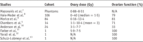
A range of evidence relating to ovarian transposition included phantom studies, single case studies and some dated clinical studies as seen in the Table 1. Two of these studiesReference Mazonakis, Damilakis, Varveris and Gourtsoyiannis 7 , Reference Haie-Meder, Mlika-Cabanne and Michel 8 used assumed or estimated positions for ovarian transposition to estimate dose. In addition some dated techniques were used including a four-field pelvic ‘brick’ and lead block shielding; modern advances including multi-leaf collimation and conformal planning may have yielded different results. Nonetheless, these studies do indicate useful prediction of the ovarian dose. Haie-Meder’s studyReference Haie-Meder, Mlika-Cabanne and Michel 8 was also affected by a wide range of doses and patients including cobalt and brachytherapy treatments. Measurements were taken using thermoluminescent dosimeters (TLDs) on the patient skin at an estimated position. Despite this, ovarian functions resumed in 79% of the patients (n=106). They concluded rather obviously that transposed ovary dose depended on the location of the ovaries but also, interestingly, that field arrangements did not affect this.
Ovarian transposition summary of evidence

Similar results with ovarian function preservation in 81% of patients (n=84) was seen in a studyReference Morice, Juncker, Rey, El-Hassan, Haie-Meder and Castaigne 9 where the ovaries were transposed bilaterally to the paracolic gutters. The dose to the ovaries in this study ranged from 0·16–13·4 Gy. Dose calculations in this study were also measured with TLDs, but there was also a good functional assessment including a routine postoperative ultrasound scan and measurement of gonadotropin and E2 levels. It was noted that 11 patients experienced menopause at a ‘physiologic age’ (>45) and therefore it was considered to be independent of the mode of treatment or their cancer. This study concluded that ovarian transposition is a safe and effective procedure for the preservation of ovarian function in patients ≤40 years old treated for cervical carcinoma by radiosurgical combination.
Site of transposition was assessed in a studyReference Chambers, Chambers, Kier and Peschel 5 into 38 International Federation of Gynaecology and Obstetrics (FIGO) Stage 1 cervical cancer patients who underwent ovarian transposition as part of their initial operative procedure. Fourteen of these patients subsequently received pelvic radiotherapy and 71% of these maintained ovarian function. A crucial finding for this study was the importance of the placement of the ovaries, with 100% of the patients developing menopause if the ovaries were placed below the iliac crest.
The remaining small sample size studies have illustrated a range of results. A 1992 studyReference Anderson, LaPolla, Turner, Chapman and Buller 10 found only 33% of patients had retained ovarian function after radiotherapy; it is not clear whether this was due to poor follow-up attendance. Three single case studiesReference Farber, Ames, Rush and Gal 11 – Reference Schulz-Lobmeyr, Schratter-Sehn, Huber and Wenzl 13 showed success, but two of these failed to note the dose received. Interestingly, Farber’s studyReference Farber, Ames, Rush and Gal 11 seemed to contradict earlier observationsReference Chambers, Chambers, Kier and Peschel 5 , Reference Wallace, Thomson, Saran and Kelsley 6 , Reference Haie-Meder, Mlika-Cabanne and Michel 8 that deemed ovarian failure to commence at doses of 5 Gy.
Although the majority of papers relating to transposition studies appear to record moderate-to-good preservation of ovarian function it is important to note that surgical transposition carries some risk including compromised blood vessels, cyst development and possible ovarian failure.Reference Falcone, Attaran, Bedaiwy and Goldburg 14 Despite this, all the data included in the review concluded that ovarian transposition is a safe and effective method used to preserve ovarian function in patients who are at risk of ovarian failure as a result of radiation induced ablation. It should be noted that these results were obtained without access to modern equipment and techniques and subsequent advance in treatment delivery may render the findings obsolete. There are also some methodological concerns about some of the studies with a range of measures and controls used. This has clearly led to a huge range in recorded ovarian doses; larger prospective studies with more rigorous control and well-defined outcome measures would yield more reliable data.
IMRT
Despite the rich evidence base surrounding IMRT use for avoidance of critical structures in treatment of a wide range of tumours, there were relatively few papers reporting its use for ovarian preservation. A 2012 paperReference D’Souza, Rumble and Fyles 15 examined the evidence for IMRT in the treatment of gynaecological cancers to quantify the potential benefits of this new technology and to make recommendations for radiation treatment programmes that considered adopting this technique. Findings were based on a review of four cohort studies including a prospective study; these included a total of 619 patients. Acute gastrointestinal toxicity was significantly reduced by IMRT, but no differences were seen in genitourinary toxicity. In the primary treatment of cervical cancer, the radiation dose (both external beam radiotherapy and brachytherapy) is higher than in the postoperative setting and so the benefit of sparing normal tissue with IMRT would probably be greater. The paper predominantly focusses on the toxicities to the rectum, bladder and small bowel and makes no mention specifically of sparing the gonads. The review did highlight how the use of IMRT for gynaecological cancers is still in its early stages and that there are no reported randomised control trials to guide clinical practice for the use of IMRT for gynaecological cancers.
A comprehensive reviewReference Fiorino, Valdagni, Rancati and Sanguineti 16 of the late and acute toxicity for the organs at risk in pelvic, mainly prostate, cancers considered toxicity to structures other than the bowel, rectum and bladder and recognised the need for established quantitative data relating to preservation of sexual function. Mention was also made of the potential role of IMRT and proton therapy for ovarian function preservation. Two other studiesReference Mundt, Lujan and Rotmensch 2 , Reference Roeske, Lujan, Rotmensch, Waggoner, Yamada and Mundt 17 evaluated the ability of IMRT to reduce the general volume of healthy tissue irradiated in women with gynaecological malignancies receiving whole pelvic radiotherapy. Both studies found a reduction in the volume of small bowel irradiated using the IMRT plans compared with 3D conformal radiotherapy which resulted in excellent planning target volume (PTV) coverage, with considerable sparing of normal tissues. D’Souza’s 2012 paperReference Roeske, Lujan, Rotmensch, Waggoner, Yamada and Mundt 17 noted that IMRT plans led to significant reduction in dose to adjacent critical structures ranging from 26% to 41% and less acute gastrointestinal toxicities than conventional whole pelvic radiotherapy. Despite the paper’s focus on gynaecological tumours there was no consideration of dose to the ovaries, although the results clearly support the use of IMRT for critical structure sparing.
The current literature for using IMRT as a means of preserving ovarian function is evidently sparse, but logically has the potential to play a useful role. IMRT planning for ovarian sparing should certainly include consideration of a International Commission on Radiation Units and Measurements (ICRU) ‘Planning Organ at Risk Volume’ margin (PRV). Like most organs, the ovaries are known to move within the pelvis every day, probably in relation to bladder and rectal distension. A 2012 studyReference Peters, Petterson, Horan, Gregory and Sala 18 assessed the extent of ovarian motion to determine a range of safety volumes to help avoid ovarian ablation and premature menopause. They did acknowledge some key limitations of their study, including small cohort, broad age range of patients (29–79) and wide variety of tumour sites. A more recent paperReference Soda, Ishiyama and Ono 19 suggested that bilateral transposed ovaries require a PRV of ~2 cm in all directions. This study produced IMRT plans with a prescribed dose of 45 Gy in 25 fractions, including an ovarian PRV, which received a maximum dose of 5 Gy. The authors also acknowledged the need for precise contouring and appropriate margins in order to compensate for the steep dose gradients associated with IMRT. Given the proven value of IMRT combined with PRV use for sparing dose to a range of structures including bone marrow and kidneysReference Hong, Alektiar and Chui 20 , Reference Lujan, Mundt, Yamada, Rotmensch and Roeske 21 there is a surprising lack of published evidence relating to in situ ovarian-sparing IMRT.
Emerging technology
Although classed as ‘experimental’ by some authorsReference Oktay and Bedoschi 22 the techniques of oocyte harvesting, cryopreservation and ovarian transplantation may have a role to play in future ovarian preservation. These techniques, however, are expensive and not every female is eligible for these procedures. With the imminent deployment of proton therapy in the United Kingdom, it is interesting to note some evidence supporting proton use for ovarian function preservation. LeeReference Lee, Bilton and Famigliettie 23 compared simple proton plans with 3D-conformal radiotherapy, electron therapy and IMRT for three paediatric disease sites, one of which included pelvic sarcoma. The results demonstrated a significant reduction in dose to the ovary with protons for the two female patients. At all dose levels (‘≥2 Gy’, ‘5 Gy’ and ‘10 Gy’), protons resulted in 0% ovarian irradiation compared with IMRT which resulted in 29% (at 5 Gy) and 0% (at 10 Gy). Despite the small sample size of this study, the results did demonstrate positive outcomes regarding ovarian function preservation with the use of protons. The use of intensity-modulated proton beams is expected to further increase the dose-shaping capabilities of proton treatment.
DISCUSSION
It should be acknowledged that many of the papers pertaining to ovarian transposition are over 10 years old with no new work being published since 2006. This might indicate that this method is outdated and has been replaced by more robust techniques in the clinical setting. Alternatively, it might equally be that such is the weight of evidence and confidence in transposition as a technique that no current studies are being pursued; it might also be that new techniques have attracted greater research funding.
In addition to the age of the evidence, it is clear that a variety of outcome measures have been used. A number of tests involving biochemical measures and ovarian imaging, so-called ovarian reserve tests, have been proposed to help predict ovarian reserve and/or reproductive potential. Although ovarian reserve tests have been applied widely, debate continues over the ability of tests currently in use to predict three related, but distinctly different, outcomes: oocyte quality, oocyte quantity and fecundity. 24 Future work should ensure that a range of measures are utilised to enable fair comparison of results.
An emerging theme from these data is that age is an important prognostic factor for transposition with ovarian failure occurring with lower doses of radiation in older patients.Reference Wallace, Thomson, Saran and Kelsley 6 , Reference Haie-Meder, Mlika-Cabanne and Michel 8 The position of the transposed ovary was also an important factor in the success of the procedure, with optimal positions reported to be laterally above the iliac crest or as far away from the radiation field edge as possible. Therefore, these factors should be considered case-by-case for each individual and whether ovarian transposition will overall benefit the patient.
When evaluating the role of emerging technologies for ovarian transposition, it is clear that the expense associated with many of these is a limiting factor. The increasingly widespread role of IMRT in clinical practice, however, does not appear to have extended to include ovarian preservation, and it is likely to present a more cost-effective use of modern technology and techniques. Despite the lack of evidence supporting use of IMRT for preservation of ovarian function, related researchReference Liu, Shiau and Lee 25 has shown that this technology can deliver highly conformal doses to target tissues while avoiding specific volumes. It is clear, therefore, that IMRT, in conjunction with a PRV margin can be used to decrease dose to ovaries without the need for transposition and there is an urgent need for dosimetric and clinical studies to investigate this further. Although beyond the scope of this review, a simple retrospective comparative planning study would easily quantify the potential gains of IMRT and provide guidance as regards margins and techniques.
CONCLUSIONS
In summary, this review documents that the available evidence concerning the preservation of ovarian function by ovarian transposition and new radiotherapy technologies is limited by small sample sizes, heterogeneity among study design, analyses and outcomes, and the lack of validated outcome measures. The evidence supporting ovarian transposition is clear, but seems to have ossified around 10 years ago. The potential value of IMRT combined with a PRV margin for ovarian function preservation has not, as yet, been determined and new studies should aim to investigate this further.
Acknowledgements
None.