I. INTRODUCTION
Despite tremendous advances in treating various musculoskeletal disorders, critical sized bone defects remain a challenge for the orthopedic surgeon. Autologous bone grafts are currently the material of choice for filling or replacing bony lesions; however, these grafts require an additional donor surgery and yield a complication rate of one in five. Reference Dimitriou, Mataliotakis, Angoules, Kanakaris and Giannoudis1 Whereas new intramedullary reamer systems may modestly drop the complication rate to one in fifteen, Reference Dimitriou, Mataliotakis, Angoules, Kanakaris and Giannoudis1 they are still invasive and associated with significant costs. Reference Nodzo, Kaplan, Hohman and Ritter2 In addition to the morbidity associated with both procedures, obtainable quantities of graft materials are limited and can pose a significant challenge for large lesions. The use of allografts over autografts eliminates the donor site morbidity and allows for larger volume harvests, but adds the risks of disease transmission 3 and increased post-operative infection rates. Reference Mankin, Hornicek and Raskin4 For these reasons, the development of graft substitutes that are safe, abundant, and effective is of significant interest.
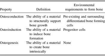
The success of the autologous bone graft seems to be dependent on its osteogenic (inherent ability to produce bone), osteoconductive (mechanical support of bone growth), and osteoinductive (induction of surrounding progenitor cells to produce bone) properties (see Table I). Reference Daculsi, Fellah, Miramond and Durand5,Reference Barradas, Yuan, Blitterswijk and Habibovic6 Graft-substitutes with inherent osteogenic properties (i.e., the ability to self-produce bone) provide an attractive avenue for bone regeneration and have seen encouraging clinical results. Reference Marcacci, Kon, Moukhachev, Lavroukov, Kutepov, Quarto, Mastrogiacomo and Cancedda7 However, osteogenic scaffolds require live cell implantation Reference Blokhuis and Arts8 and this characteristic prohibits the development of a sterile, off-the-shelf product. Alternatively, osteoinductive materials can stimulate bone production while maintaining acellularity by taking advantage of the host cellular network. Reference Miron and Zhang9 Although the exact mechanisms are only beginning to be understood, it is agreed upon that osteoinduction involves the recruitment and subsequent induction of host stem cells to form bone. Reference Urist10,Reference Song, Habibovic, Bao, Hu, Van Blitterswijk, Yuan, Chen and Xu11 In this review, we will examine the properties of graft-substitutes that induce stem cells in this regard.
Osteoconduction, osteoinduction, and osteogenesis.

II. OSTEOINDUCTION
In a series of experiments in the 1960’s, it was shown that bone grafts decalcified through treatment with hydrochloric acid, formic acid, or citric acid were more rapidly and more successfully able to bridge massive bone defects than un-decalcified bone autografts. Reference Urist10 Furthermore, these demineralized grafts promoted complete bone formation inclusive of marrow cavities when implanted subcutaneously or intramuscularly. Reference Urist10 The hunt for factors responsible for these observations led to the identification of bone morphogenetic proteins (BMPs), Reference Urist and Strates12 and the early demonstration of BMP-induced osteogenic differentiation of pericytes by Urist and colleagues Reference Sato and Urist13 prompted scientists to isolate, use, and manufacture BMPs as a means of inducing osteogenesis. Despite these advances, human trials using bone morphogenetic proteins to induce bone formation have shown mixed results, often times using costly supra-physiological doses while achieving minimal clinical effects with significant side effects. Reference Kukreja, Ahmed, Haydel, Nanda and Sin14,Reference Neovius, Lemberger, Skogh, Hilborn and Engstrand15 The risks of recombinant growth factor monotherapy at the current doses have led researchers to investigate other avenues for promoting bone formation. Particularly, materials-based systems have been examined for their abilities to either complement or replace growth factors in achieving osteoinductive effects.
III. OSTEOINDUCTIVE PROPERTIES OF MATERIALS
The observations that inorganic substrates are able to induce bone formation have led to investigations on the osteoinductive properties of these materials. Although the principal mechanisms are largely unknown, it has become increasingly clear that both physical and chemical cues influence a material’s ability to induce bone formation. Materials that have been investigated for these properties include ceramics, glass, metals, polymers, and their composites.
Some of these materials, especially ceramics, contain calcium, and investigations into calcium induced osteogenesis have provided definitive mechanisms for at least part of their osteoinductive effects. In fact, even many non-calcium based materials induce bone formation after a surface hydroxyapatite layer forms in vivo. Reference Winter16 Whether this layer is necessary or coincidentally forms in osteoinductive materials is not definitively known, especially since some non-calcium based materials induce bone formation without this preceding step. Regardless, experiments by Chai et al., 2010 suggest that osteoinduction by calcium phosphates could be attributed to the direct effect of released calcium and phosphate ions on BMP-2 expression. Reference Chai, Roberts, Schrooten and Luyten17 It has been shown that Ca2+ upregulates BMP-2 expression in differentiated osteoblasts, Reference Dvorak, Siddiqua, Ward, Carter, Dallas, Nemeth and Riccardi18 and BMP-2 expression is increased 3-fold in stem cells treated with Ca2+. Reference Barradas, Fernandes, Groen, Chai, Schrooten, van de Peppel, van Leeuwen, van Blitterswijk and de Boer19 Furthermore, González-Vázquez et al., 2014 Reference González-Vázquez, Planell and Engel20 recently blocked the osteoinductive effects of calcium on stem cells with a calcium sensing receptor (CaSR) antibody, suggesting that the CaSR is a key modulator of these cellular responses. While calcium has been described as a simple signaling molecule that can induce bone formation, Reference Cushnie, Ulery, Nelson, Deng, Sethuraman, Doty, Lo, Khan and Laurencin21 the observation of non-calcium based bone induction cannot be explained by ionic release of calcium and its effects on the calcium sensing receptor.
Whether the other non-calcium based osteoinductive materials target chemical receptors or solely rely on macro and microscopic physical properties of their structure is unknown. However, it has become clear that elements can be incorporated in some materials to increase osteoinductivity. For example, experiments have shown that silicon promotes osteoblast activity in vitro, Reference Guth, Buckland and Hing22 and animals fed with silicon-deficient diets have inadequate skeletal maturation with bony malformations compared to the controls. Reference Carlisle23 These observations have led to the hypothesis that silicon substituted ceramics may have even greater osteoinductive potential than pure calcium phosphate materials. Coathup et al., 2011 Reference Coathup, Samizadeh, Fang, Buckland, Hing and Blunn24 compared the osteoinductive potential of silicate substituted calcium phosphate with that of non-substituted calcium phosphate and showed significantly higher bone formation area at ectopic sites with the substituted implants. When applied to the clinic, a recent retrospective clinical study using silicon substituted calcium phosphate ceramics in various spinal arthrodesis procedures showed a fusion rate of 90% on a 12 month follow up. Reference Nagineni, James, Alimi, Hofstetter, Shin, Njoku, Tsiouris and Hartl25
Besides chemical properties, physical characteristics of a material can contribute to both the recruitment and induction of stem cells in bone formation. Extracellular regulated kinase proteins, influenced by RhoA GTPases, have been shown to play a mechanistic role in regulating osteogenic transcription signals that are based on extracellular substrate-mechanics. Reference Khatiwala, Kim, Peyton and Putnam26–Reference Wang, Yu, Cohen, Wozniak, Yang, Gao, Eyckmans and Chen28 Studies have shown that material porosity, Reference Coathup, Hing, Samizadeh, Chan, Fang, Campion, Buckland and Blunn29 stiffness, Reference Du, Chen, Liang, Zhang, Xu, He, Zhan, Feng, Chien and Yang30 adhesive properties, Reference Lee, Abdeen, Zhang and Kilian31 and geometry Reference Kilian, Bugarija, Lahn and Mrksich32 play independent roles in promoting bone formation through these mechanisms.
Perhaps the most notable and potent physical influence on osteoinduction is a material’s porosity. The architecture of pores in materials has been shown to influence vascularity and ultimately bone ingrowth, and both micro- and macro-pores have been shown to promote both osteoconduction and osteoinduction. It is no surprise that these porosities mimic the physiological porosities of cortical and cancellous bones (pores ranging from 1 to 1000 μm). Reference Coathup, Hing, Samizadeh, Chan, Fang, Campion, Buckland and Blunn29 Although deterministic mechanisms have not yet been elucidated, our examination of various materials and their ability to induce bone formation will be closely tied with an examination of their porosities throughout this review. There is evidence that the porous structures permit bone formation by permitting cellular attachment, angiogenesis, and diffusion of nutrients. Reference Hing, Annaz, Saeed, Revell and Buckland33 Furthermore, increasing porosity is intimately tied with increasing the solubility of the surface layer via increased surface area—perhaps also influencing osteoinduction through soluble diffuse chemical signals in addition to mechanical ones.
The layout and topography of these pores can also alter a material’s osteoinductive potential. Reference Kilian, Bugarija, Lahn and Mrksich32 As an example, Dalby et al., 2007 Reference Dalby, Gadegaard, Tare, Andar, Riehle, Herzyk, Wilkinson and Oreffo34 modified the geometrical spacing of micropores into three groups: ordered pattern (square and hexagonal), disordered square, and a random array produced on polymethylmethacrylate substrate by electron beam lithography. They observed that, in the absence of osteogenic medium, cells on random rather than symmetrical nanotopographies exhibited a more osteoblastic morphology. Likewise, topography is intimately related to the surface roughness of a material; the presence of a high number of small features on a surface indicates a greater roughness value. Zhang et al., 2015 Reference Zhang, Barbieri, ten Hoopen, de Bruijn, van Blitterswijk and Yuan35 modified this specific surface characteristic of calcium phosphate implants and found much more ectopic bone formation associated with the rougher ceramics. The development of techniques to generate such custom-designed surface nano/micro-structures has been recently proven effective in maximizing the osteoinductive potential of materials. Reference Wang, Luo, Barbieri, Bao and Yuan36
In addition, the tensile and contractile strength of a bone graft substitute has a profound effect on its ability to induce bone regeneration. Contractile forces in cells are generated by cross-bridging actin and myosin interactions. Forces generated by this process are transmitted to the substrate, and the cell responds to the resulting resistance of the substrate. Reference Discher, Janmey and Wang37 As a demonstration, Engler et al., 2006 Reference Engler, Sen, Sweeney and Discher38 reported that soft (0.1–1 kpa), medium (8–17 kpa), and hard (25–40 kpa) matrices can induce mesenchymal-stem cell lineage specification toward neurons, muscles, and osteoblasts, respectively. Coincidently, the biological stiffness of brain tissue is approximately 1.46 kPa—within the stiffness range of the neuroinductive matrices, and non-calcified osteoid is 26.12 kPa—within the stiffness range of the osteoinductive matrices. Reference Witkowska-Zimny, Walenko, Wrobel, Mrowka, Mikulska and Przybylski39 These results suggest that the ideal substrate to induce biological regeneration is one that closely mimics native tissue mechanical strength.
These mechanical signals can even prevail over the inductive capabilities of direct growth factor stimulation. To illustrate, the integration of BMP-2 with the soft gels (0.5–3.5 kPa) had no effect on osteoblastic differentiation, an effect that is expected when mesenchymal stem cells are exposed to BMP-2. Reference Zouani, Kalisky, Ibarboure and Durrieu40 Because the known chemical mechanisms for signaling molecules such as calcium and phosphate have been shown to act through BMP pathways, the implication is that physical mechanical properties may play a stronger role than their chemical counterparts. Building on this, it has recently been proposed that because cells reside in an environment in which elasticity may change spatially and temporally, materials that mimic this dynamic property may be better suited for the induction of bone formation. Reference Tse and Engler41 Although we have yet to create dynamic metals or ceramics with these changing properties, simpler techniques that create mechanical gradients in materials may enable predictable migratory patterns of cells along those gradients, which may aid in tissue regeneration. Reference Vincent, Choi, Alonso-Latorre, del Álamo and Engler42
Before contractile forces can be exchanged between a cell and a substrate, the cell must first bind to attachment sites on the material. Reference Murphy, McDevitt and Engler43 Knockdown of integrin subunits in a material can abolish the osteogenic differentiation of stem cells, suggesting their pivotal role in determining phenotype. Reference Olivares-Navarrete, Raz, Zhao, Chen, Wieland, Cochran, Chaudhri, Ornoy, Boyan and Schwartz44 The arginine-glycine-aspartic acid (RGD) sequence interaction with cellular integrins has been most characterized Reference Dalby, Gadegaard and Oreffo45 and is ubiquitously present in cell-extracellular matrix interactions involving the proteins fibronectin, vitronectin, and osteopontin. Reference Schwab, Halbig, Glenske, Wagner, Wenisch and Cavalcanti-Adam46 The modification of scaffolds by adding and modifying the RGD peptide has been shown to enhance their osteoinductive effects. Reference Wei, He, Yu, Zhao, Xing, Liu and Sun47 By changing the distribution and shape of these attachment sites on a substrate, stem cell phenotype can be altered. For example, Das et al., 2013 Reference Das, Zouani, Labrugere, Oda and Durrieu48 have demonstrated that spacing a nanoscale fibrillary network, functionalized with RGD, 63 nm apart with a helical ribbon shape induces stem cells to commit to an osteogenic lineage, while a periodicity of 100 nm with twisted ribbon shape could not. Likewise, studies have shown that cell migration along a material can be controlled by modifying the lateral spacing of RGD motifs. Reference Frith, Mills and Cooper-White49,Reference Wang, Li, Yan, Liu and Ding50 The affinities of the adhesion sites to integrin can also be changed by altering the arrangement of these sites, and this can subsequently affect stem cell differentiation. As a recent example, Kilian and Mrksich, 2012 Reference Kilian and Mrksich51 showed that RGD motifs in a cyclic pattern promoted osteogenesis while linear RGD motifs promoted myogenesis.
Before an integrin can bind to its ligand, the ligand must first be presented on the scaffold surface. Inorganic metals and ceramic scaffolds inherently do not have modifiable surface proteins, and these materials depend on a surface layer that is formed via the adsorption of serum proteins in vivo as a precursor step to integrin binding. Reference Gentleman and Gentleman52 The attraction and arrangement of these proteins onto the surface of the implanted material are influenced by both the chemical and physical properties of the material surface, and are a function of available surface binding sites. Materials with higher surface free energies have greater surface binding sites for the cationic and anionic groups of protein amino acid residue side chains. Similarly, the overall polarity of the material has a significant effect on the types of proteins it attracts and adsorbs. For example, fibronectin and vitronectin have been observed to adsorb and spread to a greater degree on hydroxyapatite surfaces than on metallic surfaces, an effect thought to be driven by the relatively greater hydrophobicity of these materials. Reference Kilpadi, Chang and Bellis53
The recruitment of stem cells is a product of cellular attraction and attachment to the material, while the induction process is a product of alteration of transcription factors. Many of the properties we have just covered may affect both the recruitment and induction processes, whereas others may only influence one of them. Both of these processes are essential for the formation of bone in vivo, and influence the results of osteoinductive tests.
IV. LITERATURE SEARCH
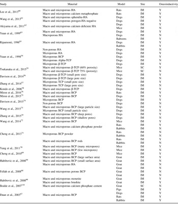
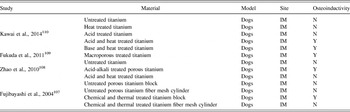
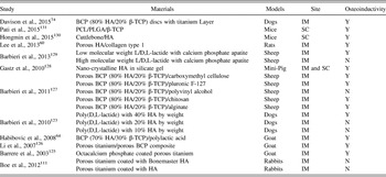
We performed a PubMed search using the keyword “osteoinduction”. Our search revealed seven hundred and thirty-eight publications using this keyword on May 20, 2016. Studies involving the implantation of a material in vivo with the aim to evaluate osteoinductivity were included. Studies using morphogenetic proteins or other organic materials or growth factors were excluded. Accordingly, we arrived at forty-nine studies investigating osteoinduction upon implantation of materials in soft tissue sites without the use of growth factors. Two of the forty-nine articles had been retracted. The remaining forty-seven articles tested various materials in rats, mice, rabbits, pigs, dogs, sheep, baboons, and goat between the years of 1996 and 2015. Some manuscripts present investigations on more than one material in the same manuscript, and, in total: twenty-four investigated calcium phosphate ceramics (Table II), seven investigated other types of ceramics including calcium-substituted ceramics, glass ceramics, titanium ceramics, and other compound ceramics (Table III), four investigated titanium metals (Table IV), and thirteen investigated composite materials (Table V).
Calcium ceramics.

Other ceramics.

Metals.

Composite materials.

V. CALCIUM-BASED CERAMICS
Ceramics are generally classified into two groups based on their chemical composition: calcium phosphates and others including stabilized zirconia (ZrO2), alumina (Al2O3), and some silicate and phosphate glasses known as glass ceramics. Of these, the ceramics most investigated for their osteoinductive properties have been calcium phosphates which include α and β-tricalcium phosphates (α and β-TCP), Reference Yuan, Yang, Li, Zhang, De Bruijn and De Groot54–Reference Kondo, Ogose, Tokunaga, Umezu, Arai, Kudo, Hoshino, Inoue, Irie and Kuroda58 hydroxyapatite (HA), Reference Yuan, Yang, Li, Zhang, De Bruijn and De Groot54,Reference Meagher, Weiss-Bilka, Best, Boerckel, Wagner and Roeder59–Reference Ripamonti65 monethite, Reference Habibovic, Gbureck, Doillon, Bassett, van Blitterswijk and Barralet66 brushite, Reference Habibovic, Gbureck, Doillon, Bassett, van Blitterswijk and Barralet66 and biphasic calcium phosphate (BCP). Reference Zhang, Barbieri, ten Hoopen, de Bruijn, van Blitterswijk and Yuan35,Reference Yuan, Yang, Li, Zhang, De Bruijn and De Groot54,Reference Habibovic, Kruyt, Juhl, Clyens, Martinetti, Dolcini, Theilgaard and van Blitterswijk64,Reference Habibovic, Gbureck, Doillon, Bassett, van Blitterswijk and Barralet66–Reference Miron, Zhang, Sculean, Buser, Pippenger, Dard, Shirakata, Chandad and Zhang76
Calcium phosphate ceramics are formed by sintering calcium phosphate crystals obtained after hydrolysis or precipitation reactions. Reference LeGeros, Lin, Rohanizadeh, Mijares and LeGeros77 Micropores with diameters less than 10 μm or macropores with diameters greater than 50 μm contribute significantly to the bioactivity of these ceramics. These microstructural features can be added to the calcium phosphate substrates during or directly before the sintering process. Reference Yuan, Kurashina, de Bruijn, Li, De Groot and Zhang63,Reference Jarcho78 Microporosity can be altered by changing the sintering temperature, Reference Yuan, Kurashina, de Bruijn, Li, De Groot and Zhang63 and macropores are introduced using the following steps: mixing appropriately sized volatile substances such as naphthalene powder or hydrogen peroxide along with the calcium phosphate powder, compacting the mixture, and subsequently removing the volatile substance via sublimation or decomposition, and sintering the final product. Reference Jarcho78
VI. HYDROXYAPATITE
Of all the calcium phosphates, hydroxyapatite (HA) [Ca5(PO4)3(OH)] most closely resembles the mineral phase of bone. Although HA is biocompatible, it is characterized with slow biodegradability that is persistent even at high surface areas of substrate. Reference Draenert, Draenert and Draenert79 Despite this and in addition to being brittle, having a low impact resistance, and displaying relatively low tensile strengths, Reference Martin and Brown80 HA has been extensively studied as an osteoconductive implant material. Reference Jarcho78 Furthermore, over two decades ago, it was discovered that certain material properties allow HA to induce bone formation at extra-skeletal sites, and thereby newly characterizing HA as an osteoinductive biomaterial. Reference Yuan, Yang, Li, Zhang, De Bruijn and De Groot54,Reference Ripamonti65,Reference van Eeden and Ripamonti81–Reference Yamasaki83
We came across six studies that tested twelve different types of pure HA for osteoinduction: six of these tests were strongly positive for osteoinduction and displayed bone formation in all of their models, Reference Yuan, Yang, Li, Zhang, De Bruijn and De Groot54,Reference Lee, Kim, Ko, Choi, Ahn, Kim and Do60–Reference Yuan, Kurashina, de Bruijn, Li, De Groot and Zhang63,Reference Ripamonti65 three demonstrated osteoinduction in some of the animal models, Reference Wang, Zhi, Lu, Li, Duan, Duan, Mu and Weng61,Reference Ripamonti65 and three showed no osteoinduction. Reference Yuan, Yang, Li, Zhang, De Bruijn and De Groot54,Reference Akiyama, Takemoto, Fujibayashi, Neo, Hirano and Nakamura62,Reference Yuan, Kurashina, de Bruijn, Li, De Groot and Zhang63 Of those HA materials that were osteoinductive, all contained both macropores and micropores. The macropores ranged from 50 μm to 700 μm in size with a majority in the 300–400 μm size range, and the micropores were in the 0.5–3 μm size range. Reference Yuan, Yang, Li, Zhang, De Bruijn and De Groot54,Reference Lee, Kim, Ko, Choi, Ahn, Kim and Do60–Reference Yuan, Kurashina, de Bruijn, Li, De Groot and Zhang63,Reference Ripamonti65
The presence and geometry of both micropores and macropores appears to influence the osteoinductive properties of HA. We found eight investigations into the osteoinductive capabilities of HA in dogs Reference Yuan, Yang, Li, Zhang, De Bruijn and De Groot54,Reference Wang, Zhi, Lu, Li, Duan, Duan, Mu and Weng61–Reference Yuan, Kurashina, de Bruijn, Li, De Groot and Zhang63,Reference Ripamonti65 ; all were positive except for two examining HA without micropores. Reference Yuan, Yang, Li, Zhang, De Bruijn and De Groot54,Reference Yuan, Kurashina, de Bruijn, Li, De Groot and Zhang63 Likewise, Yuan et al., 1999 Reference Yuan, Kurashina, de Bruijn, Li, De Groot and Zhang63 showed that whereas HA implants with macro- and micro-pores were able to induce ectopic bone formation in dogs, the same material with macropores without associated micropores was not osteoinductive in the same animal model. More complex geometrical analyses have since been carried out, and Wang et al., 2013 Reference Wang, Zhi, Lu, Li, Duan, Duan, Mu and Weng61 showed that HA can be formed which contains sphere-shaped macropores or that which contains sphere-shaped solid HA particles with complementary macropores. They showed that HA in a spherulite form inducted mature lamellar bone formation to a much greater degree than when in the porogen negative form. Reference Wang, Zhi, Lu, Li, Duan, Duan, Mu and Weng61 Besides material-based differences, animal model choice also appears to influence HA induced bone formation. For example, Akiyama et al., 2011 Reference Akiyama, Takemoto, Fujibayashi, Neo, Hirano and Nakamura62 showed positive ectopic bone induction using the same HA in dogs but not mice, and Ripamonti, 1996 Reference Ripamonti65 showed much greater bone induction using their HA in primates than in dogs and rabbits.
VII. TRICALCIUM PHOSPHATE
Tricalcium phosphates [Ca3(PO4)2] have long had many uses in orthopedic repair because of their favorable absorptive and osteoconductive abilities. Reference Liu and Lun84 TCP can be synthesized in the α- or β-polymorph depending on processing temperature, and this has an effect on the material’s resorption profile in vivo. Reference Merten, Wiltfang, Hönig, Funke and Luhr85 Although both types of TCP have been shown to resorb at a faster rate than HA, studies have shown that the resorbed calcium and phosphate are utilized and removed in a physiologically normal fashion, with no increases in serum ion levels. Reference Hollinger and Battistone86 This safety aspect has allowed for the commercialization of α- and β-TCP for bone-filling. Reference Liu and Lun84 Furthermore, it has since been shown that TCP is able to induce bone formation at ectopic sites with certain material characteristics. Reference Yuan, Yang, Li, Zhang, De Bruijn and De Groot54
Our search revealed nine investigations into the osteoinductive profile of α- or β- TCP formulations. Reference Yuan, Yang, Li, Zhang, De Bruijn and De Groot54–Reference Kondo, Ogose, Tokunaga, Umezu, Arai, Kudo, Hoshino, Inoue, Irie and Kuroda58 Whereas porous α-TCP was not osteoinductive in dogs, Reference Yuan, Yang, Li, Zhang, De Bruijn and De Groot54 it was shown to be very effective in treating calvarial defects in rabbits with resorption over 12 weeks. Reference Song, Liang, Luo, Chen and Sun87 In contrast, β-TCP did show osteoinductivity in the same experiment in dogs. Reference Yuan, Yang, Li, Zhang, De Bruijn and De Groot54 Furthermore, β-TCP has had success in treating femoral defects in rats and rabbits especially when doped with silicon and strontium. Reference Ke, Dernell, Bandyopadhyay and Bose88,Reference Fielding and Bose89 Investigations into the material properties of β-TCP showed that it was able to induce ectopic bone formation when it was microporous Reference Tsukanaka, Fujibayashi, Otsuki, Takemoto and Matsuda55,Reference Kondo, Ogose, Tokunaga, Umezu, Arai, Kudo, Hoshino, Inoue, Irie and Kuroda58 and the substrate contained micropores less than 1 µm. Reference Davison, Luo, Schoenmaker, Everts, Yuan, Barrere-de Groot and de Bruijn56,Reference Zhang, Luo, Barbieri, Barradas, de Bruijn, Van Blitterswijk and Yuan57 Whereas micropores appear to be a requirement for osteoinduction, macropores are not necessary for bone induction on the surface layer of these materials. Reference Davison, Luo, Schoenmaker, Everts, Yuan, Barrere-de Groot and de Bruijn56,Reference Zhang, Luo, Barbieri, Barradas, de Bruijn, Van Blitterswijk and Yuan57 Sub-micron micropore sizes appear to be favorable for bone induction by these materials Reference Tsukanaka, Fujibayashi, Otsuki, Takemoto and Matsuda55–Reference Zhang, Luo, Barbieri, Barradas, de Bruijn, Van Blitterswijk and Yuan57 ; this may explain why Kondo et al., 2006 Reference Kondo, Ogose, Tokunaga, Umezu, Arai, Kudo, Hoshino, Inoue, Irie and Kuroda58 did not witness bone induction until day 56 when using larger micropores in the ∼5 μm range.
β-TCP materials may require smaller micropore sizes than HA for osteoinductivity. Studies evaluating HA for osteoinduction did not historically measure or report micropore sizes, although the micropores are clearly evident in the included scanning electron microscopy images in these manuscripts. Reference Yuan, Yang, Li, Zhang, De Bruijn and De Groot54,Reference Lee, Kim, Ko, Choi, Ahn, Kim and Do60–Reference Yuan, Kurashina, de Bruijn, Li, De Groot and Zhang63,Reference Ripamonti65 After analysis of micropore sizes in these experiments, it appears that the reported sub-micron micropore sizes required to induce bone formation with β-TCP materials Reference Tsukanaka, Fujibayashi, Otsuki, Takemoto and Matsuda55–Reference Zhang, Luo, Barbieri, Barradas, de Bruijn, Van Blitterswijk and Yuan57 are smaller than those in osteoinductive HA materials. Reference Yuan, Yang, Li, Zhang, De Bruijn and De Groot54,Reference Lee, Kim, Ko, Choi, Ahn, Kim and Do60–Reference Yuan, Kurashina, de Bruijn, Li, De Groot and Zhang63,Reference Ripamonti65 This appears to be counter intuitive to the hypothesis that micropores allow for osteogenesis because they increase the overall surface area of the substrate, and thereby increase the ionic release of calcium into the surrounding environment. With this theory, it would be expected that β-TCP, a material that has a higher ionic dissolution than HA, Reference Bohner, Lemaître and Ring90 would not need as high of a surface area as HA to be osteoinductive. This does not appear to be the case.
VIII. BIPHASIC CALCIUM PHOSPHATE
Biphasic calcium phosphate is a product of sintering calcium deficient apatite, and contains varying levels of both β-TCP and HA. Reference Jarcho78,Reference LeGeros91 Because the resorption rates between HA and β-TCP differ, the bioreactivity of biphasic calcium phosphate can be controlled by varying the HA:β-TCP ratio. The sintering temperature, the presence of impurities, and the extent of calcium deficiency play an intimate role in determining the resulting HA:β-TCP ratio of the final product. Reference Jarcho78
We came across thirteen studies with twenty-four different investigations on the osteoinductive properties of BCPs in our literature search: fifteen showed osteoinductivity Reference Zhang, Barbieri, ten Hoopen, de Bruijn, van Blitterswijk and Yuan35,Reference Yuan, Yang, Li, Zhang, De Bruijn and De Groot54,Reference Habibovic, Kruyt, Juhl, Clyens, Martinetti, Dolcini, Theilgaard and van Blitterswijk64,Reference Cheng, Ye, Yang, Lu, Shi, Li, Fan and Bu69–Reference Miron, Zhang, Sculean, Buser, Pippenger, Dard, Shirakata, Chandad and Zhang76 while nine did not. Reference Zhang, Barbieri, ten Hoopen, de Bruijn, van Blitterswijk and Yuan35,Reference Habibovic, Kruyt, Juhl, Clyens, Martinetti, Dolcini, Theilgaard and van Blitterswijk64,Reference Fellah, Gauthier, Weiss, Chappard and Layrolle68,Reference Cheng, Shi, Ye and Bu71,Reference Wang, Barbieri, Zhou, de Bruijn, Bao and Yuan73,Reference Davison, Su, Yuan, van den Beucken, de Bruijn and Barrère-de Groot74 The ratio of HA:β-TCP composition ranged from 60:40 to 80:20, with more studies favoring the 80:20 ratio. Reference Zhang, Barbieri, ten Hoopen, de Bruijn, van Blitterswijk and Yuan35,Reference Yuan, Yang, Li, Zhang, De Bruijn and De Groot54,Reference Habibovic, Kruyt, Juhl, Clyens, Martinetti, Dolcini, Theilgaard and van Blitterswijk64,Reference Fellah, Gauthier, Weiss, Chappard and Layrolle68–Reference Miron, Zhang, Sculean, Buser, Pippenger, Dard, Shirakata, Chandad and Zhang76 While porous BCP with an HA:β-TCP ratio of 60:40 was able to induce ectopic bone formation in mice, Reference Cheng, Ye, Yang, Lu, Shi, Li, Fan and Bu69 it was unable to do so in goats. Reference Fellah, Gauthier, Weiss, Chappard and Layrolle68 On the other hand, ectopic bone formation was observed in goats upon implanting porous BCP with a HA:β-TCP ratio of 80:20. Reference Habibovic, Kruyt, Juhl, Clyens, Martinetti, Dolcini, Theilgaard and van Blitterswijk64
Notably, a lack of osteoinductivity for BCP materials was seen in investigations with no micropores Reference Zhang, Barbieri, ten Hoopen, de Bruijn, van Blitterswijk and Yuan35,Reference Davison, Su, Yuan, van den Beucken, de Bruijn and Barrère-de Groot74 and small particle surface areas. Reference Habibovic, Kruyt, Juhl, Clyens, Martinetti, Dolcini, Theilgaard and van Blitterswijk64 Interestingly, while macropores are not necessary for osteoinduction in pure β-TCP, Reference Davison, Luo, Schoenmaker, Everts, Yuan, Barrere-de Groot and de Bruijn56,Reference Zhang, Luo, Barbieri, Barradas, de Bruijn, Van Blitterswijk and Yuan57 a study with different particle sizes of BCP showed no osteoinductivity in the group with particles less than 45 µm in size. Reference Wang, Barbieri, Zhou, de Bruijn, Bao and Yuan73 The authors suspect this resulted because, compared to larger particles which did show osteoinduction, these small particles differed by their inability to house sufficiently sized macropores. Micropores, on the other hand are necessary for osteoinduction by BCP materials. Reference Zhang, Barbieri, ten Hoopen, de Bruijn, van Blitterswijk and Yuan35,Reference Davison, Su, Yuan, van den Beucken, de Bruijn and Barrère-de Groot74 While the diameter of the micropores has been a subject of interest by papers examining osteoinductive profiles, the depth of the micropores is seldom examined. Reference Zhang, Barbieri, ten Hoopen, de Bruijn, van Blitterswijk and Yuan35 Zhang et al., 2015 classified materials as rough if they contained deep micropores or smooth if they contained shallow micropores, and they observed osteoinduction only in the rough materials.
BCPs of varying ratios of HA/β-TCP are observed to be osteoinductive in various models when they exhibit both macropores and micropores, large surface areas, and rough surfaces. With promising results, BCPs have been translated into the clinic. After showing equal efficacy of a microstructured BCP (90% HA/10% β-TCP) to autologous bone in repairing bony defects in goats, Ruiter et al., 2014 Reference de Ruiter, Dik, van Es, van der Bilt, Janssen, Meijer, Koole and Rosenberg92,Reference de Ruiter, Meijer, Dormaar, Janssen, van der Bilt, Slootweg, de Bruijn, van Rijn and Koole93 implanted the microporous calcium phosphate graft substitute in the donor sites of 20 children with cleft palates, and showed solid, induced bone formation and almost complete resorption of the graft on 12 month follow up. The repair of these periodontal lesions is likely owed to the osteoinductive profiles of BCPs.
IX. NON-CALCIUM-BASED CERAMICS
Human bone mineral is not a pure stoichiometric compound; it has variable substitutions (<1% by weight) of calcium with sodium, potassium, magnesium, strontium, zinc, copper, aluminum, iron, chloride, and silicon. Reference Elliott94 Based on the observation that the composition of bone mineral is variable, ceramics with other chemical compositions besides TCP or HA have also been studied for creating potent osteoinductive materials. Studies adding other elements found in bone, such as zinc and magnesium to HA, showed significantly greater spreading and attachment of osteoblasts on these composite ceramics compared to HA alone. Reference Paul and Sharma95 Bone regenerative properties of magnesium calcium phosphate scaffolds have also been successfully demonstrated in vivo. Reference Wei, Jia, Wu, Wei, Zhou, Zhang, Shin and Liu96,Reference Li, Niu, Guo, Chen, Li, Zhang, Chen, Wu, Deng and Wei97 Femoral defects in rabbits treated with a porous magnesium calcium phosphate scaffold showed replacement of the scaffold with new bone formation by 3 months. Reference Wei, Jia, Wu, Wei, Zhou, Zhang, Shin and Liu96
On the other hand, some other ceramics have shown mixed results with respect to osteoinductivity. For example, whereas the use of porous titanium dioxide ceramics has promoted bone healing in rabbits, Reference Haugen, Monjo, Rubert, Verket, Lyngstadaas, Ellingsen, Rønold and Wohlfahrt98 the implantation of this ceramic in the dorsal muscle of dogs showed no bone formation even after 180 days. Reference Yuan, Yang, Li, Zhang, De Bruijn and De Groot54 Likewise, fluoride apatite ceramics appear to inhibit bone regeneration as the concentration of fluoride increases in the apatite. Reference Luo, Barbieri, Passanisi, Yuan and de Bruijn99 These data reinforce the notion that chemical signals may play both stimulatory and inhibitory roles in bone induction.
Glass-based ceramics have been investigated for their bone generating potential. The original bioglass formulation (45S5) contains many of the elements found in natural bone: 45% silica (SiO2), 24.5% calcium oxide (CaO), 24.5% sodium oxide (Na2O), and 6% phosphorous pentoxide (P2O5) by weight. 45S5 was developed to create a bioinert material that bonds to the living host, Reference Hench100 but it was shown that a strong bone implant interface forms upon implantation, thus paving the way for the clinical use of the first glass ceramics. Reference Kokubo101 Yuan et al., 2001 Reference Yuan, de Bruijn, Zhang, van Blitterswijk and de Groot102 showed that porous glass ceramic made by sintering powdered 45S5 was able to promote new bone formation when implanted at soft tissue sites. The induction of bone formation is thought to be largely due to a notable hydroxycarbonate layer that forms on the surface of 45S5 when exposed to bodily fluids, which has been demonstrated to attract osteoblasts and promote bone formation. Reference Ducheyne and Qiu103 Compared to the other discussed materials, glass ceramics hold unique properties such as the ability to generate new bone, antimicrobial effects, Reference Lindfors, Hyvönen, Nyyssönen, Kirjavainen, Kankare, Gullichsen and Salo104 and modifiable resorption rates, Reference Lindfors, Koski, Heikkilä, Mattila and Aho105 which may suite them for specific clinical situations.
X. METALS
Because of the poor mechanical properties of ceramics, metals such as titanium have been sought as alternative implant materials especially at areas of high loading. Unfortunately, it has been observed that the mismatch between the mechanically strong titanium and the weaker natural bone ultimately leads to stress shielding, bone resorption, and implant loosening. Reference Mcnamara, Toni and Taylor106 However, because of the advantageous mechanical properties, integrating the titanium bone interface has been of interest and methods have examined the induction of osteogenesis using titanium metals (see Table IV). Reference Fujibayashi, Neo, Kim, Kokubo and Nakamura107–Reference Kawai, Takemoto, Fujibayashi, Akiyama, Tanaka, Yamaguchi, Pattanayak, Doi, Matsushita and Nakamura110
The first report of titanium metal-induced bone formation at an ectopic site was in 2004 by Fujibayashi et al. Reference Fujibayashi, Neo, Kim, Kokubo and Nakamura107 who implanted chemically and thermally treated porous titanium blocks in the dorsal muscles of beagles and found lamellar bone formation upon twelve-month histological examination. Their results showed that treating a titanium metal substrate with a basic solution and then heating to 600 °C would allow titanium to induce bone formation at soft tissue sites. This treatment has been shown to result in a HA surface layer when the implant is exposed to bodily fluids in vitro, which perhaps is a precursor step to bone induction by titanium in vivo. This chemical and thermal treatment has resulted in strong osteoinductivity of titanium in a number of studies in dogs Reference Fujibayashi, Neo, Kim, Kokubo and Nakamura107–Reference Kawai, Takemoto, Fujibayashi, Akiyama, Tanaka, Yamaguchi, Pattanayak, Doi, Matsushita and Nakamura110 and in rabbits. Reference Boe, Stoen, Solberg, Reinholt, Ellingsen and Nordsletten111 Since many metal implant failures are linked to poor osteointegration of the implant, treatments or coating of these implant surfaces with osteoinductive materials has emerged as a viable strategy to improve osteointegration of the implant.
XI. POLYMERS
Polymers are a diverse class of macromolecular materials that can be composed using a wide range of synthetic or natural monomer units. Even though the first osteoinductive material observed was a synthetic sponge composed of a polymer of hydroxyethylmethacrylate, Reference Winter16 to our knowledge no other polymer has been found to be osteoinductive. Meanwhile and although a promising avenue, the controlled delivery of growth factors using polymers is beyond the scope of this review. What we believe has even more significant implications for polymer use in bone regeneration is the discovery that certain polymeric materials can sequester factors from the local environment and thereby modify their concentrations. Reference Hudalla, Kouris, Koepsel, Ogle and Murphy112 This sequestration of bioactive molecules has been shown to be effective in upregulating and downregulating growth factors that influence osteogenesis. Reference Kisiel, Klar, Ventura, Buijs, Mafina, Cool and Hilborn113 As an example, it has been shown that heparin competitively inhibits the binding activity of BMP-2, thereby limiting osteogenesis. Reference Kanzaki, Takahashi, Kanno, Ariyoshi, Shinmyouzu, Tujisawa and Nishihara114 Heparin binding domains on a polymer substrate are able to remove circulating heparin from the local environment, thereby freeing BMP-2 and increasing its active concentration. Using this knowledge, Hudalla, 2011 Reference Hudalla, Kouris, Koepsel, Ogle and Murphy112 showed that a scaffold that was chemically modified with a heparin binding protein had osteoinductive effects on stem cells even in the absence of exogenous growth factors. Our own lab has developed biologically active sintered microsphere scaffolds by using chitosan for the function of binding heparin to the scaffolds, Reference Jiang, Khan, Nair, Abdel-Fattah and Laurencin115 and have shown that this can guide bone regeneration in load bearing defects. Reference Jiang, Nukavarapu, Deng, Jabbarzadeh, Kofron, Doty, Abdel-Fattah and Laurencin116
Other techniques besides sequestering heparin have been examined; fragmented fibronectin matrices that potently bind VEGF and BMP-2 have been shown to amplify the effects of multiple growth factors to ultimately induce bone formation in the presence of very low doses of BMP-2. Reference Martino, Tortelli, Mochizuki, Traub, Ben-David, Kuhn, Muller, Livne, Eming and Hubbell117 These methods of creating and altering scaffolds to allow local manipulation of growth factor activity may eliminate the need for exogenous growth factors, proving to be suitable biomaterials for bone regeneration in themselves. In fact, the use of polymers incorporating RGD peptides, the primary binding site of fibronectin, has been successful in regenerating bone defects even without the addition of growth factors. Reference He, Ou, Zhou, Chen, Zhao, Zhao, Li, Zhu, Zhao and Jiang118
XII. COMPOSITE MATERIALS
The list of biomaterials studied for bone regeneration is broad, and one approach has been to integrate two very different materials to exploit the advantages of each. The use of a polymer and ceramic composite, for example, can improve the mechanical properties of a ceramic scaffold Reference Bhattacharyya, Kumbar, Khan, Nair, Singh, Krogman, Brown, Allcock and Laurencin119 while adding osteoinductivity to a polymer-containing implant. Polymer ceramic matrices have been developed since the initial reports of osteoinductivity by calcium phosphates. Reference Devin, Attawia and Laurencin120 Our laboratory has fashioned polyphosphazene and HA composites that match the compressive strengths of cancellous bone and promote osteoblast activity. Reference Bhattacharyya, Kumbar, Khan, Nair, Singh, Krogman, Brown, Allcock and Laurencin119,Reference Nukavarapu, Kumbar, Brown, Krogman, Weikel, Hindenlang, Nair, Allcock and Laurencin121 Polini et al., 2011 Reference Polini, Pisignano, Parodi, Quarto and Scaglione122 created polycaprolactone and TCP composite meshes and showed osteogenic differentiation of stem cells in vitro. Similarly and when applied to animal models, poly(lactic acid) and HA composites have been shown to be osteoinductive in vivo by multiple groups. Reference Barbieri, Renard, Bruijn and Yuan123,Reference Danoux, Barbieri, Yuan, de Bruijn, van Blitterswijk and Habibovic124 Overall we came across twelve studies evaluating twenty-two composite materials using various formulations of ceramics, metals, and polymers for osteoinduction (Table V). Reference Lee, Kim, Ko, Choi, Ahn, Kim and Do60,Reference Habibovic, Kruyt, Juhl, Clyens, Martinetti, Dolcini, Theilgaard and van Blitterswijk64,Reference Davison, Su, Yuan, van den Beucken, de Bruijn and Barrère-de Groot74,Reference Boe, Stoen, Solberg, Reinholt, Ellingsen and Nordsletten111,Reference Barbieri, Renard, Bruijn and Yuan123,Reference Barrere, van der Valk, Dalmeijer, Meijer, van Blitterswijk, de Groot and Layrolle125–Reference Pati, Song, Rijal, Jang, Kim and Cho131
Metal and ceramics have been combined in attempts to further improve the titanium bone interface. Reference Boe, Stoen, Solberg, Reinholt, Ellingsen and Nordsletten111,Reference Barrere, van der Valk, Dalmeijer, Meijer, van Blitterswijk, de Groot and Layrolle125,Reference Li, Habibovic, Yuan, van den Doel, Wilson, de Wijn, van Blitterswijk and de Groot126 Because the titanium in these composites is not resorbed, the indications for these implant materials are limited to permanent prosthetic applications. Regardless, Li et al., 2007 Reference Li, Habibovic, Yuan, van den Doel, Wilson, de Wijn, van Blitterswijk and de Groot126 observed osteoinductive bone formation when they machine fit BCP in titanium pores. Similarly, Barrere, 2003 Reference Barrere, van der Valk, Dalmeijer, Meijer, van Blitterswijk, de Groot and Layrolle125 observed ectopic bone formation when he implanted an octacalcium coated titanium in goat muscle, and Davison et al., 2015 Reference Davison, Su, Yuan, van den Beucken, de Bruijn and Barrère-de Groot74 coated microporous BCP discs with titanium alloy and showed that bone induction still took place as long as the surface structure remained unchanged.
Our search also revealed osteoinductive tests on a diverse array of polymer-ceramic composites. Reference Habibovic, Kruyt, Juhl, Clyens, Martinetti, Dolcini, Theilgaard and van Blitterswijk64,Reference Barbieri, Renard, Bruijn and Yuan123,Reference Barbieri, Yuan, De Groot, Walsh and De Bruijn127,Reference Barbieri, Yuan, Luo, Farè, Grijpma and de Bruijn129,Reference Pati, Song, Rijal, Jang, Kim and Cho131,Reference Mishra, Raina, Pelkonen, Lidgren, Tägil and Kumar132 When Barbieri et al., 2010 Reference Barbieri, Renard, Bruijn and Yuan123 investigated composites of poly(lactic acid) and calcium phosphate nanoparticles, they found that the composites required at minimum a 40% calcium phosphate formulation by weight to induce ectopic bone formation in dogs. The next year, with the aim to create a moldable, osteoinductive putty, the same group combined carboxymethyl cellulose, pluronic F-127, polyvinyl alcohol, chitosan, or alginate with BCP and examined the osteoinductivity of each; all but the polyvinyl alcohol/BCP composite were osteoinductive in sheep. Reference Barbieri, Yuan, De Groot, Walsh and De Bruijn127 These composites allow for the creation of materials with varying consistencies for a wide array of applications.
XIII. DISCUSSION
In this article we have focused on the inductive properties of materials used for bone regeneration. In reality, the osteoinductive ability of a material is not the only factor a surgeon must consider when choosing a suitable graft material. Each material has its own safety profile, resorption rate, and mechanical properties. An ideal biomaterial is one that has no side effects, matches natural bone in the mechanical properties, is osteoinductive, and resorbs at a rate parallel to the stimulated formation of bone.
The observations on the osteoinductive potential of many biomaterials reveal recurring patterns. From a materials standpoint, micropores are necessary, and the smaller the diameter of micropores the more effective they are in stimulating bone growth. In addition, macropores are generally required and they must possess a diameter of at least 45 µm to successfully induce bone formation, although there have been reports of bone growth on the surface of certain substrates without macropores. We have previously described the various possible modifications of these porous properties both geometrically and topographically, and while these manipulations appear to affect stem cell induction toward osteoblastic lineage in vitro, it has not been widely examined whether they affect in vivo bone induction in the studies we have come across. In vitro investigations in material properties other than porosity have also shown insights into these characteristics. It appears that the strength of the implant must closely mimic the strength of the desired induced tissue, material surfaces may also be tailored to promote stem cell attachment and subsequent induction, and the organization of topographical features has significant influence on the materials inductive properties. In contrast to these physical characteristics, from a chemical standpoint, doping a substrate with the various elements found in physiological bone mineral tends to increase the osteoinductive capabilities of that material. In addition, the chronological study of bone generation in various implants tends to show a surface HA layer formation that precedes bone formation; whether this is a prerequisite for bone induction is unclear.
What is evident is that there are numerous research groups studying osteoinduction with regards to a variety of materials in a variety of animal models in a variety of sites. Historically, the sites for implantation of experimental test-materials have been intramuscular, subcutaneous, and within the renal capsule. Reference Scott, Levi, Askarinam, Nguyen, Rackohn, Ting, Soo and James133 Each of these sites has advantages related to the presence and types of native stem cells within the tissue, ease of access during surgery, and even mechanotransduction on the implant. Likewise, different animal models harbor different reactions to similar materials, which clouds our understanding of inductive bone formation. Larger animal models may behave more closely to humans regarding kinesiology and mechanical forces across the implantation site, but may differ with regards to their biology. The mechanisms of these differences in extraskeletal bone formation observations are only beginning to be unraveled. Reference Tsukanaka, Fujibayashi, Otsuki, Takemoto and Matsuda55,Reference Akiyama, Takemoto, Fujibayashi, Neo, Hirano and Nakamura62 In this light, we do admit difficulties in interpreting the literature on osteoinduction.
To further cloud the overall understanding, when two different research groups implant similar substrates in similar animals, one group observes extra-skeletal bone induction while the other may not. One explanation for the various observations seen in similar experiments is that subtle changes in preparation of biomaterials (e.g., sintering temperatures and ionic impurities) can drastically alter the final product. Reference Jarcho78,Reference Li, Niu, Guo, Chen, Li, Zhang, Chen, Wu, Deng and Wei97,Reference Kasuga, Maeda, Kato, Nogami, Hata and Ueda134 Preparation plays a large part in determining the final chemical and structural nature of the implant, and rigorous control of implant composition is essential to achieve predictable in vivo results. Reference Jarcho78 Furthermore, the fine control of one variable material or chemical property at a time in assessing substrates is a practical challenge, but we have already seen investigators employing preparation methods that allow for such multivariable control. Reference Zhang, Barbieri, ten Hoopen, de Bruijn, van Blitterswijk and Yuan35,Reference Davison, Luo, Schoenmaker, Everts, Yuan, Barrere-de Groot and de Bruijn56,Reference Zhang, Luo, Barbieri, Barradas, de Bruijn, Van Blitterswijk and Yuan57 With these advances taking place, the investigations into osteoinduction have shifted from trials of a list of materials into the specific design of materials expected to induce bone.
We are optimistic that inorganic biomaterials may be safely and effectively added to the surgeon’s tool kit for successfully regenerating bone. Composite technologies, in particular, present solutions to vast clinical scenarios; studies suggest that the options for creating bone regenerative materials are not limited by materials that display osteoinductivity. Biocompatible materials may be mixed and matched between those that are osteoinductive and those that are not to create a bone regenerative implant that is mechanically favorable as well as osteoinductive. The different formulations ranging from injectable putties to solid load bearing blocks may be each applied to specific surgical scenarios: while putties are easily manipulated into smaller concerning lesions, a large comminuted femoral fracture may require an osteoinductive block with structural and load bearing support. In our opinion, the investigations into the ideal composite material for bone regeneration harbor a particularly exciting future.
XIV. CONCLUSION
The search for safe osteoinductive materials has led to numerous candidates such as ceramics, metals, polymers, and composites of these materials. The osteoinductive potential of materials has been shown to be influenced by both mechanical as well as chemical properties such as stiffness, porosity, topography, adhesiveness, effects of released calcium and phosphate ions, ability to form a surface hydroxyapatite layer, and more. In addition to regenerating bone, techniques to create an osteoinductive surface on implants have emerged as a viable strategy to improve osteointegration of prosthetics. Our understanding of the intimate interactions between material substrates and progenitor cells has provided clues to the development of materials with osteoinductive capabilities. As it turns out, ceramics, polymers, and metals harbor osteoinductive profiles if manufactured appropriately. These materials have intrinsic advantages that can be augmented as well as disadvantages that can be reduced by combining materials into composites. The mechanisms of material based osteoinduction are only beginning to be unraveled, and these interactions between substrates and stem cells are the bedrock of the future of bone regeneration.