Over the past two decades, percutaneous dilation of the pulmonary valve with a balloon has become widely accepted as the treatment of choice for individuals with congenital pulmonary valvar stenosis. This technique is safe and effective in patients of all ages. Currently, balloon valvoplasty is used for a wide spectrum of obstructive pulmonary valvar pathology, including valvar stenosis, dysplasia and atresia. In this review, we will address the history, indications, technique, immediate and long-term outcomes, as well as future outlook, of percutaneous balloon valvoplasty for the wide spectrum of congenital pulmonary valvar disease.
Historical review
Catheter intervention for pulmonary valvar stenosis has its origins in a dynamic technique first described by Rubio and Limon in 1956.1 Their procedure was to pull an inflated balloon from the pulmonary trunk to the right ventricle across the stenotic pulmonary valve. Although this technique was effective in relieving valve stenosis, the risk of avulsion of the valvar leaflets prevented it from gaining wide acceptance.2
A static procedure was described in 1982 by Kan et al., and first used in an 8 year old child with congenital pulmonary valvar stenosis, with unqualified success.3 This technique found its origins in the experience gained from balloon angioplasty for the treatment of stenotic peripheral and coronary arteries, and was dependant upon engineering breakthroughs that enabled the manufacture of rigid balloons on catheters of acceptable sizes. Since this first report of static balloon valvoplasty, the procedure has been performed in neonates, infants, children and adults, and has clearly replaced surgical valvotomy as the treatment of choice for the relief of congenital pulmonary valvar stenosis.4–13 Follow-up studies have documented long-term relief of obstruction in patients with typical valvar pulmonary stenosis, with important restenosis being uncommon.
The success of percutaneous balloon valvoplasty for the relief of pulmonary valvar stenosis prompted modification of the technique for application to neonates with imperforate pulmonary valves in the setting of an intact ventricular septum. Balloon dilation for pulmonary atresia, using a guide wire to perforate the atretic valve, was first described by Latson et al. in 1991.14 Since this initial description, catheters and wires utilizing laser or radiofrequency energy have been developed to perforate the atretic valve.15, 16 To date, the results for such perforations and dilations have been mixed, and somewhat difficult to assess. Even with technically successful dilation, the outcome is often adversely affected by hypoplasia of the right ventricle and tricuspid valve, which may be quite variable. Certainly, this percutaneous approach has enabled many newborns to avoid subsequent surgery, but many still require further surgical or catheter intervention.17–22
Obstructive malformations of the pulmonary valve requiring palliation can be divided into two distinct clinical entities:
- moderate to severe valvar stenosis of the infant, child or adult
- critical pulmonary valvar stenosis or atresia of the newborn.
The indications, various techniques, and outcomes will be presented separately for each of these entities.
Clinical indications
Moderate-to-severe pulmonary valvar stenosis: Traditionally, pulmonary valvar stenosis has been defined as trivial, mild, moderate, or severe on the basis of the peak resting systolic gradient across the pulmonary valve with normal cardiac output. A gradient of less than 25 millimetres of mercury is considered trivial. From 25 to 39 millimetres is considered mild, from 40 to 70 is moderate, and greater than 70 millimetres is severe.23 The peak instantaneous gradient assessed by continuous wave Doppler will accurately predict the peak systolic catheter gradient, and has become a useful noninvasive technique for serial assessment.24 Moderate-to-severe stenosis is typically progressive, and left untreated will lead to symptoms of dyspnoea, fatigue, and exercise intolerance.23, 25–27 Interventions are indicated to relieve moderate-to-severe stenosis, therefore, to reduce and prevent progressive obstruction of the right ventricular outflow tract, right ventricular hypertrophy, and fibrosis, all of these, if left untreated, leading to eventual right ventricular dysfunction. It is our recommendation and practice to perform percutaneous valvoplasty in patients with resting peak systolic gradients greater than 40 millimetres of mercury. This recommendation is a modest modification of the recommendations made by the Second Natural History Study, in light of the excellent safety of balloon interventions.28 The contraindications to performing valvoplasty for stenosis that is moderate to severe are relatively few, and are consistent with the contraindications to performing cardiac catheterization in general. Valvoplasty should not be performed in a patient with active bacteremia or untreated systemic infection, skin infection overlying the site of intended insertion of the catheter, severe haemodynamic instability, or when the orifice of the pulmonary valve is known to be severely hypoplastic.
Critical pulmonary valvar stenosis and pulmonary valvar atresia: Typically, neonates with critical pulmonary valvar stenosis or atresia with intact ventricular septum present within the first week of life with cyanosis and duct-dependant pulmonary circulation. Cyanosis results from suprasystemic right ventricular pressures, decreased right ventricular compliance, and often a degree of tricuspid regurgitation, all of which promote right-to-left shunting at the atrial level. Strategies for management of these infants continue to evolve. Presently, most centres prefer to pursue a strategy aimed at biventricular palliation, which includes decompression of the hypertensive right ventricle and restoration of adequate flow of blood to the lungs, unless there are serious contraindications to this approach. Contraindications include the presence of right ventricular-dependant coronary arterial circulation, or a severely hypoplastic right ventricle or tricuspid valve17, 19, 22 (see Fig. 1). Currently, there is no consensus regarding the degree of right ventricular or tricuspid valvar hypoplasia that precludes pursing a biventricular palliation strategy. We have, however, attempted to provide some guidelines regarding what constitutes an “adequate” right ventricle and tricuspid valve by summarizing later in our review the available data (see “Immediate and Long-term Outcomes”). For infants deemed not to be candidates for a biventricular approach to palliation a univentricular strategy, or rarely transplantation, remain the only reasonable options for treatment. In the neonate with an adequate right ventricle and tricuspid valve, decompression of the right ventricle and restoration of adequate antegrade flow of blood to the lungs can often be accomplished via percutaneous valvoplasty in the presence of a patent infundibulum.17, 19 Catheter intervention in these patients is technically challenging due to varying degrees of right ventricular hypoplasia and insufficiency of the tricuspid valve. If the staff of any given centre possess the appropriate skills at intervention, we advocate proceeding with balloon valvoplasty as the primary intervention in the newborn with critical pulmonary valvar stenosis or valve atresia unless there are important contraindications to a biventricular strategy.

Figure 1. Right ventricular angiogram in the lateral projection in a 3-day-old infant with pulmonary atresia and intact ventricular septum with right ventricular dependant coronary circulation. Prominent right ventricular fistulous connections to the right coronary artery and retrograde filling of the left coronary artery are evident.
Technique and equipment
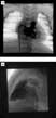
Moderate-to-severe pulmonary valvar stenosis: Balloon valvoplasty is performed using a percutaneous femoral venous approach. Typically, complete catheterization of both the right and left heart is performed to document the nature and severity of the obstruction. We prefer to obtain arterial access in order to monitor arterial pressure and blood gasses, especially in infants. A right ventricular biplane angiogram performed with straight anterior–posterior and lateral projections will demonstrate the ventricular and pulmonary valvar anatomy (see Fig. 2). The measurement of the diameter of the pulmonary valve is best determined from the lateral projection. A marker catheter is preferable to the size of the catheter in the French system for calibrating the measurements. If the valve appears appropriate for balloon dilation, an end-hole catheter is advanced across the valve, and is positioned in the distal left pulmonary artery. The position in the left pulmonary artery provides a straight course for the balloon across the valve, and also provides more stability for exchanges of wires and catheters compared to a wire positioned in the right pulmonary artery. An exchange-length guide wire is then positioned in the distal left pulmonary artery, and the end-hole catheter removed and exchanged for the balloon dilation catheter. The catheter selected for valvoplasty should have a diameter of the inflated balloon approximately one-quarter to one-third greater than the measured diameter of the pulmonary valve, and its length should be shorter than the distance from the tricuspid valve to the bifurcation of the pulmonary trunk. Longer balloons should be avoided, as they can injure the tricuspid valve or pulmonary arteries. If the diameter of the pulmonary valve exceeds 20 millimetres, or if the size of the single balloon catheter is thought to be too large to be introduced safely into the femoral vein, then it is possible to use a double balloon. This technique is performed with bilateral or unilateral venous access, depending on the size of the femoral vein. Two similar sized balloon catheters are selected, so that the sum of their inflated diameters is approximately 50–60% greater than the measured diameter of the pulmonary valve.

Figure 2. Right ventricular angiogram in the frontal (a) and lateral (b) projections in a 3-day-old infant with critical pulmonary valvar stenosis. The frontal projection demonstrates typical doming of the severely stenotic pulmonary valve. A narrow jet of contrast can be seen emanating from the severely stenotic orifice on the lateral projection.
Once selected, the catheter, or catheters, are inserted across the valve, and the balloons are centred within the orifice of the pulmonary valve. The balloon, or balloons, can be partially inflated with a mixture of half contrast and half saline to confirm appropriate positioning prior to valvoplasty. After proper positioning is confirmed, the balloon is inflated by hand until the waist produced by the pulmonary valve on the balloon disappears. Additional pressure for inflation is unnecessary once the waist is relieved. When using two balloons, both balloons are simultaneously inflated until the waist is relieved. Balloon inflation should be kept as brief as possible to reduce the period of right ventricular obstruction and resulting arterial hypotension, typical for patients with an intact atrial septum, and/or arterial hypoxemia, as seen in patients with a patent atrial foramen. Often, 3 or 4 inflations are performed to assure that the dilation is technically adequate.
Once the valvoplasty is complete, the balloon catheter is removed, and replaced with an end-hole catheter. The residual gradient across the right ventricular outflow tract, and cardiac output, are measured to assess the adequacy of the procedure. Particular attention should be given to the location of any residual gradient, as subvalvar obstruction is common following relief of severe valvar stenosis. If a significant residual gradient persists after a technically adequate dilation, a right ventricular angiogram will often document an increased, dynamic, element of infundibular obstruction. Dynamic infundibular obstruction will generally resolve with time, usually 1 to 2 months, in patients with typical valvar pulmonary stenosis, as the right ventricular hypertrophy regresses.
Critical pulmonary valvar stenosis and pulmonary valvar atresia: The newborn with critical pulmonary valvar stenosis or imperforate pulmonary valve with intact ventricular septum presents a formidable challenge to the interventional cardiologist attempting percutaneous valvoplasty. Often these infants have a variable degree of right ventricular and tricuspid valvar hypoplasia, as well as moderate-to-severe tricuspid valvar insufficiency. The challenge in these newborns is not only catheterizing the right ventricle successfully, but also maintaining precise position of the catheter in order successfully to cross the stenotic or atretic pulmonary valve.
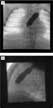
Depending on the age of the neonate, and the preference of the operator, this procedure may be performed with a femoral or umbilical arterial and a femoral venous approach. It is our preference to perform this procedure with the newborn receiving an infusion of prostaglandin E1 to maintain patency of the arterial duct. Ductal patency helps to ensure haemodynamic stability during the catheterization and inflation of the balloon, and improves the stability of the wire and catheter by providing a longer course into the descending aorta. After determining the diameter of the pulmonary valve and infundibular anatomy from a right ventricular angiogram, we prefer to position a 4 or 5 French right coronary catheter just below the stenotic or atretic pulmonary valve. The stenotic valve is then crossed with a 0.014–0.018 inch guide wire. For an atretic pulmonary valve, we use either a radiofrequency-tipped wire, or the stiff end of a 0.021 inch guide wire to perforate the valve. When utilizing a stiff guide wire, the operator must pay particular attention to the position of the guiding catheter, because it may become easily displaced anteriorly, and result in infundibular perforation. Once across the pulmonary valve, the guide wire is advanced through the duct into the descending aorta, where if necessary it can be anchored with a snare inserted from the arterial sheath. A balloon of appropriate size, usually exceeding the diameter of the valve by one-quarter to one-third, is advanced over the wire, centred within the pulmonary valve, and inflated by hand (see Fig. 3). To maintain the lowest profile prior to initial dilation, we typically do not pre-inflate or debubble the balloon. Serial dilations with balloons of increasing size, as described in the earlier literature, are now not often necessary. After the balloon catheter is deflated and withdrawn, repeat haemodynamic measurements, and right ventricular angiography, are preformed to assess the adequacy of the palliation.

Figure 3. Images in the frontal (a) and lateral (b) projections during balloon inflation across the pulmonary valve of the patient depicted in Figure 2. The balloon is inflated with complete relief of the waist in both projections. The frontal projection shows the exchange wire passing through the arterial duct and into the descending aorta for added stability during balloon inflation.
We discontinue the infusion of prostaglandin E1 within 24 hours following a technically adequate procedure. If, after 2 weeks, arterial saturations remain at or below 65%, or if the neonate develops acidosis following ductal closure, then the infant is referred for surgical creation of a systemic to pulmonary arterial shunt, and possibly a transannular patch, depending on the size of the pulmonary valve. Transcatheter ductal stenting is an option to consider in newborns with a valve of reasonable size if a surgical transannular patch is thought to be unnecessary.
Immediate and long-term outcomes
Moderate-to-severe pulmonary valvar stenosis: The large majority of infants, children and adults with isolated moderate-to-severe stenosis who undergo percutaneous valvoplasty can expect excellent short and long-term relief of their obstruction. The outstanding acute results have been well described by many investigators during the past 2 decades.5–8, 12, 13, 29, 30 The Valvuloplasty and Angioplasty of Congenital Anomalies Registry represents the largest published clinical series of percutaneous pulmonary valvoplasties available.31 The registry reported the acute results of dilations performed in 784 patients at 26 institutions between 1981 and 1986. Overall, these procedures resulted in the reduction of the peak systolic pulmonary valve gradient from 71 to 28 millimetres of mercury, with few serious complications. More severe initial obstruction, and a dysplastic pulmonary valve, were found to be the only predictors of suboptimal acute outcome, defined as a residual gradient of greater than 30 millimetres of mercury. In patients without a dysplastic valve, dynamic infundibular obstruction was identified as an important contributor to early residual outflow obstruction. The authors postulated that the infundibular obstruction would resolve as right ventricular hypertrophy regressed. Supporting evidence documenting the resolution of infundibular obstruction was reported by McCrindle et al. in 1994. They reported the mid-term follow up data for 533 of 835 patients from the registry, and noted that just over half of those with initial residual gradients of greater than 36 millimetres had gradients of less than this value at a median follow up of 33 months without any intervening procedures.32 Additional support for this phenomenon had previously been reported by Thapar and Rao.33 One quarter of their patients had infundibular gradients following pulmonary valvoplasty. At follow up evaluation 10 months after balloon dilation, the residual obstruction had resolved in half of these, and had decreased in the other half. Both these studies showed that older children, with higher initial gradients, were more likely to have obstruction after the procedure. Most of these patients did have adequate relief of their obstruction documented several months after dilation. This phenomenon of transient infundibular obstruction is important to consider when assessing the early results of pulmonary valvoplasty, especially in older children and adults.
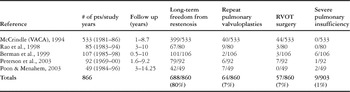
Many reports have now documented excellent long-term results in patients of all ages.11, 13, 29, 32, 34–36 Table 1 summarizes some of the largest studies reported in infants, children and adolescents. A total of 866 patients underwent dilation, and 860 were followed for 1 to 14 years after the procedure. Approximately four-fifths of patients were free from clinically significant restenosis at the end of the period of follow up. More importantly, only one-sixth of patients had required additional procedures because of clinically significant residual obstruction. A reassuring finding was that relatively few patients, about one in one hundred, developed severe pulmonary insufficiency during the follow up. Independent risk factors for a suboptimal long-term outcome which were reported in the registry cohort, as well as by many of the other investigators, include:
Table 1. Long-term tesults for moderate to severe pulmonary valve stenosis.

- higher initial pulmonary valvar gradient
- higher early residual gradient
- smaller ratio of balloon to valve
- smaller valvar diameter
- younger age at initial intervention
- dysplastic pulmonary valve.
These risk factors for poor long-term outcome identify patients with the most severe and complex valvar anatomy. In patients with more typical anatomy, the long-term data overwhelmingly demonstrate that balloon valvoplasty is effective, safe, and likely to provide long-term, if not life long, relief of moderate-to-severe pulmonary valvar stenosis.
Critical pulmonary valvar stenosis and atresia: Over the last two decades, improvements in catheterization techniques, equipment, and strategies for post-procedural management have improved the survival of neonates with critical pulmonary valvar stenosis and imperforate pulmonary valves in the setting of an intact ventricular septum. Today, most neonates with critical pulmonary valvar obstruction who undergo balloon valvoplasty will achieve excellent short and long-term relief of their obstruction with a single procedure performed in the first few days of life.19, 20, 37–40 Although less encouraging, the short and long-term survival of neonates born with imperforate pulmonary valves with intact ventricular septum also continues to improve.17, 18, 21, 22, 39, 41–44 Many of these neonates with pulmonary atresia; however, will require multiple interventional and surgical procedures prior to achieving final palliation.
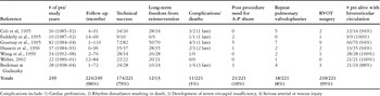
Intermediate and long-term results of balloon dilation for critical pulmonary valvar stenosis in the newborn have been reported since 1995. Table 2 summarizes the results of 221 patients previously reported in the literature, and 28 consecutive patients treated in our centre since 1998. The pulmonary valve was crossed, and an appropriately sized balloon inflated, in 224 of 249 patients, resulting in a rate of technical success of 90%. There were 12 serious complications. In addition, 13 patients died, 5 early and directly attributable to the catheterization. Of 224 patients with a technically successful procedure, 221 had documented follow up evaluations of up to 116 months. A total of 174 patients (79%) did not require any additional procedures. It was necessary to construct a systemic-to-pulmonary arterial shunt, or stent the duct, in 11 patients due to persistent cyanosis. Surgical enlargement of the right ventricular outflow tract and pulmonary valve was required in 18 patients, and repeat pulmonary valvoplasty was needed in 21. Typically, only a single additional procedure was required for the relief of critical stenosis when neonatal balloon valvoplasty proved to be inadequate. Overall, 95% of the patients who underwent technically adequate percutaneous pulmonary valvoplasty for critical pulmonary valvar stenosis as neonates were alive with a biventricular circulation at the time of their last reported follow-up evaluation.
Table 2. Long-term results for critical pulmonary valve stenosis.

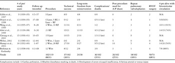
Numerous centres have reported their intermediate and long-term results for catheter perforation and balloon dilation in patients with imperforate pulmonary valves in the setting of an intact ventricular septum. The results of these studies, as well as our own data, are summarized in Table 3. A total of 119 patients underwent catheterization with the intent to dilate the atretic pulmonary valve. Multiple techniques were utilized to cross the pulmonary valve, and slightly better results were achieved with the use of laser or radiofrequency tipped wires and catheters. This finding is not surprising. It is much easier to maintain precise position of the guiding catheter while advancing a flexible laser or radiofrequency wire compared to the stiff end of the 0.021 inch guide wire. Overall, an appropriately sized balloon catheter could be advanced across the pulmonary valve and inflated in 102 of 120 patients (85%). There were 24 serious complications, which occurred during the procedures. In addition, 18 patients died following the procedures, resulting in overall rates of morbidity and mortality of 20.1% and 15.1%, respectively. Of the deaths, 13 occurred early, and were directly related to the interventional procedure, while 5 deaths occurred late, and were not related to the initial catheterization. Patients with a technically adequate dilation returned for follow up evaluation up to 87 months after the procedure. At the time of the last follow up examination, just over three-tenths of the patients had not required any additional surgical or catheter interventions. The long-term freedom from reintervention after successful balloon valvoplasty in newborns with pulmonary atresia is 36%. This figure is significantly lower than the rate which is reported for newborns with critical pulmonary valvar stenosis, albeit that it does compare favourably to the long-term results following surgical valvotomy with or without construction of a systemic-to-pulmonary arterial shunt in newborns with pulmonary atresia.21 Of the 102 patients treated with balloon dilation, 36 required a construction of a systemic-to-pulmonary arterial shunt or underwent ductal stenting for persistent cyanosis. Repeat valvoplasty was needed in 26 patients, while 18 patients underwent surgical procedures to enlarge their right ventricular outflow tract and pulmonary valve. In contrast to the newborns that remained cyanotic following a technically adequate valvoplasty for critical stenosis, many of the patients with an imperforate pulmonary valve who remained cyanotic underwent multiple additional procedures to augment the right ventricular outflow tract, often combined with creation of a systemic-to-pulmonary arterial shunt. Overall, the final circulation for 73 patients could be determined from these reports. Just over two-thirds, or 50 patients, who underwent technically adequate valvoplasty were alive with a biventricular circulation at their last follow-up.
Table 3. Long-term results for pulmonary atresia and intact ventricular septum.

Some authors have contended that critical stenosis and imperforate pulmonary valves are two district clinical entities when the ventricular septum is intact.20 We believe that they exist on a continuum, with significant overlap with regard to treatment and outcomes. In both entities, the diameters of the tricuspid and pulmonary valves, and the volume of the right ventricular volume at diagnosis, predict the ultimate success of achieving a biventricular circulation following balloon valvoplasty.10, 17–19, 21, 22 The most important anatomic factor appears to be the size of the tricuspid valve, which is often a proxy for right ventricular volume. Typically, the tricuspid valve is normal or near-normal in size in neonates with critical pulmonary valvar stenosis, and is variably hypoplastic in neonates with an imperforate pulmonary valve. The capability of the tricuspid valve to provide a haemodynamically adequate right ventricular inlet is, in our opinion, often the key factor in achieving a biventricular circulation in these children, regardless of whether the strategy for treatment is catheter-based, surgical, or a combination of the two.
In 2003, Humpl et al. provided data upon which some decisions may be made in the management of newborns with pulmonary atresia.17 They reported on 16 neonates with imperforate pulmonary valves whose initial Z scores for the tricuspid valve were −3.03 ± 2.51 who achieved a biventricular circulation, contrasting them with 12 neonates with significantly smaller tricuspid valves who died or were palliated with functionally univentricular or mixed circulations following successful valvoplasty. They also found that the Z-score for the tricuspid valve did not change significantly over time, despite adequate right ventricular decompression. Previously, Ovaert et al.18 had reported that the Z-score decreased from −0.7 to −2.3 in a similar group of 6 patients. Despite this decrease in the Z-score, all of the patients achieved a biventricular circulation. These data prompted the authors to hypothesize that “catch up” growth may not occur for the tricuspid valve after right ventricular decompression. A tricuspid valve of normal size, nonetheless, does not appear to be necessary for adequate pulmonary circulation. The intermediate and long-term data presented in our review, along with the data from the Congenital Heart Surgeons Society, form the basis of our recommendations for the management of infants with critical stenosis or imperforate pulmonary valves in the setting of an intact ventricular septum.17, 20–22
As mentioned earlier, all neonates without important contraindications to a biventricular repair, such as a severely hypoplastic right ventricle or right ventricular-dependant coronary arterial circulation, can undergo decompression of the hypertensive right ventricle with balloon pulmonary valvoplasty (Fig. 4). A Z-score of −5.0 or larger for the tricuspid valvar annulus appears potentially adequate to maintain the pulmonary circulation.17 If the neonate remains severely cyanotic after 2 weeks, or develops acidosis after discontinuation of prostaglandin E1, then construction of a systemic-to-pulmonary shunt, or ductal stenting, is indicated. Surgical enlargement of the right ventricular outflow tract and pulmonary valve may be considered in addition to shunting if the Z score for the diameter of the pulmonary valve is smaller than −2.0. For newborns with an inadequate Z score for the tricuspid valve, less than −5.0, functionally univentricular palliation is pursued with creation of a systemic-to-pulmonary arterial shunt. Right ventricular decompression can be considered to achieve a 1½ ventricle circulation assuming that there is not right ventricle-dependant coronary arterial circulation. These procedures can be carried out in the catheterization laboratory, or in the operating room, depending on the skill and preference of the interventional cardiologist and cardiothoracic surgeon.

Figure 4. Decision tree summarizing the early management strategy for neonates with an imperforate pulmonary valve in the setting of an intact ventricular septum.
Future outlook
Balloon dilation of the pulmonary valve in neonates, infants, children and adolescents has become widely accepted as the procedure of choice for relieving moderate, severe, and critical valvar stenosis. This procedure is also advocated as the initial intervention to decompress the right ventricle in neonates with an imperforate pulmonary valve in the setting of an intact ventricular septum. The next advance in the treatment of severe pulmonary valvar disease will occur with the treatment of the fetus. It is now possible to identify severe pulmonary and aortic valvar disease in a fetus as early as 20 weeks gestation. Currently, several centres have demonstrated that it is possible to relieve critical aortic and pulmonary valvar stenosis or atresia with balloon valvoplasty performed in the fetus.45, 46 Several important issues now need to be addressed as investigators move forward with fetal intervention on the pulmonary valve:
- Can a fetus at high risk of developing severe right ventricular hypoplasia be identified by fetal echocardiography or other modalities?
- Can a fetal intervention be performed to relieve critical pulmonary valvar stenosis or atresia which will prevent or reverse severe right ventricular hypoplasia?
- Can a fetal intervention be performed which is as safe and effective for the fetus and the mother when compared to a procedure performed subsequent to delivery?
The answers to these questions are currently under investigation. When known, they will determine the indications for fetal intervention on the pulmonary valve. If fetal pulmonary valvoplasty proves to be reproducibly successful, it will likely revolutionalize again the management of this disease. The possibility that severe right ventricular hypoplasia and its consequences may be prevented or reversed is certainly exciting.