Introduction
Antimicrobials provide a tremendous selective pressure on any susceptible bacterial population, whereby cells that are less susceptible are at a great advantage to survive and multiply. Depending on the mechanism of action of the antimicrobial, the severity of selective pressure and the possible genetic strategies that render bacteria either less susceptible or cause partial or full resistance, a population can shift sooner or later from ‘completely susceptible’ via intermediate stages to one that in the laboratory is determined to be ‘resistant’. This can happen in any environment, including the living host, be it human or animal.
The term ‘resistance’ can mean different things and even when the minimum inhibitory concentration (MIC) of an antimicrobial is given, this can vary according to the testing methodology. The breakpoint MIC for an antimicrobial agent can be either the threshold above which a given bacterial pathogen no longer survives or multiplies in vitro, the MIC value above which it is unlikely to respond to treatment, or a description of the wild-type population (Simjee et al., Reference Simjee, Silley, Werling and Bywater2008). These in vitro and clinical breakpoints may not be identical, due to the pharmacokinetic parameters resulting from different drug dosing regimens. An epidemiologist interested in resistance development within bacterial populations more likely uses a biological MIC or epidemiological cut-off value to separate a susceptible (wild-type) population from a less susceptible population. The latter may have obtained resistance determinants, or alternatively reduced susceptibility results from changes in expression levels of, for instance, porins or efflux pumps. This epidemiological cut-off value is often lower than the clinical breakpoint (Bywater et al., Reference Bywater, Silley and Simjee2006); it conceptually differs from a clinical breakpoint as it does not necessarily predict likelihood of treatment success or indeed clinical failure.
In this article, we do not deal with the mechanisms by which bacteria become resistant against particular antimicrobials, nor how such resistances can spread. We ignore which selection pressure is needed and is effective for a bacterial population to shift from ‘susceptible’ to ‘resistant’ and by which cut-off parameters such a shift is measured and defined. Instead, we compare three types of bacterial pathogens that differ in host specificity and zoom in at their resistances: pathogens that only occur in the human host, pathogens that are specific to particular food-producing animals and pathogens that occur in both host-types.
Resistances in pathogens specific to humans
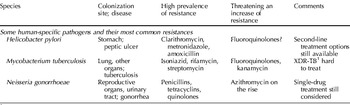
Some pathogens can only multiply in the human host, in which case any resistance observed must be the direct or indirect result of human intake of antimicrobials. We will present three such examples, each with a different route of infection. Helicobacter pylori is a colonizer of the stomach causing peptic ulcer in a minority of carriers, and related to the onset of gastric cancer. It is mainly transmitted orally from mother to child; family members usually have similar genotypes. Mycobacterium tuberculosis spreads via airborne droplets, and although infection is also asymptomatic in the majority of cases, it is so widespread that mortality rates are extremely high. Tuberculosis is treated with antimicrobials upon detection regardless of symptoms, at least in developed countries. Although M. tuberculosis is generally considered a human pathogen, transmission to animals has been described following close and prolonged contact; outbreaks in wildlife have also been described (Alexander et al., Reference Alexander, Pleydell, Williams, Lane, Nyange and Michel2002). Our third example, Neisseria gonorrhoeae, is a sexually transmitted pathogen that is treated whenever detected. The results are summarized in Table 1.
Summary of resistance patterns in various human pathogens

1 XDR-TB=extensively drug-resistant tuberculosis.
Resistance patterns in H. pylori
Discovery of this organism in 1984 came as a big surprise since the stomach epithelium was considered a sterile environment. Maybe because of this mindset, as soon as the spiral bacteria were detected, eradication was propagated. The discussion on whether and when H. pylori eradication is desired is still ongoing, but in the meantime a large number of individuals have been treated with single or multiple therapy strategies, and this has left its mark on the bacterial population. The abstract of a recent publication begins ‘The prevalence of antimicrobial drug resistance is now so high that all patients infected with H. pylori should be considered as having resistant infections’ (Graham and Shiotani, Reference Graham and Shiotani2008). As triple therapy of clarithromycin/metronidazole, amoxicillin and a proton-pump inhibitor is now ineffective in 80% of cases, quadruple therapy is considered a suitable option and quinolones are suggested as a second-line approach.
With the experience we have with Campylobacter jejuni (a gastrointestinal pathogen that colonizes both humans and animals, and which is taxonomically closely related to H. pylori), quinolones may only provide a short-term solution, as reduced susceptibility to fluoroquinolones in C. jejuni can occur with a single mutation, and H. pylori may soon learn that trick. Resistance is not restricted to developed countries, as an interesting overview presented recently illustrates (Vakil and Megraud, Reference Vakil and Megraud2007). Efforts to eradicate H. pylori by means of antimicrobials have, in approximately 25 years, clearly demonstrated that bacteria mutate and selection results in a life-saving solution, for the bacteria, that is.
Resistance patterns in M. tuberculosis
Present estimates suggest that one-third of the global population is infected with M. tuberculosis, causing approximately 3 million deaths yearly (Ducati et al., Reference Ducati, Ruffino-Netto, Basso and Santos2006). The combat of this pathogen is as old as the discovery of antibiotics, though a majority of infected people live in developing countries, where treatment is often unavailable. Compared with the short-term courses usually needed to treat H. pylori (7–10 days therapy), slow-growing M. tuberculosis can only be eradicated with months-long medication, making patient compliance a serious issue. The first-line treatment remains a quadruple therapy with isoniazid, a rifamycin, pyrazinamide and ethambutol/streptomycin (Shi et al., Reference Shi, Itagaki and Sugawari2007). Not surprisingly, resistance to each of these antimicrobials is on the rise. Second-line treatment relies on fluoroquinolones, clarithromycin and/or azithromycin. The abbreviation XDR-TB (for extensively drug-resistant tuberculosis) has been specifically coined for bacteria simultaneously resistant to at least rifampicin and isoniazid (both part of the first-line cocktail), to fluoroquinolones and to at least one of three injectable second-line drugs: amikacin, kanamycin and/or capreomycin (Jain and Mondal, Reference Jain and Mondal2008). This leaves very little treatment alternatives. XDR-TB has been found on every continent and is estimated to constitute 6% of multidrug-resistant tuberculosis cases (Jain and Mondal, Reference Jain and Mondal2008). This scary example illustrates where the story of resistance can lead to.
Resistance patterns in N. gonorrhoeae
Resistance patterns in this Gram-negative organism, the most common cause of sexually transmitted disease (STD) in a number of countries including the UK, reflect the history of antimicrobial discoveries. First, resistance to penicillins was recorded, followed by resistance to tetracyclines and now quinolones. Azithromycin resistance has also been documented to be above 5% in a European sentinel surveillance study (Martin et al., Reference Martin, Hoffmann and Ison2006). Current guidelines for first-line treatment include a third-generation cephalosporin such as cefixime or ceftriaxone (Roy et al., Reference Roy, Wang and Meltzer2005; Tyson, Reference Tyson2005). With experience from other pathogens, it may be prudent to consider multidrug medication rather than rely on single treatment regiments.
Resistances in pathogens specific to food animals
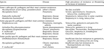
Compared with human-specific pathogens, far fewer data are available on resistance patterns in pathogens exclusively found in food animals. Some infectious diseases are controlled by culling rather than by antimicrobial treatment, with the result that there are few publications on resistance. A few examples are given for pathogens that specifically infect food-producing animals, notably cattle, pigs and poultry. Organisms were selected for the commonness of their disease, economic importance, comparable sites of infection, and possibly for taxonomic relatedness between them. The results are summarized in Table 2.
Summary of resistance patterns in various pathogens specific to food-producing animals

1 In vitro resistance to macrolides for Mycoplasma spp. does not necessarily indicate likelihood of clinical failure.
2 P. multocida and M. haemolytica are not species-specific but human infections are uncommon.
Pathogens specific to cattle
The first example of a cattle-specific disease is Johne's disease, caused by another species of the Mycobacterium genus: Mycobacterium avium subsp. paratuberculosis (MAP). (Mycobacterium bovis, perhaps a more familiar member of this genus, is not exclusively found in cattle.) Whether or not MAP is a zoonotic pathogen depends on the reader's willingness to accept the possible link to Crohn's disease. Owing to lack of data on resistance patterns, a comparison between human-specific M. tuberculosis and bovine MAP cannot be made.
For Mycoplasma bovis, a common intracellular pathogen in cattle identified in up to one-third of all pneumonia cases in calves, resistance was frequently found to tylosin, spectinomycin, lincomycin, tetracycline and oxytetracycline in a Belgian study (Thomas et al., Reference Thomas, Nicolas, Dizier, Mainil and Linden2003). It should be noted that there is no standardized procedure for determining in vitro susceptibility of Mycoplasma spp., so that various definitions of ‘in vitro resistance’ are in use. These reported resistances do not necessarily result in treatment failure and thus Mycoplasma spp. exemplify a possible conflict between in vitro MICs and clinical breakpoints. Based on the fact that Mycoplasma bovis is not replicating outside cattle, we can conclude that the observed in vitro reduced susceptibilities must result (directly or indirectly) from agricultural use of antimicrobials. Such a conclusion would be less sound for pathogens able to colonize multiple hosts.
Two major bacterial causes for bovine respiratory disease (BRD) are Pasteurella multocida and Mannheimia haemolytica. Both organisms can also infect other animals and humans so they are not strictly cattle-specific. Animal bites frequently result in P. multocida soft-tissue infections in humans; respiratory tract infections are mostly observed in elderly patients with underlying lung disease, where penicillin or doxycycline is a recommended treatment (Klein and Cunha, Reference Klein and Cunha1997). Nevertheless, P. multocida infections are far more common in cattle than in humans. German data suggest that in BRD isolates resistance to streptomycin is most common (30% in 2002/2003) followed by trimethoprim (12%), tetracycline (11%), chloramphenicol (10%) and nalidixic acid (10%) (Kaspar, 2006). A comparison between six European countries showed low prevalence (<10%) to most tested antimicrobials in most countries, with the exception of 24% tetracycline resistance in Italy and 20% in The Netherlands; streptomycin was not tested (Hendriksen et al., Reference Hendriksen, Mevius, Schroeter, Teale, Meunier, Butaye, Franco, Utinane, Amado, Moreno, Greko, Stärk, Berghold, Myllyniemi, Wasyl, Sunde and Aarestrup2008b). Japanese data identified 16% of BRD isolates exhibiting intermediate or resistant MIC values for benzylpenicillin (Yoshimura et al., Reference Yoshimura, Ishimaru, Endoh and Kojima2001). For these isolates, aminoglycosides were less active (19% resistant to dihydrostreptomycin), whereas ceftiofur and fluoroquinolones were highly active.
M. haemolytica, uncommonly found in human, is the most frequently isolated cause of BRD. In Germany, resistance is frequently found against tetracycline (14%) and streptomycin (44%) (Kaspar, Reference Kaspar2006), and these data from 2002/2003 show a sharp increase, especially for streptomycin, compared with data from 2001 (Wallmann et al., Reference Wallmann, Schröter, Wieler and Kroker2003). Tetracycline resistance was also common in some European countries, such as France (33–40%), Portugal (38%) or The Netherlands (31–51%) (Hendriksen et al., Reference Hendriksen, Mevius, Schroeter, Teale, Meunier, Butaye, Franco, Utinane, Amado, Moreno, Greko, Stärk, Berghold, Myllyniemi, Wasyl, Sunde and Aarestrup2008b). However, reported prevalences varied considerably from year to year in this study, possibly due to relatively low numbers of analysed isolates.
Pathogens specific to pigs
Resistance to tetracycline is extremely common for porcine pathogens. For Streptococcus suis, a frequent cause of pneumonia, meningitis and septicemia in pigs, a European study comprising isolates from seven countries found low persistence of resistance to gentamicin, spectinomycin and trimethoprim/sulfametoxazole but 75% resistance to tetracycline and 55% to tilmicosin. The pathogen was near to completely susceptible to fluoroquinolone, penicillin and ceftiofur (Wisselink et al., Reference Wisselink, Veldman, Van den Eede, Salmon and Mevius2006). A follow-up study with more recent isolates showed that this was changing in some countries, such as Poland (8% resistance to penicillin and 53% to gentamicin, samples from 2004), or Portugal (13 and 100% to penicillin and gentamicin, respectively) (Hendriksen et al., Reference Hendriksen, Mevius, Schroeter, Teale, Jouy, Butaye, Franco, Utinane, Amado, Moreno, Greko, Stärk, Berghold, Myllyniemi, Hoszowski, Sunde and Aarestrup2008a). German data from 2004–2006 confirm high prevalence of tetracycline resistance (70%), in addition to 17% gentamicin and 28% erythromycin resistance; penicillin-resistant S. suis was not detected, however (Schwarz et al., Reference Schwarz, Alešík, Grobbel, Lübke-Becker, Werckenthin, Wieler and Wallmann2007).
High prevalence of tetracycline resistance was also observed in Actinobacillus pleuropneumoniae, causing severe porcine pleuropneumonia with frequent mortality, for data from Spain (Gutiérrez-Martin et al., Reference Gutiérrez-Martín, del Blanco, Blanco, Navas and Rodríguez-Ferri2006). This prevalence had increased in the last decade, as had, to a lesser extent, resistance to gentamicin. Low prevalence of resistance was identified for penicillins, trimethoprim, sulfisoxazole and nalidixic acid.
For Mycoplasma hyopneumoniae, as a third example of a porcine pathogen causing pneumonia, resistance to macrolides/lincosamides and fluoroquinolones combined with susceptibility to oxytetracycline and doxycycline have been described (Vicca et al., Reference Vicca, Stakenborg, Maes, Butave, Peeters, de Kruif and Haasebrouck2004). These three porcine examples show that observed resistances are organism-dependent, even if they cause similar disease in the same host. Such differences are more likely a reflection of the genetic repertoire of the bacteria than exposure due to clinical treatment. This also illustrates that not every exposure results in resistance at the same rate for every bacterial species.
Both P. multocida and M. haemolytica can cause porcine as well as BRD, and it is interesting to compare reported resistances in these different hosts. Both organisms isolated from swine respiratory disease (SRD) in Germany were most commonly found resistant to oxacillin (32% for P. multocida and 38% for M. haemolytica) and the latter was resistant to streptomycin in 19%; low prevalences were reported for all other compounds (Wallmannn et al., Reference Wallmann, Schröter, Wieler and Kroker2003). More recent data suggest that 28% of P. multocida are resistant to trimethoprim (Kaspar, Reference Kaspar2006). Nevertheless, porcine P. multocida isolates are generally less frequently resistant to antimicrobials than bovine isolates (Kaspar, Reference Kaspar2006). A clinical breakpoint for amoxicillin was recently proposed for porcine respiratory tract pathogens (Schwarz et al., Reference Schwarz, Böttner, Goosens, Hafez, Hartmann, Kaske, Kehrenberg, Kietzmann, Klarmann, Klein, Krabisch, Luhofer, Richter, Schulz, Sigge, Waldmann, Wallmann and Werckenthin2008); however, resistance to amoxicillin is not reported as common in porcine P. multocida or M. haemolytica.
Pathogens specific to poultry
For Ornithobacterium rhinotrachialae, a poultry respiratory pathogen with important economic consequences, recent data on antimicrobial resistance are limited. One study from the US reported annual increases of prevalence between 2 and 7% for a number of antibiotics, including gentamicin, ampicillin and tetracycline (Malik et al., Reference Malik, Olsen, Kumar and Goval2003). As an example of avian Mycoplasma infection, fluoroquinolone-resistant Mycoplasma gallisepticum and Mycoplasma synoviae have been reported from Israel (Gerchman et al., Reference Gerchman, Lysnvansky, Perk and Levisohn2008). Similar results were obtained from The Netherlands for M. synoviae, which was found susceptible to doxycycline, tylosin and tilmicosin (Landman et al., Reference Landman, Mevius, Veldman and Feberwee2008). Reported tylosin resistance in this organism suffers from lack of agreement on a breakpoint and lack of standardized methods for testing. Despite such uncertainties, unpublished data suggest that macrolides can still be clinically effective against Mycoplasma in poultry even though in vitro MICs suggest otherwise, illustrating again that laboratory testing may not always give clinically relevant data (P. Silley, unpublished observations). It is interesting to compare these findings with the cattle-specific M. bovis (where resistance to tylosin was frequent but fluoroquinolones remain effective) and porcine M. hyopneumoniae (notably resistant to macrolides and fluoroquinolones) (Table 1). The observed resistance patterns of these related species are possibly reflecting the selective pressures they encounter, as a result of different medication of the host animal.
Finally, P. multocida can again be included here, as it is the cause of fowl cholera. German data suggest low prevalence of resistance in poultry isolates to all tested compounds including tetracycline (Wallmann et al., Reference Wallmann, Schröer and Kaspar2007). This is surprising, as bovine isolates have shown that the organism is quite capable of developing tetracycline resistance, and tetracycline resistance in poultry isolates of Campylobacter is common in Germany (Luber et al., Reference Luber, Wagner, Hahn and Bartelt2003). Such examples show that it is not so simple to attribute particular resistance patterns to presumable selective pressures as a consequence of usage of that antimicrobial in food animals.
Resistances in zoonotic pathogens
Since humans share a number of pathogens with food-producing animals (where they can be pathogenic or benign), it is frequently expressed that particular resistance patterns detected in human pathogens result from veterinary antibiotic use. The frequent use of antibiotics as growth promoters in agriculture was challenged and subsequently banned in Europe (Phillips, Reference Phillips2007). Even though some links between observed resistance in particular bacterial species and the use of those compounds in agriculture seemed extremely obvious, close examination of the details revealed a number of complications (Wassenaar, Reference Wassenaar2005). A detailed analysis of the apparent link between fluoroquinolone resistance in C. jejuni and its presumed increased virulence was recently published (Wassenaar et al., Reference Wassenaar, Kist and de Jong2007), illustrating that even this case may not be as clear-cut as it appeared.
We cannot easily compare antibiograms of veterinary and human isolates of the same pathogen, to assess the contribution of selection in both hosts. The literature on resistances in pathogens that can colonize multiple hosts is a plethora of publications using various MIC definitions, describing bacterial subpopulations selected for a particular disease, or host, or even for a pre-selected resistance, such as publications describing resistance patterns of methicillin-resistant Staphylococcus aureus. The variables tested are frequently different in papers written from a veterinary or a public health perspective. Even when considering one invariant organism and agent combination, there remain multiple variables that influence the outcome of monitoring studies.
Ignoring differences in methodology to determine MIC values, definitions of the term ‘resistance’ and bias in sampling data (all of which substantially influence the outcome), the most important parameters that introduce variation are country, host and year of isolation. It is oftentimes impossible to find comparable data for which only one of these parameters differs. A European initiative attempted to make comparisons within one microorganism, tested under standardized conditions, from various countries for the years 2002, 2003 and 2004 (Hendriksen et al., Reference Hendriksen, Mevius, Schroeter, Teale, Meunier, Butaye, Franco, Utinane, Amado, Moreno, Greko, Stärk, Berghold, Myllyniemi, Wasyl, Sunde and Aarestrup2008b). These results can be combined with those from Italy (Moroni et al., Reference Moroni, Pisoni, Antonini, Villa, Boettcher and Carli2006) and Germany (Kaspar, Reference Kaspar2006). The latter is a publication of the German GERM-Vet monitoring program, initiated to determine the current status of antimicrobial susceptibility of bacterial pathogens from animals in Germany. This initiative has introduced a scientific rigour to surveillance in that it has addressed many of the variables that can otherwise confound interpretation of data (Wallmann et al., Reference Wallmann, Schröter, Wieler and Kroker2003). It is hoped that these standards will be adopted in other countries. Recent publications of Hendriksen et al. (Reference Hendriksen, Mevius, Schroeter, Teale, Jouy, Butaye, Franco, Utinane, Amado, Moreno, Greko, Stärk, Berghold, Myllyniemi, Hoszowski, Sunde and Aarestrup2008a, Reference Hendriksen, Mevius, Schroeter, Teale, Meunier, Butaye, Franco, Utinane, Amado, Moreno, Greko, Stärk, Berghold, Myllyniemi, Wasyl, Sunde and Aarestrupb) surveying resistance across Europe among bacterial pathogens and indicator bacteria for pigs and cattle, although not using a harmonized methodology, are a step in the right direction, as indeed are the increasing numbers of national surveillance schemes.
S. aureus: bovine mastitis and human opportunistic infections
The problem that a subpopulation from one host species cannot be directly compared with those of other host species is most clearly illustrated by S. aureus. Mastitis in dairy cows is a frequently occurring problem that usually requires antimicrobial treatment. The bacterial causes are diverse, but S. aureus is one of the major culprits. Regional differences in reported resistances in mastitis-causing S. aureus populations can be dramatic, such as 59% prevalence of sulfonamide resistance in France but only 4% in Denmark and 0% in Spain (Hendriksen et al., Reference Hendriksen, Mevius, Schroeter, Teale, Meunier, Butaye, Franco, Utinane, Amado, Moreno, Greko, Stärk, Berghold, Myllyniemi, Wasyl, Sunde and Aarestrup2008b). In that comparative study, low levels of resistance were reported for most of the compounds tested except for 10–49% for penicillin, with Italy (43%), Latvia (49%), England (38–46% depending on the year), Denmark (23–40%) and Spain (33–44%) among the regions reporting higher prevalence, compared with only 4% in France. However, ampicillin, amoxicillin and doxycycline had not been tested, for all of which high prevalence of resistance was reported (99, 100 and 47%, respectively) in S. aureus from bovine mastitis in Italy in 2004 (Moroni et al., Reference Moroni, Pisoni, Antonini, Villa, Boettcher and Carli2006). Thus, the selection of tested compounds can give different impressions of severity of the resistance problem. Moreover, with such dramatic regional or national differences, comparisons of human and animal isolates should ideally only take place within one region.
How should we compare veterinary findings with S. aureus infections in humans? In human medicine, resistance to one compound raises red flags: methicillin, a beta-lactam whose resistance in S. aureus is usually accompanied by resistance to macrolides, aminoglycosides, clindamycin and sometimes quinolones or rifampin. Nosocomial infections of methicillin-resistant S. aureus (MRSA) are a well-known threat. The incidence of MRSA in blood cultures in various European countries differs, with highest reported incidences in the South and East. The UK is an exception, recording 44% MRSA in blood cultures, as high as Greece, Portugal or Rumania, compared with 0.6% in Denmark or 0.9% in The Netherlands (Beovic, Reference Beovic2006). In addition, community-acquired MRSA is on the rise in many countries.
S. aureus is also a common benign colonizer of the nasopharynx and skin. Surveillance programs have been targeted at MRSA, but by zooming in on this resistant subpopulation, the true variation in the overall S. aureus population is clearly not assessed. An appropriate question is to ask which of two investigated bacterial populations, nosocomial or community-acquired MRSA, would be more adequate to compare with the animal isolates causing bovine mastitis. A mastitis case in a herd of cattle may be somewhat comparable with a nosocomial human infection in a hospital, as in both settings individuals are in close contact. However, animals clearly undergo extreme environmental exposure compared with the confounded human patients, and they may be less vulnerable to opportunistic infections. Comparing S. aureus isolates from healthy human individuals (community carriers of MRSA), to use the alternative approach, would not resemble the medication pressure posed on the bacterial population that both nosocomial infective agents and veterinary pathogens may have undergone. The dilemma is solved because the bacterial populations of benign colonizers of humans are different from those causing either nosocomial infections or bovine mastitis, as established by genotyping data such as MLST (Deurenberg et al., Reference Deurenberg, Vink, Kalenic, Friedrich, Bruggeman and Stobberingh2007).
A case where both populations may overlap was identified for pig farmers in The Netherlands, who are more likely to be MRSA carriers with genotypes identical to those colonizing pigs (Khanna et al., Reference Khanna, Friendship, Dewey and Weese2008). These observations have been confirmed in other countries, and a possible epidemic has been forecasted (Wulf and Voss, Reference Wulf and Voss2008). For human medicine, treatment options are all that matter. The discovery of vancomycin-resistant S. aureus (methicillin-resistant or not) is of extreme concern. In the case of MRSA patients, treatment is often restricted to trimethoprim/sulfamethoxazole, doxycycline or linezolid (Powell and Wenzel, Reference Powell and Wenzel2008). This makes the findings of Moroni and coworkers even more disturbing (Moroni et al., Reference Moroni, Pisoni, Antonini, Villa, Boettcher and Carli2006). There is an immediate need for standardized and extended antibiogram testing of S. aureus in food animals (cattle as well as pigs) for both methicillin-susceptible and -resistant populations, in order to anticipate future treatment possibilities.
C. jejuni/coli
A lot has been said about fluoroquinolone resistance in C. jejuni/coli and the possible link to use of fluoroquinolones in poultry, and these arguments will not be repeated here. As a result of such discussions, fluoroquinolones use in poultry was banned in the US in 2005. Whether this decision was justified remains disputable. It is, however, interesting to see that this ban does not appear to have resulted in a reduction of fluoroquinolone resistance inCampylobacter isolated from poultry in the US, as deduced from data made available online by the USDA (http://www.ars.usda.gov/sp2UserFiles/Place/66120508/CampyMICAll.swf).
Escherichia coli, a complex species
E. coli, the guinea pig of microbiology, is a species with a complex epidemiology. Some strains of the species are benign to both human and animal, others are pathogenic to particular hosts, and benign strains can become opportunistic pathogens, for instance causing urinary tract infections (UTIs). Particular strains may be related to particular sites of infection, whereas other strains are more versatile. Antimicrobial resistance surveillance has mostly concentrated on particular isolation sites (mostly for one host only), frequently related to particular disease manifestations. A systematic literature review was performed by Erb and coworkers on antimicrobial resistance reported for E. coli isolated from humans (Erb et al., Reference Erb, Stürmer, Marre and Brenner2007). In absolute numbers, most antibiotic-resistant organisms are acquired in the community, not in hospitals, so that this represents the biggest reservoir of resistance. In asymptomatic populations, ampicillin resistance varied between 13% (in The Netherlands, 1994) and 100% (in Mexico, 1996). The same wide variation was seen for trimethoprim (same extremes, same countries), and ciprofloxacin resistance was already about 25% in Spain in 1999 (Erb et al., Reference Erb, Stürmer, Marre and Brenner2007). There was no difference in resistance prevalence between stool isolates from healthy individuals and blood or urine isolates from patients. This is in accordance with the view that E. coli infections are mostly caused by ascending infections by normal bowel flora.
An interesting observation is that E. coli isolated from wild animals living in the Antarctic or Gabon, believed to be unexposed to people, were completely susceptible to all antibiotics tested (Skurnik et al., Reference Skurnik, Ruimy, Andremont, Amorin, Rouquet, Picard and Denamur2006). This raises the question of what biological purpose resistance genes had and have in the absence of antimicrobial pressure. After all, resistance genes have evolved long before humans started to apply antimicrobials, and must have a role in the natural ecology of bacteria.
Antimicrobial resistance in E. coli causing UTIs in humans is of major concern, especially the increase in incidence of expanded-spectrum beta-lactamases (ESBLs) (Pitout and Laupland, Reference Pitout and Laupland2008). These pose a threat to the ‘last option’ antibiotics ceftazidime, cefepime and azteonam (Beovic, Reference Beovic2006). In that review, concern about the increase in co-trimoxazole resistance with community-acquired UTIs is also expressed. As the author states, the candidate future treatment of this common infectious disease with fluoroquinolones or beta-lactams is likely to set another vicious circle of resistance in motion. Hospital-acquired UTIs are probably even more frequently treated with antimicrobials than community acquired UTIs. A survey was conducted to investigate resistance and treatment of UTIs caused by E. coli and other organisms on one particular day in November 2003 (Europe) and November 2004 (Europe plus Asia). A total of 56% of the hospitalized urological patients were receiving antimicrobial therapy on the studied day, of which 46% were for prophylaxis, 26% for microbiologically proven UTI, 21% for clinically suspected UTI and 7% for other infections. The most commonly used antibiotics were fluoroquinolones (35%), cephalosporins (27%), penicillins (16%), aminoglycosides (15%) and co-trimoxazole (9%) (Johansen et al., Reference Johansen, Cek, Naber, Stratchounski, Svendsen and Tenke2006). Such treatment practices put the criticism of prophylactic treatment practiced in veterinary medicine into perspective.
E. coli can also cause mastitis in cattle, so that we can compare the resistance patterns observed in such a population with that of S. aureus isolated from the same niche. Amoxicillin, tetracycline and streptomycin resistances have the highest prevalence in The Netherlands reaching out to 20% in E. coli from bovine mastitis (MARAN, 2005), followed by trimethoprim/sulfamethoxazole (about 10%) and kanamycin and neomycin (below 10%). Coagulase-negative S. aureus isolated from bovine mastitis in the same country, same year, are frequently (40%) resistant to penicillin and sometimes (6%) to oxacillin. Again, the different genetic repertoire of these organisms is most likely responsible for these differences. Similar to the Dutch data, E. coli and S. aureus from bovine mastitis cases in Germany were never amoxicillin/clavulanic acid-resistant but the German isolates report lower incidences for tetracycline and streptomycin resistance in mastitis E. coli (Kaspar, Reference Kaspar2006).
E. coli isolated from cattle suffering from either mastitis or diarrhea was compared in ten European countries (Hendriksen et al., Reference Hendriksen, Mevius, Schroeter, Teale, Meunier, Butaye, Franco, Utinane, Amado, Moreno, Greko, Stärk, Berghold, Myllyniemi, Wasyl, Sunde and Aarestrup2008b). However, from the way the data are presented, it seems that in some countries mastitis isolates and in other countries diarrhea isolates were analysed. This makes comparisons somewhat difficult.
E. coli isolated in Germany from various animal sources (urinary/genital, respiratory and gastrointestinal tract infections in cats, dogs, horses or swine) were all compared under standardized conditions (Grobbel et al., Reference Grobbel, Lübke-Becker, Alešík, Schwarz, Wallmann, Werckenthin and Wieler2007). The highest incidences of resistance were reported for sulfamethoxazole (18–59%), tetracycline (14–54%) and ampicillin (14–39%), whereby isolates from cats, dogs and horses were generally more frequently susceptible.
One subpopulation of E. coli that cannot be left out here is Shiga-toxin-producing E. coli (STEC, also called VTEC for verotoxigenic E. coli), which is associated with contaminated beef and vegetables. A recent study from Ireland showed that multidrug resistance in VTEC (mainly O157 and some O111 and O26 isolates from cattle carcasses, beef and humans) is uncommon (1.4%) (Walsh et al., Reference Walsh, Duffy, O'Mahony, Fanning, Blair and McDowell2006). Nevertheless, one isolate from ground beef was resistant to ten antimicrobials belonging to seven different antimicrobial classes. One would think that such observations are of concern, and ask for careful surveillance. However, in comparison with other well-covered food-related zoonotic organisms, hardly any international comparison study is available for this important pathogen. Publications describing national data are mostly based on pre-2002 isolates, describe one isolation source only, frequently live cattle or beef at retail, or concentrate on particular resistances (ESBL-producing E. coli) without considering the pathotype. Thus, despite a vast interest in this ‘hamburger bug’, a comparison as presented for other zoonotic pathogens cannot be made due to lack of published data. In addition to this lack of data, it should be mentioned that antimicrobial treatment of human STEC infections is controversial as their usage may exacerbate symptoms, particularly the hemolytic uremic syndrome.
Salmonella enterica: serovars come and go
Important insights can be gained from an understanding of resistance in Salmonella. Salmonella resistance profiles strongly correlate to specific serovars. Fluctuations in dominant serotypes will be the most important factor determining the observed resistance levels for Salmonella. Threlfall et al. (Reference Threlfall, Day, de Pinna, Charlett and Goodyear2006) made this point very forcibly as they investigated the changes in the occurrence of antimicrobial resistance in S. enterica serotypes Enteritidis and Typhimurium from human infection in England and Wales in 2000, 2002 and 2004. They showed that the incidence of strains of Salmonella Enteritidis with resistance to nalidixic acid coupled with decreased susceptibility to ciprofloxacin had more than doubled between 2000 and 2004, whereas the overall levels of resistance in Salmonella Typhimurium had fallen by approximately 25%. In relation to published data on veterinary sales of antimicrobials in the UK, the findings demonstrated that changes in the incidence of resistance did not correlate with changes in veterinary usage (Threlfall et al., Reference Threlfall, Day, de Pinna, Charlett and Goodyear2006). For Salmonella Enteritidis, important factors in the increased incidence of resistance were foreign travel and the consumption of imported foods contaminated with drug-resistant strains. For Salmonella Typhimurium, the most important factor has been an overall decline in the occurrence of multiple drug-resistant Salmonella Typhimurium definitive phage type 104, a change in serotype prevalence. These studies have demonstrated that changes in the incidence of resistance in predominant Salmonella spp. in humans in England and Wales from 2000 to 2004 are multifactorial. The important point made here is that resistance rates were shown to be a function of serotype prevalence rather than antibiotic usage.
Concluding remarks
Resistances in a selection of pathogens specific to humans or food animals have been presented here. Clearly, multidrug resistance impairing medication is a more severe problem in those human pathogens presented. This may partly be a reflection of the selected examples; however, all presented animal pathogens are common causes of disease in farming practice, requiring antibiotic treatment.
Compared with those pathogens restricted to a single animal host, pathogens found in both human and animal hosts appear to have higher incidences of resistance. Such comparisons become difficult to properly evaluate, as the organisms vary in their available genetic repertoire of resistance. Examples have been given to illustrate that not every exposure results in resistance at the same rate for every bacterial species (e.g. different resistance patterns in pathogens causing porcine pneumonia). Differences in the available genetic repertoire are one explanation for this, but the story is more complex. Some organisms develop resistance to a particular compound in one animal host but not in another (e.g. P. multocida tetracycline resistance), although that compound is licensed for use in either host. For other pathogens, such as Salmonella, the rise and fall of particular subtypes may coincide with particular resistance patterns, so that these patterns are not necessarily a reflection of antimicrobial use.
It was not our intention to ‘name, shame and blame’ as that is not going to help solve the resistance challenge. We accept that antimicrobial use, in whatever host, provides a selective pressure that can result in less susceptible bacterial populations. Nevertheless, by looking at different pathogens with different host distributions, the frequently expressed opinion that veterinary use of antimicrobials is at the heart of many problems seems to miss the point. It is clear that the most serious and acute problems of multidrug resistance are in human medicine and it is here where solution and prevention are mostly needed. We need active research to come up with novel strategies in our fight against infectious diseases.
Some directions have already been mentioned where more data are needed. The scientific field is very active in developing new strategies beyond the next-generation compound that can give temporarily (but potentially life-saving) relief at best. Novel approaches to combat bacterial infections and their resistances include the development of inhibitors of resistance genes, a concept with proven functionality for beta-lactamases. Inhibitors of antibiotic efflux, where maybe plants can teach us a few useful lessons, and novel antibiotic strategies, such as compounds upsetting the intercellular communication on which many bacterial pathogens rely, are also possibilities worth exploring (Wright and Sutherland, Reference Wright and Sutherland2007). Finally, the exponentially increasing number of complete bacterial genomes currently being sequenced can be mined to identify novel gene candidates as antibacterial targets (Mills, Reference Mills2006).
Acknowledgment
TMW received support from Med-Vet-Net, a European Network of Excellence.