




No CrossRef data available.
Article contents
Comparative study of the presence of Trypanosoma cruzi kDNA, inflammation and denervation in chagasic patients with and without megaesophagus
Published online by Cambridge University Press: 04 July 2005
Abstract
Neuronal lesions have been considered the hallmark of chagasic megaesophagus, but the role of Trypanosoma cruzi and the participation of the inflammatory cells in this process are still debated. In the present study we counted neurons in the oesophagus from patients with and without megaesophagus and further examined these samples for the presence of parasite kDNA and cells with cytolytic potential (Natural Killer cells, cytotoxic lymphocytes and macrophages). The presence of parasite kDNA was demonstrated in 100% of cases with megaesophagus and in 60% of patients without megaesophagus. When analysed for the number of neurons, the patients without megaesophagus could be classified into 2 groups, as having normal or a decreased number of neurons. The former group did not show any inflammatory process, but interestingly, all patients without megaesophagus presenting decreased number of neurons also presented both parasite kDNA and inflammatory process in the organ. We further observed that the numbers of cytotoxic cells in the myenteric plexus region inversely correlate with the number of neurons. These data together strongly suggest that chronic lesions in chagasic megaesophagus might be a consequence of immune-mediated mechanisms, that last until the chronic phase of infection, and are dependent on the persistence of parasite in the host's tissue.
Information
- Type
- Research Article
- Information
- Copyright
- © 2005 Cambridge University Press
INTRODUCTION
Chagas' disease, a parasitic infection caused by the flagellate protozoan Trypanosoma cruzi, remains as one of the major public health problems in Latin America. Over the recent years, strong migratory trends were created by social and economic changes in Latin America, that pushed thousands of T. cruzi-infected persons from endemic areas to wealthier regions (Chapadeiro, 1999; Prata, 2001). Moreover, T. cruzi–infected persons have been encountered in increasing numbers in the United States, where they can become ill or transmit the disease by blood or organ donation (Navin et al. 1985; Kerndt et al. 1991; Smith and Wright-Kanuth, 2003).
Acute Chagas' disease is characterized by fever and myocarditis related to intracellular parasitism. Usually these symptoms subside spontaneously. Most patients then remain serologically positive, but asymptomatic for the rest of their lives. However, some patients, after a prolonged asymptomatic infection (20–30 years), exhibit cardiac and/or digestive manifestations, the latter being related to the development of megaesophagus and megacolon (Chapadeiro, 1999; Prata, 2001).
Patients with megaesophagus demonstrate decreased peristalsis and incomplete swallowing, and these abnormalities precede clinical symptoms. Pathologically, the megaesophagus exhibits striking luminal enlargement and muscular hypertrophy. Microscopically, inflammatory infiltrates are found in the muscularis mucosa, submucosa and muscularis propria. Inflammatory lesions of the intramural nervous system are associated with a striking reduction in the number of neurons. The denervation process is thought to underlie the clinical findings in chagasic megaesophagus (Rezende and Lauar, 1960; Koberle, 1968; de Oliveira et al. 1998).
The role of the parasite in the development of chronic inflammatory processes and/or in the denervation has been debated in the literature. We and other groups of researchers, have demonstrated the presence of parasite kinetoplast DNA (kDNA) in the esophagus from, respectively, 100% and 77% of patients bearing megaesophagus (Vago et al. 1996; Lages-Silva et al. 2001), and in 50% of serologically positive cases without megaesophagus (Vago et al. 2003), which suggested an association between megaesophagus development and persistence of the parasite in the chronic phase. The aim of the present study was to analyse the inflammatory process, denervation and presence of T. cruzi kDNA in chagasic patients with and without megaesophagus. The associations between these parameters were examined and discussed, giving a better understanding of the chagasic megaesophagus pathophysiology.
MATERIALS AND METHODS
Patients and tissue samples
Oesophagus tissue samples were obtained from 6 chagasic patients with megaesophagus, 8 chagasic patients without megaesophagus and 6 control individuals submitted to necropsy or surgery procedures at Faculdade de Medicina do Triângulo Mineiro (Uberaba, Minas Gerais, Brazil). The patients had never received any parasite-specific treatment. Informed consent was obtained from family members prior to tissue procurement. Serological tests indicative of Chagas disease (complement fixation, haemagglutination, and immunofluorescent tests) were positive in all patients studied. All of the patients had left the endemic area for more than 20 years before the collection procedure, and during that time, patients without megaesophagus did not present any symptom related to digestive disease. They used to live in Uberaba, MG, Brazil, where the natural transmission was interrupted more than 20 years ago, and they had never received a blood transfusion. This work was approved by UFMG Research Ethic Committee.
The control group was composed of non-infected individuals, as indicated by negative serology specific for Chagas' disease. Non-infected individuals were also from the state of Minas Gerais and had an average age of 56±22 years and a maximum oesophagus diameter of 1·4±0·3 cm.
Chagasic patients without megaesophagus did not present previously any symptom related to digestive system's compromising. The group had a medium age of 53±9 years and maximum oesophagus diameter of 1·4±0·2 cm.
Chagasic patients with megaesophagus presented a medium age of 54±10 years and the maximum oesophagus' diameter of 3·8±0·5 cm. The diagnosis of megaesophagus was established from clinical data reporting oesophagus obstruction, and from radiological studies. Manometric studies of megaesophagus demonstrated decreased peristalsis and incomplete relaxation of the lower oesophagus sphincter during swallowing. These abnormalities precede clinical symptoms and dilatation seen by radiographical studies.
Tissue samples were collected from 3 different areas of the oesophagus (upper, medium and lower). Part of each specimen was processed in 4% neutral buffered formaldehyde solution and embedded in paraffin for immunohistochemical studies. Another portion was snap-frozen in liquid nitrogen and stored at −70 °C until DNA extraction. To determine the number of ganglion cells in the intramural plexus, rings of tissues were taken from 3 different regions (upper, medium and lower) of the oesophagus, processed in formaldehyde and embedded in paraffin.
Investigation of the degree of denervation and of the presence of inflammation
After fixation in paraformaldehyde and embedding in paraffin, the oesophagus rings were cut into serial 7 μm sections, and every seventh section was selected, avoiding counting ganglion cells twice. Twenty sections were taken from each block, making a total thickness of 980 μm. Giemsa-stained sections were then analysed for the number of neurons and the presence of inflammatory processes. For each case, the number of neurons obtained was multiplied by a correction-factor, in order to compensate for any hypertrophy. The factor for each patient was calculated as following, according to the protocol described by Adad et al. (1991).
Correction-factor=perimeter of patient's oesophagus/medium perimeter of controls' oesophagus.
Immunohistochemical analysis
Paraffin-embedded oesophagus tissues from chagasic patients and controls were used for quantification and characterization of NK cells, cytotoxic lymphocytes and macrophages. Six micron-thick sections were deparaffinized through xylene, and dehydrated in graded alcohols. Endogenous peroxide activity was inhibited by incubation with 1% hydrogen peroxide and 30% absolute methanol for 30 min. The slides were then incubated with 2% normal swine serum (NSS) (Sigma) in phosphate buffered saline (PBS) for 15 min and subsequently with monoclonal antibodies specific to CD57+ NK cells (Santa Cruz Biotechnology, clone sc-6261, 1[ratio ]200), TIA-1+ cytotoxic lymphocytes (Santa Cruz Biotechnology, clone sc-1751, 1[ratio ]200), and CD68+ macrophages (DAKO, clone KP1, 1[ratio ]100), followed by a second step incubation with peroxidase-conjugated rabbit anti-mouse antibodies (Dako) for 45 min. Peroxidase activity was demonstrated by incubation with 3–3′-diaminobenzidine (Sigma) and hydrogen peroxidase for 10 min. Slides were counterstained with Gill's haematoxylin (Sigma), dehydrated in graded alcohols, and mounted in synthetic mounting media. Appropriate negative control slides were run for each case.
Enumeration of NK cells and cytotoxic lymphocytes was performed by counting cells on 20 fields (total area of 53·333 μm2) of myenteric plexus, on a single slide per patient. Morphometric studies of macrophages were performed by image analysis (Kontron KS300 v. 2.0), by measuring CD68+ areas on 20 fields (total area of 53·333 μm2) of myenteric plexus, on a single slide per patient.
Statistical analysis
The significance of differences in the number of CD57+ NK cells, TIA-1+ cytotoxic lymphocytes, and macrophages among the different groups was analysed by ANOVA one-way test. Differences were considered statistically significant at P<0·05.
DNA isolation and hot-start PCR
To test for the presence of T. cruzi kDNA we used the polymerase chain reaction (PCR) with the S35 (5′AAATAATGTACGGGTGAGATGGCATGA3′), and S36 (5′GGTTCGATTGGGGTTGGTGTAATATA 3′) primers (Sturm et al. 1989) flanking a 330 bp variable segment of the kDNA minicircles, a naturally amplified target which was expected to permit high sensitivity in the PCR reaction, using a hot-start PCR protocol. For the DNA extraction step, oesophagus sections from the 3 anatomic regions of each case were used. Oesophagus sections of approximately 5×5 mm were minced, subjected to alkaline lysis with 50 mM NaOH, neutralized with 130 mM Tris-HCl (pH 7·0) and used directly in the PCR reaction after 10-fold dilution in twice distilled water. As negative control of the DNA-extraction step, samples of amphibian muscle were used.
The hot-start PCR is a 2-step procedure which consists of preparing 2 distinct PCR mix layers. The lower layer contained 10 mM Tris-HCl, pH 8·4, 75 mM KCl, 3·5 mM MgCl2, 0·1% Triton X-100, 200 μM of each dATP, dCTP, dTTP and dGTP (Sigma, Inc.) and 10 pmol of S35 and S36 primers. The upper layer was then added to each reaction tube, which consisted of 10 mM Tris-HCl, pH 8·4, 75 mM KCl, 3·5 mM MgCl2, 0·1% Triton X-100, 0·25 UI of Taq DNA Polymerase (Phoneutria Inc., Brazil) and double-distilled water. Two μl of the DNA diluted 1[ratio ]10 were used as PCR template. For the amplification of the 330 bp kDNA fragment, the PCR program consisted of an initial cycle of denaturation at 94 °C for 5 min, followed by 34 cycles of denaturation at 94 °C for 1 min, annealing at 64 °C for 1 min and extension at 72 °C for 1 min. The final extension step was equivalent to 72 °C for 5 min. As negative control for the PCR, tubes containing PCR mix without DNA were used. Five μl of the PCR products were analysed by electrophoresis on a silver stained 6% polyacrylamide gel (Vago et al. 1996).
To test the quality of the DNA samples extracted, we amplified a 380 bp fragment of the variable region from the human mitochondrial DNA. The PCR conditions were 10 mM Tris-HCl (pH 8·5); 50 mM KCl; 0·1% Triton X–100; 1·5 mM MgCl2; 200 μM of dNTPs; 1 UI of Taq DNA polymerase (Phoneutria Inc., Brazil) and 5 pmol of each primer L15926 (5′ CGGAGATGAAAACCTTTC 3′) and H16246 (5′ TGAGGGGTGGCTTTGGAGTT 3′) in a final volume of 25 μl. The reactions were amplified for 30 cycles using denaturation at 94 °C for 1 min, annealing at 55 °C for 30 sec and extension at 72 °C for 1 min (Sturm et al. 1989).
RESULTS
Table 1 shows the results of neuronal counting in the intramural nervous system of oesophagus from chagasic patients with and without megaesophagus, and control individuals. The number of neurons in 980 μm of oesophagus length ranged from 0 to 4 neurons in chagasic patients with megaesophagus, and from 347 to 459 in control individuals. Chagasic patients without megaesophagus presented extremely heterogeneous neuronal counting, a fact that instigated us to reclassify this group into 2 subgroups: group IIa, composed of patients with numbers of neurons ranging from 196 to 351/980 μm; and group IIb composed of patients with 20 to 46 neurons/980 μm.

Histological analysis of oesophagus sections from patients without megaesophagus was coherent with the previously proposed reclassification of this group. Patients from group IIa did not show any inflammation, while those with low neuronal counting, group IIb, presented discrete inflammatory process composed of mononuclear cells (Table 1). Histological analysis of oesophagus from patients with megaesophagus showed an intense focal and diffuse inflammatory process, composed mainly of mononuclear cells, and in some cases, eosinophils and mast cells (Fig. 1A and B). Ganglionitis, periganglionitis and vasculitis were frequently observed.

Fig. 1. Aspects of the inflammatory process in myenteric plexus of oesophagus representative of patients of group IIb (A, C, E, and G) and patients with megaesophagus (B, D, F, and H). (A) Chronic inflammatory process with mononuclear cells; connective and vascular neo-formation. Neurons are not visible. Haematoxylin and eosin staining, objective 20X. (B) Intense and nodular mononuclear inflammatory infiltration, with presence of eosinophils (inset, arrowhead) and mast cells. (C and D) Rare CD57+ natural killer cells (arrow-head) are seen in inflammatory foci in patients of group IIb (C) compared to patients with megaesophagus (D). (E and F) The same is observed for TIA-1+ cytotoxic lymphocytes. (G and H) CD68+ positive cells.
Analysis of immunostained sections with anti-CD57 mAb and anti-TIA-1 mAb, revealed the presence of NK cells (Fig. 1C and D) and cytotoxic lymphocytes (Fig. 1E and F) in the myenteric plexus of oesophagus from patients with megaesophagus, and also in the group IIb of individuals without digestive disease. Patients without megaesophagus (group IIb) presented NK cells and cytotoxic lymphocytes in a lower frequency than patients with megaesophagus (Fig. 2). Image analysis of CD68+ stained sections demonstrated that the concentration of macrophages in the myenteric plexus of the oesophagus is correlated with the development of the disease (Fig. 1G and H, and Fig. 3).

Fig. 2. Morphometric analysis of TIA-1+ cytotoxic lymphocytes and CD57+ NK cells in patients of group IIb and patients with megaesophagus. Values for the controls are not detectable. The values are expressed as means±S.D. * Statistically significant differences between the two groups were observed for both analyses.

Fig. 3. Morphometric analyses of CD68+ cells in patients without megaesophagus (groups IIa and IIb), and patients with megaesophagus. The values are expressed as means of CD68+ areas±S.D. * Statiscally significant differences between this group and control individuals. ** Statiscally significant difference between group IIb and group of patients with megaesophagus.
All of the analysed patients with megaesophagus demonstrated parasite kDNA in the oesophagus, while 60% of patients without megaesophagus presented T. cruzi kDNA in the organ (Fig. 4 and Table 2). Interestingly, all of the patients without megaesophagus, from group IIb, presenting decreased numbers of neurons, also had parasite kDNA, while only 2 patients from group IIa, with no inflammatory process, presented positive PCR amplification (Table 2).

Fig. 4. (A) Amplification of a 330 bp Trypanosoma cruzi kDNA fragment from human oesophagus samples by using a Hot – start protocol. Five μl of PCR products were loaded in each lane of a 6% silver-stained polyacrylamide gel. Lane 1, molecular size marker (1 Kb DNA Ladder, BRL). Lanes 2–6, chagasic patients with megaesophagus: E08, E11, E12, E21, E24. Lanes 7–9, chagasic patients without megaesophagus (group IIb): E04, E07, E10. Lanes 10–11, chagasic patients without megaesophagus (group IIa): E13, E18. Lanes 12 and 13: Control individuals without Chagas' disease: E05, E17. Lane 14: negative control of the DNA extraction (amphibian muscle). Lane 15: T. cruzi genomic DNA used as positive control of PCR. Lane 16: negative control of the PCR amplification steps (PCR mix without DNA). (B) Amplification of the 380 bp fragment from human mitochondrial DNA from the same human tissues. Five μl of PCR products were loaded in each lane of a 6% silver-stained polyacrylamide gel. Lane 1, molecular size marker (1 Kb DNA Ladder, BRL). Lanes 2–14 are the same patients cited above.

DISCUSSION
In the present work, we demonstrated the PCR detection of T. cruzi kDNA in 60% of oesophageal samples obtained from chagasic patients without megaesophagus. Although the intensity of T. cruzi DNA amplification could be ascertained by the abundance of the obtained PCR products, we could not determine the exact amount of parasite present in the analysed samples. In a previous study we showed that our PCR protocol was able to detect an amount of parasite DNA equivalent to 10 fg in blood samples (Gomes et al. 1998). Many other reports have described the use of T. cruzi-specific quantitative (Benavides et al. 1995; Graefe et al. 2003; Schijman et al. 2003) or semi-quantitative (Hoft and Eickhoff, 2002) PCR. Although all those methods were very sensitive and reliable to determine the amount of parasite present in biological samples, their use have been restricted to the parasite quantification in blood (Gomes et al. 1998; Schijman et al. 2003) or in tissues from acute-infected mice (Hoft and Eickhoff, 2002; Graefe et al. 2003). A method capable of quantifying T. cruzi parasites directly from chronic chagasic human tissues remains to be developed.
It is well accepted in the literature on Chagas' disease that the acute inflammation is elicited by the presence of parasites in the tissues (Toledo et al. 2004; Fuenmayor et al. 2005). However, the specificity of inflammatory cells that are present in the chronic lesions is still debated (Pontes-de-Carvalho et al. 2002; Kierszenbaum, 2003; Mahler, Hoebeke and Levin, 2004). In this study we strongly support the hypothesis that, also in the chronic phase, the inflammatory process is closely associated with the presence of parasite. All of the chagasic patients with inflammatory process analysed here, even those without megaesophagus, also presented T. cruzi kDNA in oesophagus samples. We further showed that all those cases with parasite kDNA and inflammation also presented decreased neuronal counting. These results suggest that the denervation process in the oesophagus of chagasic patients could be, at least in part, a consequence of immune cytotoxic mechanisms, which by themselves are dependent on the persistence of the parasite in the tissues. We do not eliminate with these data, however, the participation of autoimmune reactions in the pathogenesis of chagasic megaesophagus, as has been proposed in the literature for chagasic cardiomyopathy (Cossio et al. 1974; Levitus et al. 1991; Kalil and Cunha-Neto, 1996; Pontes-de-Carvalho et al. 2002).
The inflammation could be also a consequence of apoptosis triggered directly by parasites. In this case, the persistence of cells with cytolytic potential in the tissue could intensify the process of neuronal lesions in the chronic phase. Macrophages, as well as NK cells and cytotoxic lymphocytes, are cells of the immune system with cytolytic potential. We previously reported the presence of these cells in the muscularis propria of oesophagus from chagasic patients bearing megaesophagus (d'Avila Reis et al. 2001). In order to investigate further the association of these cells with neuronal loss, we quantified CD57+ NK cells, TIA-1+ cytotoxic lymphocytes and the area of CD68+ macrophages in the myenteric plexus of organs from patients with and without chagasic megaesophagus. NK cells and cytotoxic lymphocytes were observed on the myenteric plexus of patients with megaesophagus and also, in small amount, on the oesophagus of group IIb, without megaesophagus, which presented a decreased number of neurons and discreet inflammation. In contrast, they were absent in the oesophagus from patients of group IIa. These data presented here strongly suggest the participation of NK cells and cytotoxic lymphocytes in the neuronal loss that seems to precede the development of chagasic megaesophagus. The presence of cytotoxic lymphocytes (Reis et al. 1993) and NK cells (Corbett et al. 2001) has been previously demonstrated in hearts and colon, respectively, of chagasic patients with cardiomiopathy or megacolon, but the association between those cells and neuronal lesions in human Chagas disease had never been described before.
We also presented evidence that macrophages are involved in the development of megaesophagus, since in the oesophagus of chagasic patients analysed here, CD68+ areas were always inversely correlated with the number of neurons. Histological studies developed on hearts of T. cruzi infected rats (Melo and Machado, 2001), demonstrated that macrophages could be associated with lesions of cardiac autonomic nervous system and could thus contribute to the development of chronic chagasic cardiomiopathy. We believe that different cytotoxic mechanisms are involved in the development of chronic chagasic megaesophagus. Further studies on the functional characterization of the inflammatory cells will elucidate such mechanisms.
This work was supported in part by Fundação de Amparo à Pesquisa do Estado de Minas Gerais, FAPEMIG, Brazil and The World Health Organization.
References
REFERENCES
Adad, S. J.,
Andrade, D. C.,
Lopes, E. R. and
Chapadeiro, E. (
1991).
Contribuição ao estudo da anatomia patológica do megaesôfago chagásico.
Revista do Instituto de Medicina Tropical de São Paulo
33,
443–450.CrossRefGoogle Scholar
Benavides, G. R.,
Hubby, B.,
Grosse, W. M.,
McGraw, R. A. and
Tarleton, R. L. (
1995).
Construction and use of a multi-competitor gene for quantitative RT-PCR using existing primer sets.
Journal of Immunological Methods
181,
145–156.CrossRefGoogle Scholar
Chapadeiro, E. (
1999).
Clinical evolution and morbi-mortality in Chagas disease.
Memórias do Instituto Oswaldo Cruz
94,
309–310.CrossRefGoogle Scholar
Corbett, C. E.,
Ribeiro, U. Jr.,
Prianti, M. G.,
Habr-Gama, A.,
Okumura, M. and
Gama-Rodrigues, J. (
2001).
Cell-mediated immune response in megacolon from patients with chronic Chagas' disease.
Diseases of the Colon and Rectum
44,
993–998.CrossRefGoogle Scholar
Cossio, P. M.,
Diez, C.,
Szarfman, A.,
Kreutzer, E.,
Candiolo, B. and
Arana, R. M. (
1974).
Chagasic cardiopathy. Demonstration of a serum gammaglobulin factor which reacts with endocardium and vascular structures.
Circulation
49,
13–21.Google Scholar
d'Avila Reis, D.,
Lemos, E. M.,
Silva, G. C.,
Adad, S. J.,
McCurley, T.,
Correa-Oliveira, R. and
Machado, C. R. (
2001).
Phenotypic characterization of the inflammatory cells in chagasic megaesophagus.
Transactions of the Royal Society of Tropical Medicine and Hygiene
95,
177–178.CrossRefGoogle Scholar
de Oliveira, R. B.,
Troncon, L. E.,
Dantas, R. O. and
Menghelli, U. G. (
1998).
Gastrointestinal manifestations of Chagas' disease.
American Journal of Gastroenterology
93,
884–889.CrossRefGoogle Scholar
Fuenmayor, C.,
Higuchi, M. L.,
Carrasco, H.,
Parada, H.,
Gutierrez, P.,
Aiello, V. and
Palomino, S. (
2005).
Acute Chagas' disease: immunohistochemical characteristics of T cell infiltrate and its relationship with Trypanosoma cruzi parasitic antigens.
Acta Cardiologica
60,
33–37.CrossRefGoogle Scholar
Gomes, M. L.,
Macedo, A. M.,
Vago, A. R.,
Pena, S. D. J.,
Galvão, L. M. C. and
Chiari, E. (
1998).
Trypanosoma cruzi: Optimization of Polymerase Chain Reaction for detection in human blood.
Experimental Parasitology
88,
28–33.CrossRefGoogle Scholar
Graefe, S. E. B.,
Jacobs, T.,
Gaworski, I.,
Klauenberg, U.,
Steeg, C. and
Fleischer, B. (
2003).
Interleukin – 12 but not interleukin – 18 is required for immunity to Trypanosoma cruzi in mice.
Microbes and Infection
5,
833–839.CrossRefGoogle Scholar
Hoft, D. F. and
Eickhoff, C. S. (
2002).
Type 1 immunity provides optimal protection against both mucosal and systemic Trypanosoma cruzi challenges.
Infection and Immunity
70,
6715–6725.CrossRefGoogle Scholar
Kalil, J. and
Cunha-Neto, E. (
1996).
Autoimmunity in Chagas disease cardiomiopathy: fulfilling the criteria at last?
Parasitology Today
12,
396–399.Google Scholar
Kerndt, P. R.,
Waskin, H. A.,
Kirchhoff, L. V.,
Steurer, F.,
Waterman, S. H.,
Nelson, J. M.,
Gellert, G. A. and
Shulman, I. A. (
1991).
Prevalence of antibody to Trypanosoma cruzi among blood donors in Los Angeles, California.
Transfusion
31,
814–818.CrossRefGoogle Scholar
Kierszenbaum, F. (
2003).
Views on the autoimmunity hypothesis for Chagas disease pathogenesis.
FEMS Immunology and Medical Microbiology
37,
1–11.CrossRefGoogle Scholar
Koberle, F. (
1968).
Chagas' disease and Chagas' syndromes. The pathology of american trypanosomiasis.
Advances in Parasitology
6,
63–116.Google Scholar
Lages-Silva, E.,
Crema, E.,
Ramirez, L. E.,
Macedo, A. M.,
Pena, S. D. and
Chiari, E. (
2001).
Relationship between Trypanosoma cruzi and human chagasic megaesophagus: blood and tissue parasitism.
The American Journal of Tropical Medicine and Hygiene
65,
435–441.CrossRefGoogle Scholar
Levitus, G.,
Hontebeyrie-Joskowicz, M.,
Van Regenmortel, M. H. and
Levin, M. J. (
1991).
Humoral autoimune response to ribosomal P proteins in chronic Chagas heart disease.
Clinical and Experimental Immunology
85,
413–417.Google Scholar
Mahler, E.,
Hoebeke, J. and
Levin, M. J. (
2004).
Structural and functional complexity of the humoral response against the Trypanosoma cruzi ribosomal P2 beta protein in patients with chronic Chagas' heart disease.
Clinical and Experimental Immunology
136,
527–534.CrossRefGoogle Scholar
Melo, R. C. and
Machado, C. R. (
2001).
Trypanosoma cruzi: peripheral blood monocytes and heart macrophages in the resistance to acute experimental infection in rats.
Experimental Parasitology
97,
15–23.CrossRefGoogle Scholar
Navin, T. R.,
Roberto, R. R.,
Juranek, D. D.,
Limpakarnjanarat, K.,
Mortenson, E. W.,
Clover, J. R.,
Yescott, R. E.,
Taclindo, C.,
Steurer, F. and
Allain, D. (
1985).
Human and sylvatic Trypanosoma cruzi infection in California.
American Journal of Public Health
75,
366–369.CrossRefGoogle Scholar
Pontes-de-Carvalho, L.,
Santana, C. C.,
Soares, M. B.,
Oliveira, G. G.,
Cunha-Neto, E. and
Ribeiro-dos-Santos, R. (
2002).
Experimental chronic Chagas' disease myocarditis is an autoimmune disease preventable by induction of immunological tolerance to myocardial antigens.
Journal of Autoimmunity
18,
131–138.CrossRefGoogle Scholar
Prata, A. (
2001).
Clinical and epidemiological aspects of Chagas disease.
The Lancet Infectious Diseases
1,
92–100.CrossRefGoogle Scholar
Reis, D. D.,
Jones, E. M.,
Tostes, S. Jr.,
Lopes, E. R.,
Gazzinelli, G.,
Colley, D. G. and
McCurley, T. L. (
1993).
Characterization of inflammatory infiltrate in chronic chagasic myocardial lesions: presence of tumor necrosis factor cells and dominace of granzyme A, CD8+ lymphocytes.
The American Journal of Tropical Medicine and Hygiene
48,
637–644.CrossRefGoogle Scholar
Rezende, J. M. and
Lauar, K. M. (
1960).
Aspectos clínicos e radiológicos da aperistálsis do esôfago.
Revista Brasileira de Gastroenterologia
12,
247–262.Google Scholar
Schijman, A. G.,
Altcheh, J.,
Burgos, J. M.,
Biancardi, M.,
Bisio, M.,
Levin, M. J. and
Freilij, H. (
2003).
Aetiological treatment of congenital Chagas' disease diagnosed and monitored by the polymerase chain reaction.
Journal of Antimicrobial Chemotherapy
52,
441–449.CrossRefGoogle Scholar
Smith, L. A. and
Wright-Kanuth, M. S. (
2003).
Transfusion-transmitted parasites.
Clinical Laboratory Sciences
16,
239–245.Google Scholar
Sturm, N. R.,
Degrave, W.,
Morel, C. and
Simpson, L. (
1989).
Sensitive detection and schizodeme classification of Trypanosoma cruzi cells by amplification of kinetoplast minicircle DNA sequences: use in diagnosis of Chagas disease.
Molecular and Biochemical Parasitology
33,
205–214.CrossRefGoogle Scholar
Toledo, M. J.,
Bahia, M. T.,
Veloso, V. M.,
Carneiro, C. M.,
Machado-Coelho, G. L.,
Alves, C. F.,
Martins, H. R.,
Cruz, R. E.,
Tafuri, W. L. and
Lana, M. (
2004).
Effects of specific treatment on parasitological and histopathological parameters in mice infected with different Trypanosoma cruzi clonal genotypes.
Journal of Antimicrobial Chemotherapy
53,
1045–1053.CrossRefGoogle Scholar
Vago, A. R.,
Macedo, A. M.,
Adad, S. J.,
Reis, D. D. and
Correa-Oliveira, R. (
1996).
PCR detection of Trypanosoma cruzi DNA in esophagus tissues of patients with chronic digestive Chagas' disease.
Lancet
348,
891–892.CrossRefGoogle Scholar
Vago, A. R.,
Silva, D. M.,
Adad, S. J.,
Correa-Oliveira, R. and
d'Avila Reis, D. (
2003).
Chronic Chagas disease: presence of parasite DNA in esophagus without megaesophagus.
Transactions of the Royal Society of Tropical Medicine and Hygiene
97,
308–309.CrossRefGoogle Scholar

Table 1. Counting of neurons and presence of inflammatory cells in the oesophagus of chagasic patients and control individuals
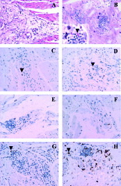
Fig. 1. Aspects of the inflammatory process in myenteric plexus of oesophagus representative of patients of group IIb (A, C, E, and G) and patients with megaesophagus (B, D, F, and H). (A) Chronic inflammatory process with mononuclear cells; connective and vascular neo-formation. Neurons are not visible. Haematoxylin and eosin staining, objective 20X. (B) Intense and nodular mononuclear inflammatory infiltration, with presence of eosinophils (inset, arrowhead) and mast cells. (C and D) Rare CD57+ natural killer cells (arrow-head) are seen in inflammatory foci in patients of group IIb (C) compared to patients with megaesophagus (D). (E and F) The same is observed for TIA-1+ cytotoxic lymphocytes. (G and H) CD68+ positive cells.

Fig. 2. Morphometric analysis of TIA-1+ cytotoxic lymphocytes and CD57+ NK cells in patients of group IIb and patients with megaesophagus. Values for the controls are not detectable. The values are expressed as means±S.D. * Statistically significant differences between the two groups were observed for both analyses.

Fig. 3. Morphometric analyses of CD68+ cells in patients without megaesophagus (groups IIa and IIb), and patients with megaesophagus. The values are expressed as means of CD68+ areas±S.D. * Statiscally significant differences between this group and control individuals. ** Statiscally significant difference between group IIb and group of patients with megaesophagus.

Fig. 4. (A) Amplification of a 330 bp Trypanosoma cruzi kDNA fragment from human oesophagus samples by using a Hot – start protocol. Five μl of PCR products were loaded in each lane of a 6% silver-stained polyacrylamide gel. Lane 1, molecular size marker (1 Kb DNA Ladder, BRL). Lanes 2–6, chagasic patients with megaesophagus: E08, E11, E12, E21, E24. Lanes 7–9, chagasic patients without megaesophagus (group IIb): E04, E07, E10. Lanes 10–11, chagasic patients without megaesophagus (group IIa): E13, E18. Lanes 12 and 13: Control individuals without Chagas' disease: E05, E17. Lane 14: negative control of the DNA extraction (amphibian muscle). Lane 15: T. cruzi genomic DNA used as positive control of PCR. Lane 16: negative control of the PCR amplification steps (PCR mix without DNA). (B) Amplification of the 380 bp fragment from human mitochondrial DNA from the same human tissues. Five μl of PCR products were loaded in each lane of a 6% silver-stained polyacrylamide gel. Lane 1, molecular size marker (1 Kb DNA Ladder, BRL). Lanes 2–14 are the same patients cited above.
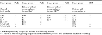
Table 2. Amplification of a 330 bp Trypanosoma cruzi kDNA fragment in samples of oesophagus from chagasic patients and control individuals
 You have
Access
- 51
- Cited by