Introduction
According to the National Sexually Transmitted Disease Surveillance System and Sentinel Site Network of China (Chinese Center for Disease Control and Prevention, www.chinacdc.cn), there were 464,280 syphilis cases in 2012, which was an increase of 10.72% over the same period in 2011. If left untreated, 30% of syphilis patients may develop cardiovascular disease and neurosyphilis (NS; Friedrich et al., Reference Friedrich, Geusau, Greisenegger, Ossege and Aigner2009). Syphilitic infection of the central nervous system can occur early or late in the course of the disease (Marra, Reference Marra2009). According to the clinical manifestations, NS can be divided into the following types: asymptomatic NS, syphilitic meningitis, meningovascular NS, and parenchymal syphilis (including general paresis, tabes dorsalis, and gummatous NS) (Friedrich et al., Reference Friedrich, Geusau, Greisenegger, Ossege and Aigner2009). Parenchymal syphilis is progressive and largely irreversible. General paresis (also known as “general paresis of the insane”) can occur at any time after the first year of the untreated infection and has a peak incidence at 10–20 years, which often results in the development of personality disorders, dementia, irritability, and weakness. Argyll-Robertson and hyper-reflexia are common characteristics found in general paresis (Shah and Lang, Reference Shah and Lang2012). These variable nonspecific presentations not only create diagnostic problems but can also result in incorrect therapeutic decisions. Clinicians, including general physicians, geriatricians, neurologists, psychiatrists, or venereologists, and particularly psychiatrists, need to have an increased awareness of NS, which may present as a psychiatric illness. Without such awareness on the part of the clinicians, these illnesses may result in undue delayed diagnoses and serious consequences (Zheng et al., Reference Zheng2011). Thus far, very few studies have focused on neuropsychiatric disorders secondary to NS in elderly people (age >65 years). In this study, we report ten elderly patients with psychiatric manifestations secondary to NS at Zhongshan Hospital, Medical College of Xiamen University between June 2005 and February 2012.
Methods
Participants
A retrospective review of the patient records at Zhongshan Hospital, Medical College of Xiamen University, was performed (except for return-visit patients). A total of 59 hospitalized patients (48 males and 11 females) were recruited between June 2005 and February 2012 with an average age of 50 years (range: 33–75 years). These patients were diagnosed with general paresis on the basis of their clinical evaluations, serological tests, and cerebrospinal fluid examinations. HIV infection was excluded from all of the patients using ELISA with HIV 1+2 antigens/antibodies (Beijing Wantai Biological Pharmacy Enterprise Co. Ltd., China). The ethics committees of the Medical College of Xiamen University approved this study. An informed consent was also obtained from all of the subjects.
Diagnostic criteria
The diagnosis of syphilis was established using treponemal tests including enzyme-linked immunosorbent assay and Treponema pallidum particle agglutination (TPPA; French et al., Reference French, Gomberg, Janier, Schmidt, van Voorst Vader and Young2009). Patients identified with syphilis were evaluated for neurological findings as previously described (Tong et al., Reference Tong2013a). These examinations included positive serologies and one or more of the following: (a) positive CSF Venereal Disease Research Laboratory (VDRL)/rapid plasma reagin (RPR); (b) positive CSF TPPA and increased CSF protein (protein > 500 mg/L) or white blood cells (WBC > 10 × 106 cells/L), and an otherwise unexplained neurological manifestation consistent with NS. The criteria used for excluding NS were similar to those used in a previous study (Liu et al., Reference Liu2012). The psychiatric diagnostic procedures were performed according to the Diagnostic and Statistical Manual of Mental Disorders, text revision (American Psychiatric Association, 2000).
Syphilitic serological and CSF tests
The syphilitic serological and CSF tests for each sample were performed using RPR (InTec, Xiamen, China), TPPA tests (Fujirebio, Tokyo, Japan), and enzyme-linked immunosorbent assay (InTec, Xiamen, China) according to the manufacturer's instructions and previous studies (Lin et al., Reference Lin2010; Reference Lin2011).
CSF protein and WBC examinations
CSF protein and WBC examinations were performed according to our previous studies (Liu et al., Reference Liu2012): approximately 2 mL of the CSF samples was collected in plain sterile tubes and analyzed within 1 h to determine the protein level using a Roche–Hitachi Modular P800 analyzer (Roche Diagnostics, F. Hoffmann-La Roche Ltd., Basel, Switzerland) and CSF WBC using an automatic blood cell XE5000 analyzer (Sysmex International Reagents Co. Ltd., Japan).
Magnetic resonance imaging (MRI), magnetic resonance angiography (MRA), and electroencephalograph (EEG) assessment
Within seven days of assessment, the patients underwent multislice MRI of the brain. The images were acquired using a 3.0 T superconducting magnet (Simens Company, Germany). The imaging sequences included coronal T1 spin echo (time to recovery 650 ms; time to echo 20 ms, field of view 30 mm; slice thickness 5 mm and interslice gap 1 mm) and axial T2-weighted spin echo (time to recovery 2,000 ms; time to echo 20 ms, 90 ms; field of view 26 mm; slice thickness 6 mm and interslice gap 2 mm). MRA imaging was performed after administration of 20 cc of IV Gd-DTPA at an injection rate of 5 cc per second by first utilizing a 3D spoiled gradient echo sequence. The imaging protocol for imaging vascular stenosis was follows: TR = 2.95, TE = 1.07, 384 × 36 matrix, slices 88, and GRAPPA ×3. The acquisition was 0.6 s per 3D slab; the first acquisition was in the axial plane and the second acquisition was in either the sagittal or coronal plane. EEG data were recorded using a 19-channel MR compatible EEG system (Nicolet BIMEDICAL, USA). The electrodes were attached to the scalp according to the international 10/20 system (time constant: 0.3 s; high-frequency response: 70 Hz; low-frequency response: 1.6 Hz; sensitivity: 10 μV/mm) with the subjects lying relaxed with their eyes closed in an electrically shielded room.
Results
Clinical data for the elder patients with psychiatric symptoms secondary to NS
We retrospectively review 59 diagnosed cases of general paresis between June 2005 and February 2012 at Zhongshan Hospital, Medical College of Xiamen University. Overall, 52 of the 59 general paresis patients presented with psychiatric manifestations; 44 males and eight females with a median age of 50 years (range: 33–75 years). Among these 52 patients, ten elderly patients (age >65 years) were included; eight males and two females, with an average age of 70.5 years (range: 66–75 years). None of the ten elderly patients had a previous history of other psychiatric disorders, substance abuse, or a family history of psychiatric disorders in their first-degree relatives. They were first treated in the neurology department of the hospital, and none of the patients were initially suspected of having NS. In these cases, patients were all diagnosed with NS during their follow-up treatment and were originally diagnosed with Alzheimer's disease (four patients), hypertensive arteriosclerotic intracerebral ischemic stroke (two patients), Parkinsonism (two patients), epilepsy (one patient), and subcortical arteriosclerotic encephalopathy (one patient). With the exception of patient 9, who provided a history of unprotected sexual activity in the absence of any skin lesions 18 years ago, the other nine patients and their spouses all denied having any history of unprotected sexual activity.
Symptoms and signs presentation in elder patients with psychiatric manifestations secondary to NS
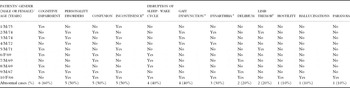
Among the ten elderly patients with psychiatric symptoms secondary to NS, a large number of the patients demonstrated features of more than one syndrome. The more common presenting symptoms were a wide variety of psychiatric manifestations. Cognitive impairment was the most frequent symptom, where six patients (60%, 6/10) showed different degrees of decline in intelligence and memory. Personality disorders (50%, 5/10), confusion (patient presented with disorientation, whilst he or she has one or both symptoms of bewildered and difficulty following commands) (50%, 5/10), disruption of the sleep-wake cycle (40%, 4/10), and delirium (patient presented with disorientation, and one or more symptoms of restless, decline in cognition, hallucinating, sometimes delusional) (30%, 3/10) were also common symptoms. Less common (≤10%) psychiatric manifestations included hostility, hallucinations, and paranoia. The other more frequent neurological symptoms were incontinence of feces and urine (50%, 5/10), gait dysfunction (40%, 4/10), dysarthria (30%, 3/10), and limb tremor (20%, 2/10) (Table 1).
Clinical symptoms from ten elderly psychiatric patients secondary to NS

a These were neurological symptoms.
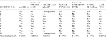
In our study, many patients displayed more than one psychiatric sign, and there was an overlap in the patients’ major signs. Among the ten elderly patients with psychiatric manifestations secondary to NS, amnesia (80%, 8/10) was the most common sign. A positive Babinski sign (50%, 5/10) was also a common sign, which indicated effects in the left or right limbs. Another common sign was cerebellar ataxia (30%, 3/10). During the physical and neurological examinations about cerebellar ataxia, two patients were uncooperative. Less common (≤10%) clinical signs included muscle weakness (10%, 1/10), Argyll-Robertson pupil, hyper-reflexia, and a masked face (Table 2).
Clinical signs from ten elderly psychiatric patients secondary to NS

Laboratory findings in elderly patients with psychiatric manifestations secondary to NS
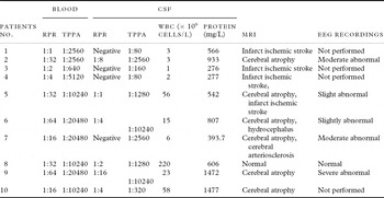
Among the ten elderly patients with psychiatric symptoms secondary to NS, both the RPR and TPPA tests were positive for the serum samples. All of the patients’ TPPA titers were within a high range dilution, where the overall titer was ≥1:2560, except for patient 3 who had an overall titer of 1:640. As for the CSF samples, the TPPA was positive in all of the patients (100%, 10/10), while the RPR was positive in only six patients (60%, 6/10). Assuming that the CSF-WBC ≤ 10 × 106 cells/L and CSF-protein ≤ 500 mg/L were within normal reference intervals (Control and Prevention, 1998), five patients (50%, 5/10) had CSF pleocytosis and seven patients (70%, 7/10) had elevated levels of CSF protein. Five patients (50%, 5/10) demonstrated both CSF pleocytosis and elevated CSF protein levels, while three patients (30%, 3/10) had normal CSF WBC and protein levels (Table 3).
Laboratory findings, MRI and EEG recordings from ten elderly psychiatric patients secondary to NS

Neuroimaging findings for elderly patients with psychiatric symptoms secondary to NS
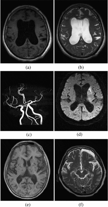
All of the ten elderly patients with psychiatric symptoms secondary to NS underwent brain MRI studies. Nine patients (90%) demonstrated abnormal imaging, and three patients had more than one imaging results (Table 3). Six cases (60%, 6/10) displayed cerebral atrophy. The MRI of patient 5 showed cerebral atrophy with widened sulci and cisterns with enlarged ventricles, which were prominent in the lateral and third ventricle, and particularly in the fifth and sixth ventricles (on T1-weighted and T2-weighted images; Figures 1a and b). The imaging of patient 7 not only showed cerebral atrophy but also cerebral arteriosclerosis in second part of the middle cerebral artery, and the degree of stenosis reached 25% (on MRA; Figure 1c). Four cases (40%, 4/10) showed an infarct ischemic stroke, including one or multiple lacunar infarctions. The MRI of patient 1 showed hyperintense signal abnormalities in the left lateral ventricle (Figure 1d). In addition, the T1-weighted MRI of patient 4 showed multiple lacunar infarctions in the left lateral ventricle as well as old lacunar infarctions in the bilateral basal ganglia and brainstem (Figure 1e). One case (patient 6; 10%, 1/10) had hydrocephalus of the left frontal lobe (Figure 1f).
Brain imaging findings of elderly psychiatric patients secondary to NS. (a, b) Axial T1-weighted and T2-weighted MRI of patient 5 showing cerebral atrophy with widened sulci and cisterns with enlarged ventricles, which were prominent in the lateral and third ventricle, and particularly in the fifth and sixth ventricles. (c) MRA of patient 7 showed cerebral arteriosclerosis in second part of the middle cerebral artery, and the degree of stenosis reached 25%. (d) Axial DWI MRI of patient 1 showing hyperintense signal abnormalities on the left lateral ventricle. (e) Axial T1-weighted MRI of patient 4 showing multiple lacunar infarctions in the left lateral ventricle, old lacunar infarctions in the bilateral basal ganglia and brainstem. (f) T2-weighted MRI of patient 6 showing hydrocephalus of the left frontal lobe.

Brain EEG for elderly patients with psychiatric symptoms secondary to NS
Six out of ten elderly patients with psychiatric symptoms secondary to NS were examined using EEG. One case (patient 8) showed normal EEG recordings. Two patients (33.3%, 2/6) showed slightly abnormal activity with a slowing of background activity; two patients (33.3%, 2/6) showed moderate EEG activity with increased ө and δ waves; one patient (patient 9) showed severely abnormal activity, including low- and high-voltage waves, sharp- and slow-wave complexes, and a triphasic-like wave (Table 3).
Discussion
We characterized 59 general paresis inpatients from Zhongshan Hospital, Medical College of Xiamen University, who were admitted between June 2005 and February 2012. Among these patients, there were ten elderly people aged >65 years, who presented with psychiatric manifestations and behavior disorders. None of these patients were initially suspected of having NS when they were first treated in the neurology department of the hospital. Initially, Alzheimer's disease was suspected in these patients because psychotic symptoms in elder age are mostly due to incipient dementia (Broadway and Mintzer, Reference Broadway and Mintzer2007). The incidence of Alzheimer's disease in the older than 65 years age group was approximately 6% and was steeply rising to 20% or more in people older than 80 years (Copeland et al., Reference Copeland1992). Alzheimer’s-type dementia has been characterized by temporoparietal neuropsychiatric features such as amnesia for recent events, apraxia, aphasia, and visuospatial difficulties (Hilton, Reference Hilton1998). These cognitive findings differed from patients with NS, which mainly manifested as cognitive impairment and amnesia in the elderly patients. Thus, in the absence of a relevant history or laboratory results, neurological features and specific cognitive impairment, general paresis was unlikely to be confused with the classical presentation of Alzheimer’s-type dementia. Furthermore, cerebrovascular disease (such as hypertensive arteriosclerotic intracerebral ischemic stroke) may include neurological symptoms that frequently coexist with dementia, and may cause difficulty in diagnosis because they may demonstrate similar signs as advanced dementia, where cognitive features become more generalized and neurological abnormalities are more common (Hilton, Reference Hilton1998). Thus, it is difficult for clinicians to diagnose NS presented with psychiatric manifestations and behavior disorders immediately when the patients were admitted into the hospital.
In our study, psychiatric manifestations presented as the primary symptom in ten elderly psychiatric patients secondary to NS, and mainly demonstrated cognitive impairment, personality disorders, confusion, disruption of the sleep–wake cycle, delirium, hostility, hallucinations, and paranoia. Moreover, psychotic symptoms and cognitive impairment were present as a consequence of extensively damaged parenchyma in the cortical regions of the brain (Kent and Romanelli, Reference Kent and Romanelli2008). Cerebellar ataxia, muscle weakness, Argyll-Robertson pupils and hyper-reflexia were the most common characteristic findings in patients secondary to general paresis. However, the extrapyramidal symptoms and signs of NS (e.g., gait dysfunction, dysarthria, limb tremor, positive Babinski sign, and mask-like face) were observed in the tabes dorsalis (Shah and Lang, Reference Shah and Lang2012). Thus, we inferred that our patients might have general paresis combined with tabes dorsalis. In our study, the incontinence of the feces and urine was a frequent neurological symptom (50%) in elderly patients. Luo et al. Reference Luo, Ouyang, Xu, Chen, Ding and Zhang(2008) and Zheng et al. Reference Zheng(2011) reported that the incidence of incontinence in general paresis was approximately 35%–40%. Moreover, it may display hypocompliant detrusor hyper-reflexia with detrusor–sphincter dyssynergia and postmicturition residual urine (Luo et al., Reference Luo, Ouyang, Xu, Chen, Ding and Zhang2008).
In our study, patients with neuropsychiatric disorders secondary to NS showed no specificity of their clinical symptoms and signs. In addition, elderly people demonstrated characteristics of aging degeneration of the brain. Thus, fewer clinicians considered the diagnosis of NS. The laboratory confirmation of NS was an important basis for the diagnosis of NS. Generally, a combination of several CSF abnormalities including pleocytosis, elevated protein, VDRL (or RPR), and TPPA must be evaluated (Larsen and Schultz, Reference Larsen and Schultz1993; Luger et al., Reference Luger, Schmidt and Kaulich2000). In our study, of the ten elderly patients with general paresis and psychiatric manifestations, all had positive peripheral blood TPPA and RPR tests. For the CSF samples, a positive rate of TPPA was 100%, whereas a positive rate of RPR was 60% with less sensitivity. There were five patients who had both CSF pleocytosis and elevated CSF protein levels.
The current literature (Berbel-Garcia et al., Reference Berbel-Garcia2004; Fadil et al., Reference Fadil, Gonzalez-Toledo, Kelley and Kelley2006) indicates that brain MRI scans of most NS patients were normal or showed nonspecific changes, which did not suggest a diagnosis of NS. In our study, nine patients had an abnormal brain MRI out of the ten elderly psychiatric patients secondary to NS, including cerebral atrophy, infarct ischemic stroke, and hydrocephalus. However, other reports have indicated brain MRI findings of cerebral atrophy (Kodama et al., Reference Kodama2000; Yu et al., Reference Yu2010) or vasculitis-related infarcts, which may support a diagnosis of NS (Kearney et al., Reference Kearney, Mallon, Kavanagh, Lawler, Kelly and O’Rourke2010). Such cerebral atrophy has been correlated with the destruction of the cerebral cortex and neuronal loss. An altered mental status and disturbed cognition are likely to be related to cerebral atrophy (Russouw et al., Reference Russouw, Roberts, Emsley and Truter1997; Yu et al., Reference Yu2010). The brain MRI of four patients exhibited an infarct ischemic stroke, including one or multiple lacunar infarction in the bilateral basal ganglia and centrum semiovale. Furthermore, four patients (cases 2, 3, 4, and 10) presented with gait dysfunction and neuropsychiatric disorders; thus, we inferred that the patients might have general paresis combined with meningovascular syphilis (Zheng et al., Reference Zheng2011). In our study, only the MRI of patient 6 presented hydrocephalus, in which meningovascular syphilis-associated chronic basal meningitis may result in uncommon general paresis in patients (Long and Mulley, Reference Long and Mulley2001).
NS is commonly known as “the great imitator” or “the great impostor,” because of its various clinical manifestations, laboratory findings, MRI and EEG findings, but all studies lack specificity, so that it is usually not easily diagnosed (Shah and Lang, Reference Shah and Lang2012). We advocate that every patient with neurological or psychiatric symptoms without unambiguous causes should have a blood test for syphilis. When serology proves positive, patients should undergo CSF examination (Tong et al., Reference Tong2013b). Of course, as Cleare stated, this screening method could increase extra charges for patients (Cleare et al., Reference Cleare, Jacoby, Tovey and Bergmann1993). But without the screening method, NS could not be diagnosed at early stage; thus, it would prevent patients from receiving the most appropriate treatment, resulting in more severe neurological damage and the later nerve damage induced by NS is usually an irreversible process. Moreover, NS is not rare in many areas of the world, including our country (Mitsonis et al., Reference Mitsonis2008; Liu et al., Reference Liu2012). The problem becomes a public concern.
The limitations of our study should be acknowledged. As a retrospective study, patients’ symptoms and signs were not systematically or uniformly assessed, and some information is missing, uniform access is the inevitable risk. In addition, only ten inpatients were studied in our study, and larger sample sizes are needed to confirm this conclusion. Meanwhile, during the past few years, SPECT and PET have become an important aid in diagnosing the central nervous system diseases, especially patients with normal MRI or CT test. It has also been documented that NS patients can have abnormal SPECT and PET (Ide et al., Reference Ide, Mizukami, Fujita, Ashizawa and Asada2004; Sun et al., Reference Sun, Cheng, Shi, Xu and Liu2010; Omer et al., Reference Omer, Fitzgerald, Sheehy and Doherty2012). Unfortunately, in this research we lacked this information, which we need to study further.
In conclusion, the importance of a rapid diagnosis of NS is emphasized because early and effective treatment may not only prevent further disease progression but may also allow for a complete recovery (Hooshmand et al., Reference Hooshmand, Escobar and Kopf1972). It is recommended that every patient with clinically evident neurological or psychiatric symptoms should have a blood examination for syphilis. If the serology proves positive, the patient should undergo CSF examination. Elderly psychiatric illness secondary to NS is rarely diagnosed in elderly patients, but deserves medical attention.
Conflict of interest
We confirm that all the authors have satisfied the conditions required for authorship. We also confirm that there is no potential conflict of interest as described in the instructions for authors. All the authors have read and approved the manuscript.
Description of authors’ roles
Tian-Ci Yang was responsible for designing the study, formulating the research questions, and carrying it out. Wei-Hong Zheng took part in designing the study. Yan-Li Zeng and Hui-Lin Zhang operated the experiment, carried it out, and wrote the paper. Wen-Jie Wang analyzed the data and wrote the paper. Song-Jie Huang, Gui-Li Liu, Ya Xi, and Xiao-Jing Guo assisted with operating the experiment and collected the data. Fu-Yi Chen was responsible for analyzing the data.
Acknowledgments
The current study was supported by the National Natural Science Foundation's Major Research Planning (grant no. 91029729), the National Natural Science Foundation (grant nos. 81171625, 81271335, 81101324, and 81201360), the Technology Foundation's Major Project of Social Development in Fujian Province (grant no. 2011Y4009), and the Natural Science Foundation of Fujian Province (grant no. 2012D039). These organizations had no role in the study design, data collection and analysis, decision to publish, or preparation of the manuscript.