Antiphospholipid syndrome (APS) is defined as an autoimmune and multisystem disorder of recurrent thrombosis, pregnancy loss, and thrombocytopaenia (low platelet count) associated with the presence of antiphospholipid antibodies (aPL Abs) and persistently positive anticardiolipin (aCL) and/or lupus anticoagulant (LA) tests (Refs Reference Harris1, Reference Wilson2). It is now well established that aPL Abs are heterogeneous and that they bind to various proteins that associate with phospholipids, among them the plasma proteins β2glycoprotein I (β2GPI) and prothrombin. β2GPI is an abundant plasma glycoprotein that consists of five homologous domains of approximately 60 amino acids each (Ref Reference McNeil3, Reference Atsumi and Koike4). Other antigenic targets have been identified in APS patients, including tissue plasminogen activator (tPA), plasmin, annexin A2 and thrombin (Refs Reference Lin5, Reference Chen6, Reference Cesarman-Maus7). Thus, it seems reasonable to speculate that the pathophysiological mechanisms in APS may involve Abs directed to these target antigens.
Clinical aspects of APS
APS was first described in patients with systemic lupus erythematosus (SLE), and more specifically in a subset of patients with SLE that had an abnormal LA test (Ref. Reference Boey8). APS was then classified as ‘secondary’ (SAPS) in the presence of SLE, and ‘primary’ (PAPS) in the absence of SLE or other autoimmune disorders. In the general population, PAPS is the most common cause of acquired thrombophilia and accounts for 15–20% of all episodes of deep-vein thrombosis with or without pulmonary embolism, a third of new strokes occurring in patients under the age of 50, and 10–15% of women with recurrent fetal loss (Ref. Reference Ginsburg9). It has been estimated that 2–5% of the general population have experienced an episode of deep-vein thrombosis, suggesting that the prevalence of venous thrombosis associated with PAPS may be as high as 0.3–1% of the general population (Ref. Reference Ginsburg9). SAPS also accounts for a significant proportion of thromboembolic disease and recurrent fetal loss in patients with SLE. aPL Abs are present in 30–40% of SLE patients, and approximately a third of those with Abs, or 10–15% of all SLE patients, have clinical manifestations of APS (Ref. Reference McClain10).
In addition to arterial and venous thrombotic events, including cerebral infarction and strokes, for which this syndrome is well recognised, APS can also present clinically with several features not necessarily stemming from a thrombotic tendency (Refs Reference Moroni11, Reference Moroni12). These include heart valve lesions, livedo reticularis, thrombocytopaenia, nephropathy, and neurological manifestations such as chorea, epilepsy and transverse myelopathy (Refs Reference Miyakis13, Reference Gleason14, Reference Toubi15, Reference Atsumi16, Reference Levine17, Reference Sherer18, Reference Al-Matar, Jaimes and Malleson19, Reference Hughes20, Reference Mesa21, Reference Shoenfeld22). In addition, as indicated above, a range of additional Abs may be detected in laboratory tests, albeit uncommonly: positive IgA aCL Abs, IgA anti-β2GPI Abs, antiphosphatidylserine Abs, antiphosphatidylethanolamine Abs, Abs against prothrombin alone, and Abs to the phosphatidylserine–prothrombin complex (Ref. Reference Miyakis13). Of note, these clinical and laboratory findings are not included in the revised criteria for diagnosis of APS (so-called Sydney criteria as opposed to the original Sapporo criteria) because, although sometimes seen, they are nonspecific and in some cases the tests to detect them are not completely developed or standardised (Ref. Reference Miyakis13).
A rare complication of APS is catastrophic APS (CAPS). CAPS is a life-threatening condition, which is often difficult to distinguish clinically from thrombotic thrombocytopaenic purpura (TTP), characterised by a widespread thrombotic microangiopathy, disseminated intravascular coagulation (DIC), pulmonary alveolar haemorrhage, and the acute respiratory distress syndrome (ARDS) (Refs Reference Asherson23, Reference Wiedermann24). Other unusual complications of APS arise from venous occlusions in organs such as the adrenal glands, resulting in adrenal insufficiency (Ref. Reference Asherson and Cervera25), and in the kidneys (Ref. Reference Nzerue26) and liver (Budd–Chiari syndrome) (Ref. Reference Asherson and Cervera25). Indeed, any vessel can be involved, such as the femoral, iliac and retinal veins, and the inferior vena cava – as well as the pancreatic and mesenteric vasculature, with thrombotic pancreatitis and bowel infarction with colonic perforation, respectively. An association of APS with osteonecrosis, although reported, remains controversial (Ref. Reference Tektonidou27).
Treatment
The treatment of thrombosis in APS involves anticoagulation, initially either with intravenous therapeutic heparin or with the low molecular weight heparins (such as subcutaneous enoxaparin), followed typically by oral warfarin (Coumadin®) for life (Refs Reference Khamashta28, Reference Krnic-Barrie29, Reference Crowther30, Reference Finazzi31). When using oral anticoagulation with warfarin, most clinicians favour keeping the international normalised ratio (INR) between 2.0 and 3.0, to avoid haemorrhagic complications (Refs Reference Khamashta28, Reference Krnic-Barrie29, Reference Crowther30, Reference Finazzi31, Reference Garcia, Khamashta and Crowther32). Hydroxychloroquine (Plaquenil®) is normally also an integral component of the treatment, particularly if the patient also suffers from concomitant SLE (Ref. Reference Erkan33). In patients with recurrent thrombotic events despite the above-mentioned anticoagulation regimen, low-dose aspirin is often added. Cytotoxic agents are usually avoided in the treatment of pure APS unless the patient is experiencing an exacerbation of concomitant SLE or related autoimmune disease. The presence of potential additional causes of thrombophilia should be investigated in APS patients, such as factor V Leiden or prothrombin gene mutations. APS patients should also avoid prothrombotic lifestyle factors, such as cigarette smoking, the use of hormone replacement therapy (HRT) containing oestrogens, and the use of illicit drugs such as cocaine.
Many patients with PAPS or SAPS may have ‘subclinical’ or asymptomatic APS – that is, positive blood Abs without overt clinical thrombosis. However, if additional procoagulant factors come into play, clinical clotting may ensue. The approach to managing patients with aPL Abs without a previous thrombotic event is still debated. Some clinicians recommend prophylaxis with low-dose aspirin, others advise more aggressive treatment, and still others recommend no treatment at all. The efficacy of prophylaxis with low-dose aspirin is not certain; it may not be effective in preventing venous events (Ref. Reference Erkan34).
For CAPS, plasma exchange is often used, although there have not been controlled studies to validate this approach (Ref. Reference Asherson23). Other interventions used include intravenous Ig and/or immunosuppressive agents. Recently, rituximab (Rituxan®), a monoclonal Ab that selectively depletes CD20+ B cells, has been successfully employed to treat this life-threatening syndrome in a small number of patients with resistant APS (Ref. Reference Rubenstein35).
Thrombosis per se is a devastating consequence in PAPS and SAPS and may affect any organ. The recurrence of thrombosis in patients with aPL Abs is high. APS occurs predominantly in young women, and the recurrence of the symptoms combined with high morbidity calls for an adequate treatment. In addition, this disease is associated with a significant socioeconomic impact, often involving long-term disability and costly treatments. As indicated, in the case of thrombotic manifestations, treatment has focused on preventing thromboembolic events, utilising antithrombotic medications, or modulating the immune response itself. Recurrences in spite of treatment have been reported and the use of oral anticoagulation at a relative high INR for a long period of time has also been associated with a high risk of bleeding, with the need for frequent monitoring and patient compliance with diet and lifestyle to optimise the therapy. A serious need therefore exists for new and more-efficient modalities of treatment for thrombosis in APS.
Pathogenic mechanisms induced by aPL Abs
Studies utilising animal models of thrombosis, endothelial cell (EC) activation, and pregnancy loss have provided strong evidence demonstrating that aPL Abs are pathogenic in vivo (Refs Reference Branch36, Reference Blank37, Reference Pierangeli38, Reference Jankowski39). APL Abs are heterogeneous, and more than one mechanism may be involved in causing thrombosis. In fact, in vitro studies have shown that aPL Abs may cause thrombosis by interfering with activation of protein C or inactivation of factor V by activated protein C, inhibiting endothelial prostacyclin production, impairing fibrinolysis, activating ECs and monocytes, and exerting stimulatory effects on platelet function (Ref. Reference Roubey40). In addition, convincing data now indicate that activation of complement mediates aPL-induced fetal loss, thrombosis and EC activation (Refs Reference Holers41, Reference Salmon, Girardi and Holers42, Reference Pierangeli43, Reference Fischetti44). This article focuses first on the effects of aPL Abs on platelets and then discusses effects on ECs and monocytes.
Effects of aPL Abs on platelets
Thrombocytopaenia is a frequent feature of APS, giving rise to speculation that aPL Abs may bind platelets and cause aggregation and thrombosis. Indeed, aPL Abs have been shown to increase platelet activation and aggregation in both in vitro and in vivo studies. The intracellular events that aPL Abs trigger in platelets and the target receptor(s) are not completely known.
Evidence that aPL Abs cause platelet activation and aggregation
Initial studies showed binding of aPL Abs did not occur in intact platelets but when platelets were exposing negatively charged phospholipids (i.e. phosphatidylserine) after repeated freezing and thawing (Ref. Reference Khamashta45). Agonist stimulation, which also leads to the exposure of phosphatidylserine on the cell outer membrane, seems to be required for aPL Ab effects on platelets. One study, using affinity-purified aPL Abs derived from six patients with APS, showed an increase in platelet aggregation and activation in the presence of low concentrations of thrombin, ADP or collagen, compared with normal IgG (Ref. Reference Campbell46). In addition, submaximal prestimulation of platelets by agonists such as thrombin or collagen in vitro appears to be a prerequisite for β2GPI-reactive Abs in the presence of β2GPI to be able to potentiate activation (Ref. Reference Campbell46).
aPL Abs can enhance the expression of platelet membrane glycoproteins, particularly GPIIb–IIIa (a fibrinogen receptor, platelet-activation marker and mediator of platelet aggregation induced by all physiological agonists) and GPIIIa when platelets are pretreated with suboptimal doses of TRAP (thrombin-receptor-agonist peptide) (Ref. Reference Espinola47). Furthermore, thrombus formation in vivo induced by aPL Abs has been shown to involve platelet activation, via GPIIb–IIIa: thrombus formation was not increased by aPL Abs in GPIIb–IIIa-deficient (β3 null) mice and aPL-Ab-mediated enhanced thrombus formation in vivo was reduced by pretreatment of the mice with a monoclonal anti-GPIIb–IIIa Ab (1B5) (S. Pierangeli et al., unpublished). The data underscore the possibility of using GPIIb–IIIa inhibitors to treat thrombotic events in patients with APS (see later). The effects of aPL Abs on glycoprotein expression as well as the enhanced thrombus formation in mice can also be significantly reduced by hydroxychloroquine (Refs Reference Espinola47, Reference Edwards48). In vivo platelet activation by aPL Abs was also shown recently by other investigators, who demonstrated that a monoclonal Ab with anti-β2GPI and LA activity produced a platelet-rich thrombus in a rat model of thrombosis, concomitant with an increase in platelet aggregation, when platelets were treated with a low concentration of ADP (Ref. Reference Jankowski39). Hence, data support the notion that aPL Abs can activate platelets, providing an explanation for aPL-mediated thrombosis (particularly arterial thromboses).
Intracellular mechanisms
Thromboxane A2 (TXA2) is the major eicosanoid produced in platelets and has a potent platelet-aggregatory and vasoconstrictor activity. Elevated levels of TX metabolic breakdown products in the urine of APS patients provides further support for dysregulated platelet activation contributing to thrombosis in APS, and sheds light on the intracellular events that aPL Abs trigger in platelets. A significant correlation was found between the levels of anti-β2GPI IgG and urinary excretion of 11-DH-thromboxane B2 (11-DHTXB2) in 34 patients with aPL Abs (Ref. Reference Martinuzzo49). In addition, urinary excretion of 11-DHTXB2 was significantly increased in six patients with LA and aPL Abs compared with healthy controls (Ref. Reference Forastiero50); furthermore, TXB2 production was increased when normal platelets were incubated with low doses of thrombin and F(ab′)2 isolated from patients with aPL Abs (Ref. Reference Forastiero50). Similarly, independent work showed a significant increase in the urinary excretion of 11-DHTXB2 in seven patients with aPL Abs compared with controls (Ref. Reference Robbins51). An increase in cyclic AMP (cAMP), through agonists such as PCI2 or inhibitors of the phosphodiesterase such as theophylline, abrogated aPL-Ab-mediated TX production in platelets (Ref. Reference Opara, Robbins and Ziboh52).
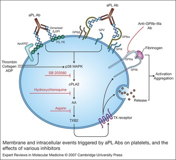
In platelets, p38 MAPK (mitogen-activated kinase) is activated by thrombin, collagen and a TX analogue, and is involved in cytosolic phospholipase A2 (cPLA2) phosphorylation, with subsequent production of TXB2 (Fig. 1). Treatment of normal platelets with aPL Abs or with their F(ab′)2 fragments in the presence of subactivating doses of thrombin resulted in a significant increase in phosphorylation of p38 MAPK. Furthermore, pretreatment of the platelets with a specific p38 MAPK inhibitor – SB203580 – significantly abrogated aPL-Ab-mediated platelet aggregation (Ref. Reference Vega-Ostertag, Harris and Pierangeli53). The data indicate that aPL Abs induce TXB2 production mainly through the activation of p38 MAPK and subsequent phosphorylation of cPLA2 (Ref. Reference Vega-Ostertag, Harris and Pierangeli53) (Fig. 1).
Membrane and intracellular events triggered by aPL Abs on platelets, and the effects of various inhibitors. APL Abs bound to dimerised β2GPI may interact with apoER2′ or GPIbα on the surface of platelets. Interaction with exposed phosphatidylserine or phosphatidylethanolamine also seems to be required. Intracellular signalling is triggered and involves phosphorylation of p38 MAPK, cPLA2 and release of TXB2 when platelets are primed with low doses of agonists (e.g. thrombin, collagen, ADP); steps of the pathway inhibited by SB 203580, hydroxychloroquine and aspirin are shown. APL Abs also induce enhanced expression of GPIIb–IIIa on the surface of platelets, which can be effectively blocked by an anti-GPIIb–IIIa monoclonal Ab. Abbreviations: AA, arachidonic acid; Ab, antibody; aPL Ab, antiphospholipid antibody; apoE2R′, apolipoprotein E2 receptor; GP, glycoprotein; TX, thromboxane; PLA2, phospholipase A2; ADP, adenosine diphosphate; PS, phosphatidylserine; PE, phosphatidylethanolamine; β2GPI, β2-glycoprotein I; p38 MAPK, p38 mitogen-activated protein kinase.

Receptor for aPL Abs on platelets
The apolipoprotein E receptor 2 (apoER2′), the only receptor of the low-density lipoprotein family described on platelets, is thought to play a role in the activation of platelets by aPL Abs. When apoER2′ was blocked on platelets using receptor-associated protein (RAP), the increased adhesion by platelets to collagen induced by the anti-β2GPI–β2GPI complex was lost (Ref. Reference Lutters54). In addition, the apoER2′ receptor coprecipitates with dimerised β2GPI, providing evidence for a direct interaction between β2GPI and the receptor (Ref. Reference Lutters54) (Fig. 1).
Another recent study, using a novel method in which the Fc portion of the β2GPI Ab complex was immobilised with protein A coated onto a microtitre plate, demonstrated that β2GPI is able to directly bind GPIbα, which is a subunit of the GPIb–IX–V platelet receptor – a receptor for multiple ligands, including von Willebrand factor. This would also enable anti-β2GPI to activate platelets (Ref. Reference Shi55) (Fig. 1).
The anti-β2GPI–β2GPI complex might initially form on exposed negatively charged phospholipid (e.g. phosphatidylserine) before interacting with specific platelet receptors to potentiate activation (Fig. 1). When the phosphatidylserine-binding site was blocked, potentiation of platelet activation via apoER2′ by dimers of β2GPI did not occur (Ref. Reference Lutters54).
Effects of aPL Abs on ECs and monocytes
ECs express significantly higher amounts of cellular adhesion molecules such as intercellular adhesion molecule (ICAM)-1, vascular cell adhesion molecule (VCAM)-1, and E-selectin when incubated with aPL Abs and β2GPI in vitro (Ref. Reference Simantov56). Furthermore, aPL Abs were shown to activate endothelium in vitro and in mouse models and this correlated with enhancement of thrombus formation in vivo (Ref. Reference Pierangeli38). The use of ICAM-1, E-selectin and P-selectin knockout mice and specific anti-VCAM-1 monoclonal Abs demonstrated that the pathogenic effects of aPL Abs are mediated by these CAMs (Refs Reference Pierangeli57, Reference Espinola58). In accordance with these findings, increased levels of soluble adhesion molecules such as VCAM-1 and P-selectin have been detected in patients with aPL Abs and thrombosis (Ref. Reference Kaplanski59).
Tissue factor (TF), a transmembrane protein that is a member of the class II cytokine and haematopoietic growth factor receptor family, is present upon activation on the surface of several cell types, primarily monocytes, vascular ECs, and smooth muscle cells (Ref. Reference Nemerson60). In ECs, it is upregulated by pro-inflammatory cytokines such as tumour necrosis factor (TNF)-α and bacterial lipopolysaccharide (LPS) during vascular injury and repair (Ref. Reference Nemerson60), probably via translocation of nuclear factor (NF)-κB to the nucleus of the cell (see below) (Ref. Reference Semerato and Colucci61). TF upregulation has been advocated as an important mechanism to explain the prothrombotic effects of aPL Abs. TF expression and function is upregulated in ECs and monocytes treated with aPL Abs (Refs Reference Kornberg62, Reference Cuadrado63, Reference Amengual64, Reference Dobado-Barrios65, Reference Martini66, Reference Reverter67); moreover, higher plasma levels of TF (sTF) occur in APS patients than in controls (Ref. Reference Forastiero, Martinuzzo and De Larranaga68). Recently, IgG from patients with APS was shown to significantly increase TF function and transcription in monocytes and these effects were inhibited by the antiplatelet agent and adenosine-uptake inhibitor dilazep (Ref. Reference Zhou, Wolberg and Roubey69). Vascular endothelial growth factor (VEGF) may stimulate TF expression in monocytes through its receptor tyrosine kinase Flt-1. Interestingly, VEGF and Flt-1 expression are increased in monocytes of APS patients (Refs Reference Cuadrado70, Reference Williams71). Hence, convincing evidence exists that aPL Abs induce EC and monocyte activation and a procoagulant and pro-inflammatory phenotype in vitro and in vivo.
Intracellular events induced by aPL Abs in ECs and monocytes
Although convincing evidence demonstrates that aPL Abs can stimulate monocytes and ECs, as for platelets knowledge of the cell-surface receptors and intracellular signalling pathways involved is limited.
Cell activation through cytokine stimulation, engagement of Toll-like receptors (TLRs), or stress initiates a host-defence signalling pathway that converges on IκB kinases, which results in degradation of the inhibitors that normally sequester the NF-κB transcription factor. NF-κB mediates transcription of many cytokine genes [including interleukin (IL)-6, IL-8, TNF-α and IL-1β], EC adhesion molecules (VCAM-1, E-selectin and ICAM-1) and TF – actions that result in recruitment of inflammatory cells to extravascular sites (Refs Reference Xie, Ray and Short72, Reference Morgan73, Reference Chen74, Reference Saccani, Pantano and Natoli75). Activation of NF-κB has been shown to be a critical mediator in some autoimmune diseases such as rheumatoid arthritis (Ref. Reference Firestein76).
p38 MAPK is an important component of intracellular signalling cascades that initiate various inflammatory cellular responses. For example, p38 MAPK has been implicated as an important regulator of the coordinated release of cytokines by immunocompetent cells and the functional response of neutrophils to inflammatory stimuli (Ref. Reference Cohen77). Different stimuli can activate p38 MAPK, including LPS and other bacterial products, TNF-α and IL-1, growth factors, and stresses such as heat shock, hypoxia and ischaemia/reperfusion. In addition, p38 MAPK positively regulates a variety of genes involved in inflammation, such as TNF-α, IL-1, IL-6, IL-8, cyclooxygenase 2 and collagenase (Ref. Reference McGilvray78). p38 MAPK also activates transcription factors such as activating transcription factor 2, which forms a heterodimer with JUN family transcriptional factors and associates with the activator protein 1 (AP1)-binding site. The promoter region of the TF gene contains two AP1-binding sites and one NF-κB binding site, and these transcription factors are required for maximal induction of TF gene transcription (Refs Reference Cohen77, Reference McGilvray78).
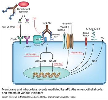
Evidence from several research groups has demonstrated a role for p38 MAPK and NF-κB signalling in mediating the effects of aPL Abs on ECs and monocytes (Fig. 2). Initial work using monocytes and ECs indicated a role for NF-κB (Refs Reference Espinola58, Reference Dunoyer-Geindre79, Reference Bohgaki80). Further in vitro work showed aPL Abs induced significant TF transcription, function and expression on ECs, a pronounced increase in pro-inflammatory cytokines (IL-6 and IL-8) and phosphorylation of p38 MAPK, and use of the specific p38 MAPK inhibitor SB 203580 [4-(4 fluorophenyl)-2 (4methylsulfinylphenyl)-(4pyridyl) 1 imidazole] and the specific NF-κB inhibitor MG132 (carbobenzoxyl-leucinyl leucinylleucinal) showed both p38 MAPK phosphorylation and NF-κB activation are required for aPL-Ab-induced TF upregulation (Ref. Reference Vega-Ostertag81). Similar results have been reported for monocytes by others (Refs Reference Bohgaki80, Reference López-Pedrera82). Subsequent independent work showed that IgG from 12 patients with APS caused a large and sustained increase in reactive oxygen species (ROS) that activated p38 MAPK and its subsequent target, the stress-related activating transcription factor 2, thereby upregulating VCAM-1 expression by human umbilical vein endothelial cells (HUVECs) and increasing monocytic cell adhesion (Ref. Reference Simoncini83). More recently, in two separate studies, investigators demonstrated that aPL Abs upregulated TF in vivo in carotid artery and in monocytes, and VCAM-1 was significantly upregulated in aorta of mice; these effects correlated with enhanced thrombosis and were inhibited by SB203580 and MG132 in vivo (Refs Reference Vega-Ostertag84, Reference Montiel-Manzano85) (Table 1). Interestingly, aPL-Ab-mediated activation of ECs in vitro (upregulation of adhesion molecules and TF) and in vivo, as well as thrombogenic effects of aPL Abs in mice, can be inhibited by pretreatment with fluvastatin (Refs Reference Meroni86, Reference Ferrara87, Reference Ferrara88) (Fig. 2; Table 1).
Membrane and intracellular events mediated by aPL Abs on endothelial cells, and effects of various inhibitors. aPL Abs are shown binding to the surface of ECs through TLR-4 and/or annexin A2, and trigger intracellular signalling involving phosphorylation of p38 MAPK and activation of NF-κB. These processes lead to upregulation of cellular adhesion molecules (VCAM-1, E-selectin and ICAM-1) and tissue factor. iNOS is also induced and cytokine production (IL-1, IL-6 and IL-8) is increased. These effects can be effectively blocked by MG132, SB 203580 or by fluvastatin. The binding of aPL/anti-˙β2GPI Ab to β2GPI on the surface of ECs can be effectively blocked by TIFI, a peptide that mimics part of domain V of β2GPI. aPL/anti-β2GPI Abs may activate complement, and its split products [i.e. C3a, C5a or C5b-9MAC (latter not shown)] may bind to specific receptors on ECs and further enhance the pro-inflammatory effects of aPL Abs on ECs. These effects can be diminished by an anti-C5 mAb. Abbreviations: aPL Abs, antiphospholipid antibodies; C5aRc, C5a receptor; C3aRc, C3a receptor; EC, endothelial cell; β2GPI, β2-glycoprotein I; ICAM, intercellular cell adhesion molecule; IL, interleukin; mAb, monoclonal antibody; MAC, membrane attack complex; MAPK, mitogen-activated protein kinase; iNOS, inducible nitric oxide synthase; NF, nuclear factor; NO, nitric oxide; TLR-4, Toll-like receptor 4; VCAM, vascular cell adhesion molecule.

Possible new targeted therapies for aPL-Ab-mediated thrombosis

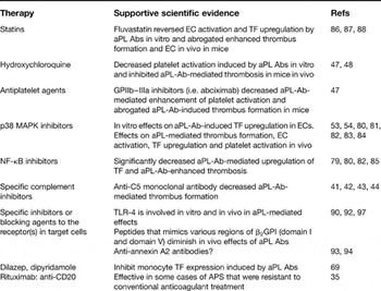
Abbreviations: aPL Abs, antiphospholipid antibodies; APS, antiphospholipid syndrome; β2GPI, β2glycoprotein l; NF-κB: nuclear factor κB; p38 MAPK, p38 mitogen-activated protein kinase; TF, tissue factor; TLR-4, Toll-like receptor 4.
Receptor for β2GPI in ECs
APL/anti-β2GPI Abs bind to β2GPI through domain I of β2GPI (Ref. Reference Bouma89); interestingly, in a recent study the thrombogenic effects of aPL/anti-β2GPI Abs in mice were significantly diminished when mice were injected with a recombinant form of this domain (Ref. Reference Ioannou90). Domain V of β2GPI is unique in its high content of lysine residues, which contribute to the formation of a positively charged phospholipid-binding region (Refs Reference Bouma89, Reference Del Papa91). Studies with β2GPI mutants and synthetic peptides show that the molecule binds to EC membranes through this putative phospholipid-binding site, possibly through negatively charged heparan sulphate (Ref. Reference Del Papa91). It has been suggested that such interaction may facilitate the binding of β2GPI to the putative receptor(s), and that aPL Abs may, in turn, increase the binding affinity to the receptor(s). TIFI, a 20 amino acid synthetic peptide that shares similarity with the phospholipid-binding region of β2GPI inhibits the thrombogenic properties of human aPL Abs in mice by competing with β2GPI and by preventing its binding to target cells (ECs and monocytes) (Ref. Reference Vega Ostertag92) (Fig. 2).
EC structures other than heparan sulphate might also be responsible for the binding of β2GPI. For example, annexin A2 mediates EC activation by aPL Abs after binding to β2GPI (Refs Reference Ma93, Reference Zhang and McCrae94). Because annexin A2 does not span the cell membrane, this interaction may require an ‘adaptor’ protein able to transduce intracellular signalling. One candidate coreceptor is TLR-4, discussed further below (Ref. Reference Akira, Takeda and Kaisho95) (Fig. 2). ApoER2′ might also act as a β2GPI receptor; as discussed above for platelets, apoER2′ binds dimers of β2GPI (which mimic anti-β2GPI Abs) leading to phosphorylation of p38 MAPK, TX production and cell activation (Ref. Reference Lutters54) (Fig. 1).
Upon ligation of LPS to TLR-4, translocation of NF-κB, phosphorylation of p38 MAPK and other intracellular events lead to the upregulation of pro-inflammatory cytokines, CAMs and TF. Myeloid differentiation factor 88 (MyD88), a common adaptor protein for TLRs, mediates intracellular signal transduction after TLR-4 activation has been shown to be triggered by aPL/β2GPI Abs on human ECs in vitro (Ref. Reference Raschi96). The role of TLR-4 in aPL-mediated EC activation/thrombosis in vivo has been investigated in LPS-nonresponsive and LPS-responsive mice. [The nonresponsive mice have a point mutation of the Tlr4 gene leading to the expression of a TLR-4 that does not recognise LPS (Ref. Reference Pierangeli97).] The LPS-nonresponsive mice are protected from IgG-APS-induced thrombophilia and EC activation in vivo (Ref. Reference Pierangeli97).
Activation of the complement cascade and aPL Abs
Complement components and their activation products [i.e. C3a, C5a and C5b-9 or membrane attack complex (MAC)] interact with ECs by binding to specific receptors expressed on these cells. Recent studies have suggested that activation of the complement cascade is necessary for aPL-Ab-mediated thrombophilia and fetal loss. First, inhibition of the complement cascade in vivo, using the C3 convertase inhibitor complement receptor-1-related gene protein y (Crry)-Ig, blocks aPL-Ab-induced fetal loss and growth retardation and inhibits aPL-Ab-mediated thrombosis (Ref. Reference Holers41). Second, mice deficient in complement C3 and C5 (C3−/− and C5−/−, respectively) were shown to be resistant to thrombosis, EC activation, and fetal loss induced by aPL Abs (Refs Reference Salmon, Girardi and Holers42, Reference Pierangeli43). Third, anti-C5 monoclonal Abs reversed thrombogenic properties of aPL Abs in vivo, confirming involvement of C5 complement activation in aPL-Ab-induced thrombosis (Ref. Reference Pierangeli43) (Fig. 2; Table 1).
In patients with APS, aPL Abs tend to be of the IgG isotype although the IgM and/or IgA isotypes can be present concurrently and have also been shown to be pathogenic in vivo (Ref. Reference Pierangeli98). The IgG subclasses can be IgG1, IgG2 and IgG4, with the IgG2 subclass more prevalent in APS (Ref. Reference Sammaritano99). It is known that IgG2 and IgG4 Ab subclasses are weak in their ability to fix complement via the classical pathway in humans. A recent study showed that C5a-receptor-deficient (C5aR−/−) mice were protected from aPL-Ab-mediated thrombophilia when mice were injected with an IgM isolated from a patient with APS but not with an IgG preparation from another patient that did not have the ability to fix complement (Ref. Reference Romay-Penabad100). This argues for the necessity to consider additional mechanisms to the classical complement pathway by which aPL/anti-β2GPI Abs enhance thrombosis in APS patients. More specifically, the data indicate that the use of C5 and C5a inhibitors or antagonists for the C5aR may be useful in reversing pathogenic effects of aPL Abs (Table 1). These findings were recently confirmed in rats by another group, who showed that thrombus formation induced by Abs to β2GPI requires a priming factor such as bacterial LPS and is complement dependent (Ref. Reference Fischetti44). Interestingly, hypocomplementemia has been found in a significant proportion of patients with PAPS, and it was associated with thrombosis in one study and with livedo reticularis and thrombocytopaenia in another publication (Refs Reference Munakata101, Reference Carbone102, Reference Davis and Brey103). Hence complement activation may also be a pathogenic mechanism in patients with APS.
Proposed mechanisms for aPL Abs
In summary, the following mechanism for the pathogenic effects of aPL Abs on thrombosis may be proposed. First, binding of aPL Abs to ECs may directly induce their activation and a procoagulant state, as demonstrated in vivo and in vitro, by upregulating adhesion molecules and TF expression. APL Abs may also induce platelet activation. Furthermore, APL Abs may interact with elements of the coagulation cascade. This may amplify the direct effects on ECs and platelets by generation of potent mediators of platelet and EC activation, including C3a and C5a and the C5b-9 MAC (Fig. 2). We hypothesise that in APS patients with aPL Abs targeted to the endothelium, complement activation may be increased locally and may overwhelm normally adequate inhibitory mechanisms. Hence, activation of complement may be a critical proximal effector mechanism in aPL-associated thrombosis.
Conclusions
It has been suggested that aPL Abs may lower the threshold for activation in ECs and platelets (first hit) and that the clinical event (i.e. thrombosis or ‘second hit’) happens sporadically, possibly associated with another triggering event (infection, trauma, surgery and so on). Treatments that modulate early effects of aPL Abs on target cells, such as EC activation, would be more beneficial and potentially less harmful than current options. Furthermore, agents able to inhibit the binding of aPL Abs to the receptor or the cell signalling activation or complement activation may act directly on the putative ‘first hit’, reducing the risk for developing the clinical events in the case that a ‘second hit’ does occur. This is particularly important given that ‘second hits’ (such as common infectious processes) cannot be easily prevented. Knowing the molecular events induced by aPL Abs and the nature of the receptor and its interaction with β2GPI and specific Abs may help to identify useful surrogate markers (biomarkers) of thrombotic risk and to devise new targeted, more-specific modalities for treatment and prevention of thrombosis with fewer adverse effects in patients with aPL Abs (Table 1). Interestingly, gene expression patterns that characterise APS and predict patients with APS at high risk for thrombotic events have recently been reported, and these findings may help in the management of APS-related thrombosis as well as in targeting antithrombotic therapy in these patients (Ref. Reference Potti104).
The potential new targeted therapies listed in Table 1 encompass agents acting on intracellular events and interactions at the cell surface. Intracellular events induced by aPL Abs in ECs, monocytes and platelets can be diminished by using p38 MAPK and NF-κB inhibitors in vitro and in animal models, and also by hydroxychloroquine in platelets; in addition statins seem to ameliorate procoagulant and pro-inflammatory effects of aPL Abs possibly by exerting anti-inflammatory effects (Figs 1 and 2). Events at the cell surface might be modulated by anti-GPII–IIIa antagonists (such as the drug abciximab) and possibly by blocking binding of aPL Abs to their receptor(s) on target cells (e.g. by TIFI) (Figs 1 and 2). It is possible to speculate that inhibition of complement activation should ameliorate vascular thrombosis in individuals with APS. Clinical trials will need to be done to validate these findings in humans and to examine the efficacy and effectiveness of these proposed targeted therapies to prevent/treat thrombosis and possible other complications in patients with aPL Abs.
Acknowledgments and funding
These studies were partially funded by a Research Centers in Minority Institutions – National Institutes of Health grant (G12-RR03034), a Minority Biomedical Research Support Grant from the National Institutes of Health (GM58268-02) and a NIAMS MIH Multidisciplinary Research Center grant (2P60 AR047785-06). The authors thank the reviewers of the manuscript for their helpful comments.