INTRODUCTION
Donkeys are important for the economy of Africa, Asia and the Arabian Peninsula, and their popularity is increasing in Europe. Sarcocysts have been found in donkeys from Egypt, Austria, Russia (former USSR) and Great Britain, but the parasite has not been adequately described. In general, there is considerable confusion concerning the validity of different species of Sarcocystis in equids, especially donkey (Dubey et al. Reference Dubey, Calero-Bernal, Rosenthal, Speer and Fayer2015). It is believed that all equids (horses, mules, donkeys, zebra and others) share common Sarcocystis species, but definitive studies are lacking.
Gadaev (Reference Gadaev1978) found sarcocysts in 8 of 20 donkeys from the former USSR and named a new species, Sarcocystis asinus from the donkey, based on a very cryptic morphological description of the sarcocyst (more on this in discussion section).
Hinaidy and Loupal (Reference Hinaidy and Loupal1982) found sarcocysts in 1 of 2 donkeys in Austria. However, they did not provide any detail and remarked that the sarcocysts in the donkey were similar to those of Sarcocystis bertrami in the horse. Similarly, Edwards (Reference Edwards1984) found sarcocysts in 1 of 2 donkeys from a slaughter house in Cheshire, England but did not elaborate on the morphology of the parasite. Kirmse (Reference Kirmse1986) found sarcocysts in histological sections of 9 of 41 donkeys slaughterd at Zoological Gardens of Rabat, Morocco for feeding captive carnivores; the morphology of sarcocysts was not described. Zayed and El-Ghaysh (Reference Zayed and El-Ghaysh1998) reported that 4 dogs fed meat from 11 donkeys slaughtered at National Circus, Giza, Egypt excreted Sarcocystis sporocysts; the sarcocysts were assumed to be S. bertrami.
Hilali and Nasser (Reference Hilali and Nasser1987) found sarcocysts in 18 of 20 donkeys in Egypt. In histologic sections, sarcocysts were microscopic, up to 410 µm long and 50·2 µm wide. By transmission electron microscopy (TEM) the sarcocyst wall had villar protrusions (vp) that were up to 3·7 µm long and up to 1 µm wide (Hilali and Nasser, Reference Hilali and Nasser1987). The bradyzoites were 16·2–16·9 × 4·1–4·2 µm in size. They considered the parasite same as S. bertrami in the horse. We now provide a full morphological description of sarcocysts in donkey.
MATERIALS AND METHODS
An adult donkey was obtained live on 14 January 2015 from El Fayoum Governorate, Egypt for teaching post mortem technique to veterinary students. The donkey was euthanized by the Pathology Department, Faculty of Veterinary Medicine, Cairo University, Giza, Egypt.
For the present study, tongue muscle was fixed in glutaraldehyde (GF) and formalin. The formalin-fixed (FF) tissue was processed for paraffin embedding. The paraffin blocks and the GF samples were transported to the Faculty of Veterinary Science, University of Pretoria, Onderstepoort, South Africa for light and electron microscopic examinations. For light microscopy (LM), paraffin-embedded sections were cut at 5 µm thick and examined after staining with haematoxylin and eosin (H and E). For TEM, GF tissue was processed using standard techniques. Briefly, the samples were post-fixed in 1% osmium tetroxide in Millonig's buffer (pH 7·4), dehydrated through a series of graded ethanols, infiltrated with an epoxy resin/propylene oxide mixture before being embedded in absolute resin, and polymerized at 60 °C overnight. Two cysts were detected in the GF sample. A further 17 tissue cysts, located in paraffin blocks (by matching with H and E sections) were deparaffinised (Van den Berg Weermans and Dingemans, Reference Van den Berg Weermans and Dingemans1984).
RESULTS
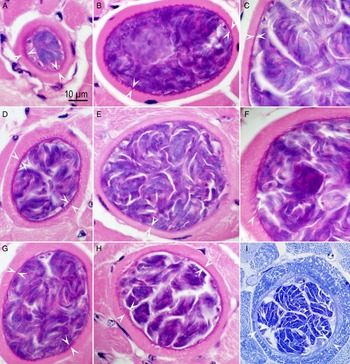
In total, 19 sarcocysts were studied ultrastructurally; 2 from GF and 17 from paraffin blocks. Sarcocysts were microscopic, and up to 110 µm wide. By LM of 5 µm histological sections, the sarcocyst wall thickness varied from <1 to 5 µm, depending on the individual sarcocyst and the plane of section (Fig. 1). Some sarcocysts had protrusions that varied in shape and size. Examples from 9 sarcocysts are shown in Fig. 1.
Sarcocysts in histological sections of tongue of a donkey in Egypt. A–H, haematoxylin and eosin stain, I, Toluidine blue stain. Bar applies to all images. The sarcocyst wall appears smooth or striated (opposing arrowheads).

By TEM of GF sarcocysts, the parasitophorous vacuolar membrane (pvm) was undulating, and lined by an electron dense layer (edl). The edl was thin at irregular intervals and invaginated giving the pvm a vacuolated appearance (Figs 2 and 3). The pvm was folded into vp that varied in shape and size, within the same cyst (Fig. 2B and C). In some cysts, vp were adjacent to each other, while in others they were up to 2 µm apart. Villar protrusions were mostly elongated, up to 6 µm long and up to 1 µm wide, some were conical in shape. Electron dense, evenly distributed hair-like structures were seen on vp tips, both in GF and FF vp (Fig. 3A). The vp contained microtubules (mt) that varied ultrastructurally. A tuft of highly electron dense mt1 extended from the tip of the villus deep into the interior of the sarcocyst reaching the pellicle of the zoites (Fig. 3A). Another group of less electron dense mt2 existed parallel to the electron dense mt1 (Fig. 3A). Additionally, fine mt3 were located towards the tips of the vp. Microtubules lacked electron dense granules. Dense granules, however were present in the distal portion of vp, juxtaposed with host myocyte. The ground substance layer (gs) was up to 2·5 µm thick and was relatively homogenous, except for a few dense granules. In tangentially cut sarcocysts, the gs sections of electron dense mt were scattered throughout the gs (Fig. 3B).
TEM of S. bertrami sarcocyst walls from 2 cysts. Glutarladehyde-fixed. Note parasitophorous vacuolar membrane (pvm) lined by electron dense layer, villar protrusions (vp), ground substance layers (gs), microtubules (mt), bradyzoites (br), metrocytes (me) and host cell (hc). (A) The vp are folded on the sarcocyst wall. The gs is thin. Cyst #1. (B) The vp are angled or folded over the sarcocyst wall. The gs appears thick and has portions of microtubules (mt). (C) Same sarcocyst as in B but the vp are conical, the gs is thick. The mt extend from the tip of the vp to the pellicle of metrocytes (arrowheads). Cyst #2.

TEM of S. bertrami sarcocyst walls from cyst #2. (A) Tip of a vp showing undulating pvm, lined by electron dense layer (edl), that is thinned out or absent at places (arrowheads). The pvm has outpocketing of protrusions (pr). Three types of mt are visible: (i) a thick tuft of electron dense microtubules (mt1), (ii) another tuft of medium electron dense microtubules (mt2), and (iii) fine mt that appear to crisscross towards the villar tips (mt3). Note numerous dense granules (gr) in the villar tips. A collar of hair-like (arrow) projections is present on vp. The gs is relatively smooth with few granules. (B) Cross-section of vp showing protrusions (pr) on the pvm and mt.

All sarcocysts contained few metrocytes and numerous mature bradyzoites. Both metrocytes and bradyzoites were arranged in groups (Fig. 2). The bradyzoites were 11–15 µm long and 2·5–3·0 µm wide. They had the same structures as seen in other Sarcocystis bradyzoites, including micronemes, 2 rhoptries, a micropore, several dense granules, a nucleus, a mitochondrion, and numerous amylopectin granules (Fig. 4). The micronemes were in the conoidal third of the bradyzoite. Most micronemes were arranged in rows and were approximately 300 × 50 nm in size with tapering or round ends (Fig. 4). The micropore was located 2·5 µm from the tip of conoid. Granular material was seen below the micropore (Fig. 4C).
TEM of S. bertrami bradyzoites. Glutarladehyde-fixed. Note conoid (co), numerous micronemes (mn), several dense granules (dg), a nucleus (nu) and rhoptries (rh) with long slender neck. (A) A longitudinally cut bradyzoite. (B) Longitudinal section of conoidal part of a bradyzoite. Note conoid (co) with two rhoptries (rh1, rh2) with slender neck. The micronemes (mn) are arranged in rows. (C) Note a micropore (mp) with a collared rim (rm), dense granules (dg), and electron dense secretory material surrounds below the mp.

DISCUSSION
As stated earlier, there is considerable confusion concerning the species of Sarcocystis in equids. Rommel and Geisel (Reference Rommel and Geisel1975) in reviewing the earlier literature stated that Siedamgrotzky (Reference Siedamgrotzky1872) first reported sarcocysts in horses in Germany. He noticed cilia-like 2 µm long protrusions on the sarcocyst wall. Doflein (Reference Doflein1901) named the parasite, Sarcocystis bertrami; he noticed cone-like structures on the sarcocyst wall. Rommel and Geisel (Reference Rommel and Geisel1975) found thin-walled sarcocysts that lacked protrusions; they named the parasite Sarcocystis equicanis following the nomenclature proposed by Heydorn et al. (Reference Heydorn, Gestrich, Mehlhorn and Rommel1975), combining the names of intermediate and definitive hosts. Göbel and Rommel (Reference Göbel and Rommel1980) described the ultrastructure of S. equicanis and reported 2–3 µm long hairy protrusions on the sarcocyst wall. Odening et al. (Reference Odening, Wesemeier, Walter and Bockhardt1995) expanded on the ultrastructure of sarcocysts in wild Equidae, and concluded that the structure broadly resembled the description of S. equicanis from the domestic horse.
Experimental evidence suggests the similarity of a Sarcocystis species in the horse and the donkey (Matuschka, Reference Matuschka1983). Tissues from 20 naturally infected horses in Germany were fed to a dog, and those of 10 donkeys (also from Germany) were fed to another dog. Both dogs excreted sporocysts. Experimental infections were conducted with 4 ponies (#1–4) raised in captivity using the sporocysts derived from the infected donkeys (inoculum A) and sporocysts derived from the inocula from horses (inoculum B). The foal (#1) fed donkey-derived sporocysts became febrile on days 10 and 11 and 19–21 but sarcocysts were not found when muscle biopsies were obtained on 44 and 59 days post-inoculation (DPI). The same foal (#1) was then fed horse-derived sporocysts on day 117 and killed 21 days later (138 days after feeding donkey-derived sporocysts). Sarcocysts were detected histologically in muscles of 3 foals when killed 138 (#1), 122 (#2) and 221(#3) DPI but no parasites were detected in the foal (#4) killed 21 DPI. Sporocysts were structurally similar in dogs that excreted sporocysts after ingesting donkey or horse muscles; the sporocysts were 12·2–13·8 × 9·2–9·9 µm in size and the prepatent period was 9–10 days. Sarcocysts and sporocysts appeared similar to those of S. equicanis/S. bertrami (Rommel and Geisel, Reference Rommel and Geisel1975). Although this experiment is not definitive (transmission based on 1 foal fed donkey-derived sporocysts), it is suggestive of possible transmission of the parasite in horse and donkey.
Two dog transmitted Sarcocystis species, S. bertrami (S. equicanis) and Sarcocystis fayeri are known to infect horses. Sarcocystis fayeri, orginally described from horses in the USA has been also found in Germany (Dubey et al. Reference Dubey, Streitel, Stromberg and Toussant1977; Erber and Geisel, Reference Erber and Geisel1981). However, S. bertrami has not been reported in horses in the USA, and S. fayeri has not been reported from other equids, including the donkey. Dubey et al. (Reference Dubey, Calero-Bernal, Rosenthal, Speer and Fayer2015) reviewed the literature on Sarcocystis species in equids and synonmysed S. equicanis with S. bertrami. Illustrations in Fig. 1 in the present study from sarcocysts in donkey may explain why the confusion arose concerning the thin-walled S. equicanis described by Rommel and Geisel (Reference Rommel and Geisel1975) and the hairy to thick-walled sarcocysts described by Doflein (Reference Doflein1901). In the present study, both smooth and hairy portions were seen in the same sarcocyst.
As stated earlier, Gadaev (Reference Gadaev1978) named S. asinus, solely based on the size, shape and staining of bradyzoites in smears. He said that bradyzoites of S. asinus were 1–3 µm larger (12·3–17·5 × 4·5–6·2 µm for S. asinus and 11·5–14·5 × 3·5–4·0 µm for S. bertrami) than S. bertrami of horses, and the nuclear chromatin was pale red in S. asinus vs crimson in S. bertrami (Gadaev, Reference Gadaev1978). These differences are untenable for separating Sarcocystis species, because these differences in bradyzoite morphology could be related to techniques used. Therefore, we support the contention that S. asinus is invalid (Levine and Tadros, Reference Levine and Tadros1980; Odening, Reference Odening1998; Dubey et al. Reference Dubey, Calero-Bernal, Rosenthal, Speer and Fayer2015).
All sarcocysts in tongue of the donkey in the present study were microscopic. Hinaidy and Loupal (Reference Hinaidy and Loupal1982) mentioned that sarcocysts in a donkey oesophagus were 1·7–14·1 mm long. Macroscopic sarcocysts, however, were never found in donkeys in Egypt (Hilali, personal observations). The structure of the sarcocyst wall is useful in taxonomy of Sarcocystis species in a given host. Dubey et al. (Reference Dubey, Calero-Bernal, Rosenthal, Speer and Fayer2015) grouped sarcocysts in more than 40 morphologic types. Sarcocysts in the present study were type 11, found in sarcocysts in equids and also in some avian species. One characteristic of type 11 sarcocysts is mt that extend from the villar tips to the zoite pellicle. Here, we have described in detail the structure of vp of the sarcocyst wall of the sarcocyst in the donkey, and it is distinct from vp in S. fayeri. Villar protrusions in S. fayeri are slender, upright, and contain 1 group of mt. The vp in S. bertrami are folded over the cyst wall and have mt of 3 types.
The description of the S. bertrami sarcocysts described here from the donkey should assist in the recognition of this parasite in horses and other equids. The parasite described in the present study in donkey was considered S. bertrami, based on literature. As stated earlier, there are no archived specimens of Sarcocystis species named from equids before 1975. A critical comparison of the molecular characteristics of Sarcocystis species in the horse vs sarcocysts from other equids will help in reaching a final conclusion.
ACKNOWLEDGEMENTS
We thank Dr Petras Prakas for translation of the Gadaev (Reference Gadaev1978) paper and Dr Gereon Schares for a copy of the paper by Siedamgrotzky (Reference Siedamgrotzky1872).
FINANCIAL SUPPORT
This research received no specific grant from any funding agency, commercial or not-for-profit sectors.