About 130 years ago, Elie Metchnikoff put forward the idea of macrophage-like cells that, as part of mammalian immunity, patrol the body and take up foreign material (Ref. Reference Gordon1). Nowadays, the macrophage is known as a versatile cell playing central roles in inflammation, wound healing, tissue homeostasis and tissue remodelling. Macrophages are professional phagocytes, clearing dying cells and pathogens. They can also function as antigen-presenting cells and are able to secrete a variety of cytokines that modulate migration and activation of other immune and nonimmune cells (Ref. Reference Gordon2).
The intestinal immune system has to remain tolerant to beneficial flora and encountered food antigens while dealing with invading pathogens. This suggests that immune responses in the intestine need to be tightly regulated. Together with dendritic cells, the strategically located macrophages contain pattern-recognition receptors (PRRs) to recognise conserved microbial molecules, and provide immune-modulating functions that are of major importance in this regulation. Continuous activation of intestinal immune responses by normal intestinal flora is believed to cause inflammatory bowel diseases, which are characterised by abdominal pain, diarrhoea, rectal bleeding, weight loss, malnutrition and fever (Ref. Reference Podolsky3). The inflamed mucosal tissues display a large influx of granulocytes and monocytes, and the inflammatory responses lead to further influx of inflammatory cells and destruction of the mucosal structure.
Two major forms of inflammatory bowel disease are Crohn disease and ulcerative colitis, which are distinguished by the affected area: ulcerative colitis exclusively affects the colon, whereas Crohn disease can affect the whole gastrointestinal tract but is mainly localised to the colon and ileum (Ref. Reference Podolsky3). These two diseases have also been suggested to have different cytokine profiles, with a T helper 1 (Th1) phenotype for Crohn disease and a Th2 phenotype for ulcerative colitis. The incidence of inflammatory bowel diseases in Europe is estimated at 2.2 million (Ref. Reference Loftus4). As a result of limited understanding of the disease pathogenesis, treatment is symptomatic and mainly relies on nonspecific immune suppression. Patients are thus chronically dependent on medication, and have significant morbidity.
The role for immune cells in inflammatory bowel diseases has been extensively studied, and the influences on intestinal immune responses of dendritic cells, T cells, B cells and mast cells have been reviewed (Refs Reference Coombes and Powrie5, Reference Ahern6, Reference Brandtzaeg7 and Reference Rijnierse8, respectively). The role for macrophages in inflammatory bowel diseases is underlined by genetic studies that link macrophage-specific molecules with disease (Refs Reference Hugot9, Reference Ogura10), and experimental colitis models where a lack of macrophages in the intestine increases inflammation (Ref. Reference Qualls11), as discussed further in Box 1. In this review, we focus on the intestinal macrophage, discuss its specific phenotype, and highlight new insights into its role in intestinal inflammation.
Box 1 Methods for studying macrophage function in inflammatory bowel disease
Various tools are available to study mechanisms and cells involved in inflammatory bowel disease. Animal models have proved extremely useful. These can be divided into three categories depending on the cells involved (Ref. Reference Wirtz and Neurath150): models that depend on a defect in the epithelial barrier function, models with a defect in innate immunity, and models caused by a defect in adaptive immunity. For most of these models, transgenic mice or mice with a specific gene deficiency are needed, which limits their use.
Widely used models are dextran sodium sulphate (DSS)- and trinitrobenzene sulphonic acid (TNBS)- induced colitis, which are easy to use and low in cost. DSS damages the epithelial barrier, whereas TNBS haptenises self-molecules making them immunogenic. DSS-induced colitis is particularly interesting for the study of innate immune function since T- and B-cell-deficient mice also develop disease in this model, suggesting it is mainly driven by innate immune responses (Ref. Reference Wirtz and Neurath150). Using a DSS-induced colitis model it was shown that depletion of macrophages causes more severe intestinal inflammation (Ref. Reference Qualls11). However, it has also been shown that macrophage depletion reduced spontaneous colitis in interleukin (IL)-10-deficient mice (Ref. Reference Watanabe151). The difference in outcome of macrophage depletion is most likely due to the different mechanisms of induced colitis, and this may underline the dual function of intestinal macrophages. DSS-induced colitis disrupts the mucosal barrier leading to microbial contact with the underlying lamina propria; depleting macrophages here would lead to partial loss of tolerance and increased inflammation. IL-10-deficient mice develop chronic colitis as a result of a lack of tolerance, which leads to an influx of inflammatory macrophages and depleting these will reduce inflammation.
The downside of using animal models of inflammatory bowel disease is that no one model represents all aspects of the human disease, and so studies with patient material are needed to supplement and validate data. Patient material is mainly used for genetic studies and many genes have been implicated in inflammatory bowel disease, as indicated in the main text and summarised in Table 2.
Studying intestinal macrophage function is complicated by the fact that isolation and culture methods change macrophage phenotype and have low yields. Many researchers use blood-derived monocytes instead, which also lack the intestine-specific resident phenotype. Attempts to create intestinal macrophage cell lines have failed because of a lack of knowledge of environmental factors needed to induce a lamina propria phenotype.
Monocyte development into intestinal macrophages: modulation of phenotype
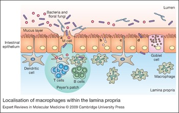
The intestine is lined with a layer of enterocytes (intestinal epithelium), which form the first barrier to the flora; some enterocytes specialise into goblet cells that secrete mucus. Macrophages lie strategically located underneath the enterocytes, and their location suggests that they will be the first immune cells in contact with invading pathogens (Fig. 1). The lamina propria (loose connective tissue under the epithelium) of the large and small intestine has the largest number of resident macrophages in the body (Ref. Reference Lee12). Other cells in the lamina propria include mast and dendritic cells. T and B cells are mainly located within Peyer's patches, which also contain macrophages in the dome region. Peyer's patches contain specialised epithelial cells, called M-cells, that take up large samples of the intestinal lumen. To control tissue homeostasis, the macrophage interacts with epithelial, vascular and other immune cells as well as neurons.
Localisation of macrophages within the lamina propria. Possible routes for microbial uptake in the intestine are indicated: (a) translocation through M-cells; (b) endocytosis by enterocytes for microbes <0.5 µm; (c) direct sampling of the gut lumen by dendritic cells that can form protrusions between enterocytes; (d) invasion of the lamina propria by pathogenic micobes.

In general, macrophages develop from granulocyte/monocyte precursors in the bone marrow. These precursor cells develop into monocytes under the influence of macrophage colony-stimulating factor (M-CSF; also know as CSF-1), granulocyte–macrophage colony-stimulating factor (GM-CSF; also known as CSF-2) and interleukin (IL)-3. Monocytes leave the bone marrow, enter the bloodstream and after approximately one day migrate into tissues where they differentiate into macrophages. Little is known about how resident lamina propria macrophages become localised to the intestine. Mucosal IL-8 and transforming growth factor β (TGF-β) have been shown to be chemoattractants for blood monocytes (Refs Reference Wahl13, Reference Smythies14), but it is unclear if this is sufficient to attract monocytes to the healthy intestine. Some tissue macrophages are able to proliferate locally, including Kupffer cells (Ref. Reference Crofton15) and alveolar macrophages (Ref. Reference Tarling16), but it has been shown that lamina propria macrophages are unable to replicate (Ref. Reference Smythies14), which suggests intestinal macrophages are replenished solely by blood monocytes.
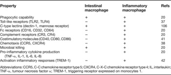
Interestingly, lamina propria macrophages are phenotypically distinct both from blood monocytes and from other tissue macrophages (Table 1). Inflammatory macrophages and tissue macrophages develop from distinct monocyte subsets: inflammatory cells develop from CX3CR1loGR1+ monocytes and tissue macrophages from CX3CR1hiGR− monocytes (Ref. Reference Geissmann17). This supports the general idea that resident lamina propria macrophages differ in phenotype and function from recruited macrophages during inflammatory bowel disease. Furthermore, development into tissue macrophages is mediated by factors specific to the local environment, which renders tissue macrophages heterogeneous in function, receptor expression and inflammatory responses (Ref. Reference Gordon and Taylor18).
Macrophage phenotype

Abbreviations: CCR5, C-C chemokine receptor type 5; CXCR4, C-X-C chemokine receptor type 4; IL, interleukin; TNF-α, tumour necrosis factor α; TREM-1, triggering receptor expressed on monocytes 1.
The noninflammatory nature of lamina propria resident macrophages
Differentiation into intestinal macrophages results in an anti-inflammatory phenotype although bactericidal activity is retained (Ref. Reference Smythies19). The exact factors involved in such differentiation are unclear, but cytokines such as TGF-β and IL-10 are able to induce at least some of the intestinal macrophage features (Ref. Reference Platt and Mowat20). Both these cytokines are able to inhibit NF-κB activation mediated by PRRs and downregulate secretion of the inflammatory cytokine tumour necrosis factor α (TNF-α) (Refs Reference Smythies19, Reference Kuwata21, Reference Randow22, Reference Schottelius23, Reference Naiki24). Deficiency of TGF-β and IL-10 leads to development of spontaneous inflammatory diseases (Refs Reference Kohn25, Reference Shull26). Unlike other resident macrophages, the majority of lamina propria macrophages lack TREM-1 (triggering receptor expressed on myeloid cells 1), a receptor involved in amplification of inflammatory responses induced by microbial products. TGF-β and IL-10 downregulate TREM-1 expression, supporting the idea that these cytokines are essential for stimulating a lamina propria phenotype (Ref. Reference Schenk27). TGF-β alone downregulates expression of the immunoglobulin A (IgA) Fc receptor (CD89) and the complement receptor CR3 (Refs Reference Reterink28, Reference Hogasen29), while IL-10 affects expression of C-type lectin PPRs (Refs Reference Willment30, Reference Martinez-Pomares31). Even though IL-10 and TGF-β render macrophages anti-inflammatory, this seems not sufficient to produce a complete intestinal macrophage phenotype, and other factors such as direct interaction with intestinal epithelial cells and interactions with extracellular matrix proteins such as collagen have been suggested to play a role (Refs Reference Spottl32, Reference Jacob33). It is also likely that continuous exposure of lamina propria macrophages to microbial ligands influences the macrophage phenotype directly, via stimulation of PRRs; for instance, prolonged muramyl dipeptide (MDP) stimulation via the PRR NOD2 (nucleotide-binding oligomerisation domain-containing protein 2) mediates tolerance to subsequent stimulation with ligands for the Toll-like receptors TLR2 and TLR4 (Ref. Reference Hedl34).
Human lamina propria macrophages lack expression of various PRRs, including the lipopolysaccharide (LPS) receptor CD14, the CR3 component CD11b and Ig receptors CD16, CD32 and CD64, supporting a tolerant phenotype towards the intestinal flora (Refs Reference Smythies19, Reference Rogler35, Reference Hausmann36). Furthermore, intestinal macrophages also lack receptors for IL-2 and IL-3 as well as the chemokine receptors CCR5 and CXCR4, and the integrin LFA-1 (leukocyte-function-associated antigen 1), which probably leads to a reduced response to certain immune stimuli (Refs Reference Smythies19, Reference Meng37). Lack of the costimulatory molecules CD80 and CD86 renders them unable to activate T-cell-mediated immune responses (Ref. Reference Rogler35). Together, this phenotype results in a tolerant macrophage that is highly phagocytic and bactericidal without subsequent secretion of pro-inflammatory cytokines.
The small and large intestine are distinct with respect to function, amount of flora and the microbial species they contain, and so it is not surprising that there are differences between macrophages isolated from these sites. For instance, jejunal macrophages express major histocompatibility complex class II antigen (MHCII), TLR2 and TLR4, but these seem to be absent from colonic macrophages (Refs Reference Smythies19, Reference Rogler35, Reference Hausmann36, Reference Smith38). Nevertheless, it is generally clear that intestinal macrophages have an unusually tolerant phenotype, although underlying mechanisms are only starting to be clarified. Signalling components and macrophage receptors that inhibit immune-stimulatory responses including SOCS (suppressor of cytokine signalling) proteins, IRAK-M (IL-1-receptor-associated kinase), the CD200 receptor, and SHPS1 (SHP substrate 1; also known as signal-regulatory protein α, Sirpα) are still largely unexplored in the intestine, but are likely to play important roles in maintaining intestinal tolerance (Ref. Reference Barclay39).
Inflammatory properties of recruited monocytes and macrophages
During intestinal inflammation, blood monocytes are recruited and differentiate into activated macrophages that produce pro-inflammatory cytokines (Refs Reference Wahl13, Reference Rugtveit40, Reference Burgio41, Reference Grimm42). This pro-inflammatory response has detrimental effects, leading to epithelial apoptosis, barrier dysfunction, necrosis, granuloma formation and fibrosis. Radiolabelled CD14+ monocytes injected back into patients with active inflammatory bowel disease located predominantly to inflamed parts of the intestine (Ref. Reference Grimm42). Lamina propria macrophages from patients with inflammatory bowel disease have been shown to upregulate expression of CD14, TREM-1, CD80 and CD86 (Refs Reference Rugtveit43, Reference Schenk44), and have increased NF-κB activation (Ref. Reference Neurath45). Furthermore, a recent study of Crohn disease patients revealed an increased number of CD14-expressing macrophages in the intestine that stimulate higher levels of interferon γ (IFN-γ) and IL-23 production (Ref. Reference Kamada46). These cells also produced more TNF-α, in keeping with findings from other studies of lamina propria macrophages of Crohn disease patients (Refs Reference Reinecker47, Reference Reimund48).
TNF-α is a key cytokine in inflammatory bowel disease, and neutralising anti-TNF-α antibodies are effective in treating many patients. However, besides its potent effect on inflammatory cells, TNF-α also regulates epithelial cell growth and apoptosis, intestinal permeability and matrix metalloproteinase production. Therefore, it is not surprising that a complete lack of TNF-α leads to more severe inflammation in colitis models (Ref. Reference Naito49). A chemokine receptor antagonist that reduces influx of inflammatory macrophages lowered pro-inflammatory cytokines and inflammation in experimentally induced colitis (Ref. Reference Tokuyama50), and some drugs used to treat inflammatory bowel disease indeed reduce monocyte recruitment to the inflamed intestine.
Interaction with microbes
Intestinal flora
Currently it is still unclear if inflammatory bowel diseases are caused by changes in intestinal flora or by changes in immune responses towards the flora, but it is generally accepted that the intestinal flora plays a major role; this is supported by experiments where IL-10-deficient mice, which develop spontaneous colitis, stay healthy when kept under sterile conditions, and, furthermore, antibiotics reduce inflammation in animal models and patients with inflammatory bowel disease (Ref. Reference Sellon51). Bacteria make up 99% of the intestinal flora; the remainder is mainly fungi and possibly protozoa. The highest number of bacteria is found in the colon, which may harbour more that 400 different, predominantly anaerobic, species (Ref. Reference Tannock52). Besides the important role in protection against colonisation by pathogens and production of co-enzymes, the intestinal flora is also essential for intestinal homeostasis (Ref. Reference Rakoff-Nahoum53). Furthermore, a recent study has shown that bacterial ATP in the lumen could be responsible for the presence of Th17 cells in the lamina propria (Ref. Reference Atarashi54).
According to the hygiene hypothesis, disorders such as inflammatory bowel diseases and allergies, which are typical of the developed world, arise as a result of a lack of microbes to shape the immune system; in particular, it has been proposed that a lack of helminth infection is related to predisposition to inflammatory bowel diseases (Ref. Reference Elliott55). This is supported by a substantial amount of data from studies in both experimental models and Crohn disease patients that suggest that helminth infection shifts the predominantly Th1 immune response to a Th2 response (Ref. Reference Wang56). Macrophages have also been shown to play a role in helminth-induced tolerance. Helminth infection increased the numbers of immunosuppressive intestinal macrophages, which reduced experimentally induced colitis (Ref. Reference Smith57). Together these data form substantial evidence to suggest that the flora is essential for shaping intestinal immunity.
Differences in flora composition and metabolic activity have been found in patients with inflammatory bowel diseases compared with healthy controls, including a reduction in numbers of potentially beneficial Bifidobacteria and Lactobacillus species (Refs Reference Othman58, Reference Linskens59). Different viruses have been associated with inflammatory bowel disease, including Paramyxoviruses and herpes family viruses; however, available data are largely contradictory (Ref. Reference Barclay39). The innate immune system may also influence microbial flora composition as is shown by an elegant study using Rag2−/− T-bet-deficient mice: such mice, lacking adaptive immune responses and deficient in the inflammation-regulating transcription factor T-bet, develop spontaneous colitis, and, interestingly, bedding material transferred disease to healthy wild-type mice (Ref. Reference Garrett60).
In the normal healthy intestine, epithelial cells form a barrier to the microbial flora; however, several ways have been described through which the microbial flora are able to cross this barrier (Ref. Reference Mowat61) (Fig. 1). First, enterocytes are able to mediate endocytosis of antigens that have diffused through a thick mucus layer, and these antigens can then be presented to macrophages and dendritic cells underneath the basement membrane. Second, professional endocytic and phagocytic M-cells take up large amounts of microbial and food antigens, which are thereby presented to underlying dendritic cells and macrophages. M-cells are solely associated with Peyer's patches and the large number of internalised microbes is enhanced by a reduction in mucus covering these cells. Third, dendritic cells are able to form protrusions through the epithelial layer to take up luminal bacteria, although it is not known whether macrophages can also achieve this (Ref. Reference Rescigno62). Last, some pathogenic microbes have developed ways to invade via intestinal epithelial cells, including Salmonella and Shigella species; furthermore, functional alterations in commensal microbes may cause invasion by species generally seen as nonpathogenic (Ref. Reference Sartor63).
Pattern-recognition receptors
Recognition of microbial flora is essential for regulating immune responses and this is mediated by PRRs. PRRs recognise highly conserved molecules expressed by microbes referred to as pathogen-associated molecular patterns (PAMPs). These PAMPs are generally important for survival of microbes and include LPS (part of the cell wall of Gram-negative bacteria), lipoteichoic acid (LTA) (part of Gram-positive bacteria), mannans and β-glucans (found on fungi), and viral double-stranded RNA or foreign DNA (Ref. Reference Janeway and Medzhitov64). PRRs expressed by macrophages include TLRs (which are membrane-bound molecules), C-type lectin(-like) molecules (which are mainly membrane bound but can also be secreted), and cytosolic PRR-like molecules containing NODs, which recognise intracellular pathogens (Ref. Reference Janeway and Medzhitov64). Recognition by PRRs can lead to phagocytosis, secretion of pro-inflammatory cytokines, and induction of apoptosis and immunity.
Toll-like receptors
The Toll receptor, previously shown to be important in development, was first found to be involved in immune defence against fungi in Drosophila (Ref. Reference Lemaitre65). Soon after, mammalian homologues were characterised and to date the TLR family is known to contain at least 11 members, with each of them recognising specific ligands such as LPS, peptidoglycan, flagellin (a component of microbe flagella), DNA and RNA. TLRs are type I transmembrane glycoproteins and are part of the IL-1 receptor family. The cytoplasmic signalling domain is similar to the IL-1 receptor signalling domain and is called the Toll/IL-1R (TIR) domain; it is characterised by three homologous regions that are essential for signalling. The extracellular domain of TLRs is very different from the Ig-like domain of IL-1Rs, consisting of 19–25 tandem-repeat copies of leucine-rich repeats (LRRs), which mediate ligand binding. Upon ligand recognition, TLRs dimerise and change conformation, which recruits downstream signalling molecules. The majority of TLRs signal via a pathway dependent on Myd88 (myeloid differentiation primary response protein) that leads to NF-κB activation and translocation to the nucleus, where it triggers expression of various genes that can induce many processes, including cytokine production (e.g. TNF-α and IL-12), nitric oxide (NO) production, actin reorganisation, phagocytosis, phagosome maturation and induction of apoptosis (Refs Reference Doyle66, Reference West67, Reference Blander and Medzhitov68, Reference Shiratsuchi69). TLR3 is the only TLR that is unable to signal via Myd88 and instead signals via TRIF (Tir-domain-containing adaptor), while TLR4 is able to stimulate both Myd88- and TRIF-dependent pathways (Ref. Reference Akira and Sato70).
Monocytes and macrophages express mRNA for most TLRs, except perhaps TLR3 (Ref. Reference Muzio71). TLR1, TLR2 and TLR4 can be found on the surface of cells, while TLR7, TLR8 and TLR9 are expressed on intracellular membrane compartments, including endosomes and endoplasmic reticulum (Refs Reference Ahmad-Nejad72, Reference Heil73, Reference Latz74, Reference Matsumoto75). It has been proposed that intracellular compartments may be the main site for TLR recognition of microbial components, including surface-expressed molecules (Refs Reference Takeda and Akira76, Reference Underhill77). TLRs are able to collaborate with other PRRs, to produce a specifically tailored microbial response.
Mice deficient in TLR2, 4 or 9, which recognise peptidoglycan, LPS and microbial DNA, respectively, have shown contradictory results in colitis studies, with some studies reporting susceptibility to disease and others showing disease protection (Ref. Reference Cario78). Interestingly, patient studies have shown association only of TLR4 with inflammatory bowel diseases (Refs Reference Torok79, Reference Franchimont80). TLR5 is the main receptor for flagellin, and since Crohn disease patients have elevated levels of antiflagellin antibodies in their serum it is likely this receptor plays a role in colitis (Ref. Reference Lodes81). Consistent with this, TLR5-deficient mice develop spontaneous colitis (Ref. Reference Vijay-Kumar82); however, currently no genetic linkage has been found between TLR5 and Crohn disease in patients. The effects of various TLR agonists and antagonists on the course of experimental colitis have been studied but results remain controversial, showing that we still know too little about this complex interaction (Ref. Reference Cario78).
NOD-like receptors
The NOD-like receptor (NLR) family comprises cytoplasmic PRRs recognising both microbial and endogenous ligands. In humans the family contains 23 members, which all contain C-terminal LRR domains for ligand recognition, a central NACHT domain that mediates oligomerisation, and an N-terminal effector domain that generates downstream signalling. Two main groups of NLRs are the NODs and the NALPs (NACHT/LRR/PYD-containing proteins).
NOD1 and NOD2 are the best studied. NOD1 is ubiquitously expressed, while NOD2 is expressed by monocytes, macrophages, dendritic cells and Paneth cells (Ref. Reference Shaw83). NOD1 recognises meso-DAP (γ-d-glutamyl-meso-diaminopimelic acid), which is found on all Gram-negative and some Gram-positive bacteria as part of cell-wall peptidoglycan. NOD2 recognises MDP, which is present on peptidoglycan of all bacteria. Upon ligand recognition, both NOD1 and NOD2 undergo conformational changes and form self-oligomers. Both have a caspase-recruitment domain (CARD) through which they recruit and activate RIPK2 (receptor-interacting serine/threonine-protein kinase), which mediates subsequent downstream signalling activating NF-κB and mitogen-activated protein kinases (MAPKs), leading to production of inflammatory mediators IL-1β and IL-8 (Ref. Reference Shaw83). NOD2-deficient mice have been shown to be more susceptible to bacterial infection, especially via the oral route (Ref. Reference Kobayashi84).
NOD2 lies on the IBD1 susceptibility locus (an area on chromosome 16 that is strongly associated with inflammatory bowel disease), and a frameshift mutation, an insertion mutation and two missense mutations have been associated with Crohn disease (Refs Reference Hugot9, Reference Ogura85, Reference Hampe86). These mutations affect the LRRs and are thought to influence microbial recognition, subsequent NF-κB signalling and IL-1β secretion (Refs Reference Inohara87, Reference Netea88, Reference Maeda89, Reference Li90). Mice with the insertion mutation associated with Crohn disease had higher levels of NF-κB activation and IL-1β production upon MDP stimulation and showed more-severe inflammation in a colitis model, with increased macrophage apoptosis (Ref. Reference Maeda91). Polymorphisms in NOD1 have also been screened for association with inflammatory bowel diseases; one insertion/deletion polymorphism shows association although contradictory data exist (Refs Reference Tremelling92, Reference McGovern93, Reference Franke94, Reference Zouali95, Reference Van Limbergen96).
NALPs are expressed in immune cells but also on mucosal epithelial cells and neurons (Ref. Reference Kummer97). These molecules contain a PYD (pyrine domain), which mediates signalling upon ligand recognition. This leads to formation of the ‘inflammasome’, a molecular scaffold for caspase-1 activation that mediates IL-1β and IL-18 signalling and can lead to programmed cell death. NALP1 and NALP3 are both reported to form inflammasome complexes. NALP1 recognises MDP and Bacillus anthracis toxin (Refs Reference Boyden and Dietrich98, Reference Faustin99). NALP3 signalling is activated by a variety of different ligands and stimuli including bacterial RNA, LPS, changes in potassium efflux, and uric acid from dying cells associated with gout (Refs Reference Boyden and Dietrich98, Reference Faustin99, Reference Mariathasan100). IPAF (ICE protease-activating factor), which has an N-terminal CARD domain, is also able to induce caspase-1 activation upon recognition of flagellin in the cytoplasm (Ref. Reference Mariathasan and Monack101). Inflammasome-inducing NLRs have been implicated in autoimmune diseases including Muckle–Wells syndrome and familial cold autoinflammatory syndrome (Refs Reference Hoffman102, Reference Agostini103), and recently it was shown that single-nucleotide polymorphisms (SNPs) in NALP3 contribute to Crohn disease susceptibility (Ref. Reference Villani104).
C-type-lectin-like receptors
The C-type-lectin-like superfamily of receptors recognise proteins or carbohydrates and include molecules with a classical carbohydrate-recognition domain that depends on calcium ions (Ca2+) for ligand binding and receptors with similar structure that bind their ligands independently of Ca2+ (Ref. Reference Zelensky and Gready105). C-type lectin receptors can have various functions and only a few play a role as PRRs; these include dectin-1 (dendritic-cell-associated C-type lectin-1), dectin-2, DC-SIGN (dendritic-cell-specific intercellular-adhesion-molecule-3-grabbing non-integrin) and mannose receptor (MR) (Ref. Reference Zelensky and Gready105).
Dectin-1 was first identified on dendritic cells (Ref. Reference Ariizumi106). This receptor is a type II transmembrane receptor that consists of a carbohydrate-recognition domain, stalk region, transmembrane domain, and signalling domain that contains an immunoreceptor tyrosine-based activation motif (ITAM)-like region. Dectin-1 binds β-glucans found on fungi, plants and some bacteria, and binding is independent of Ca2+ (Ref. Reference Brown and Gordon107). Dectin-1 is expressed by monocytes, macrophages, neutrophils, dendritic cells, eosinophils (in humans), B cells (in humans) and subpopulations of T cells, and its expression is particularly high in tissues at portals of entry such as lung and intestine but low or absent in other tissues, such as heart and brain (Refs Reference Reid108, Reference Brown109). Dectin-1 expression is regulated by cytokines and immune stimuli (Ref. Reference Willment30). Following β-glucan recognition, the dectin-1 ITAM motif is phosphorylated and signalling pathways dependent and independent of the tyrosine protein kinase SYK mediate phagocytosis and stimulate TNF-α, CXCL2 (MIP-2), IL-10 and IL-12 production (Ref. Reference Brown109). Recently it has been shown that dectin-1 signalling can induce dendritic cell maturation and direct Th17 responses linking innate and adaptive immunity (Ref. Reference LeibundGut-Landmann110).
Dectin-2 is a type II transmembrane receptor that contains a C-type lectin domain and a small cytoplasmic domain (Ref. Reference Ariizumi111). The receptor is expressed by macrophages, dendritic cells and Langerhans cells and expression is upregulated during inflammation (Ref. Reference Taylor112). It recognises high-mannose structures in the presence of Ca2+ and has been shown to mediate fungal recognition, upon which it interacts with the Fc receptor γ chain for stimulation of TNF-α and IL-1ra (IL-1-receptor antagonist protein) production (Refs Reference McGreal113, Reference Sato114).
DC-SIGN was first identified as a lectin able to bind intercellular adhesion molecule 3 (ICAM-3) expressed by T cells and to support T cell proliferation. This C-type lectin is a type II transmembrane receptor with a cytoplasmic tail (containing internalisation motifs and an incomplete ITAM), a transmembrane region, a stalk that is required for oligomerisation, and a C-type lectin domain that mediates carbohydrate-selective binding in a Ca2+-dependent manner (Ref. Reference van Kooyk and Geijtenbeek115). DC-SIGN is expressed by dendritic cells and tissue macrophages, and expression can be upregulated by the Th2 cytokine IL-13. DC-SIGN recognises mannose-containing oligosaccharides on endogenous ligands ICAM-2 (on endothelial cells) and ICAM-3, and exogenous ligands including viruses, bacteria, parasites and fungi (Ref. Reference van Kooyk and Geijtenbeek115). DC-SIGN has been shown to mediate endocytosis and phagocytosis, leading to ligand targeting to endosomes and lysosomes (Ref. Reference van Kooyk and Geijtenbeek115). It is thought to be used by pathogens to evade the immune system. For instance, pathogenic Mycobacterium tuberculosis enters dendritic cells via DC-SIGN and uses this receptor to downregulate dendritic-cell-mediated immune responses (Ref. Reference Geijtenbeek116). Furthermore, human immunodeficiency virus (HIV) can use DC-SIGN to infect T cells (Ref. Reference van Kooyk and Geijtenbeek115). One polymorphism in the gene encoding DC-SIGN has been shown to be mildly associated with ulcerative colitis but not with Crohn disease (Ref. Reference Nunez117).
MR is a recycling receptor expressed by macrophages and selected dendritic cell populations (e.g. inflamed-skin dendritic cells), but not monocytes. Expression is regulated by cytokines, growth factors and glucocorticoids (Ref. Reference Gazi and Martinez-Pomares118). MR is a type I transmembrane glycoprotein and contains a cytoplasmic tail, transmembrane domain, eight carbohydrate-recognition domains, a domain with fibronectin type II repeats, and a cysteine-rich domain. Both the carbohydrate-recognition domain and the cysteine-rich domain are able to bind ligands, enabling the MR to recognise a large range of endogenous and exogenous carbohydrate ligands, including fungi, protozoa, bacteria, parasites and viruses (Ref. Reference Gazi and Martinez-Pomares118). MR has been suggested to mediate phagocytosis and has been shown to stimulate pro-inflammatory cytokine secretion upon ligand recognition (Refs Reference Heinsbroek119, Reference Netea120). Mannose-binding receptors may play a role in inflammatory bowel diseases since blocking experiments with mannan reduced inflammation in colitis induced by trinitrobenzene sulphonic acid (TNBS) (Ref. Reference Liu121).
Microbial killing
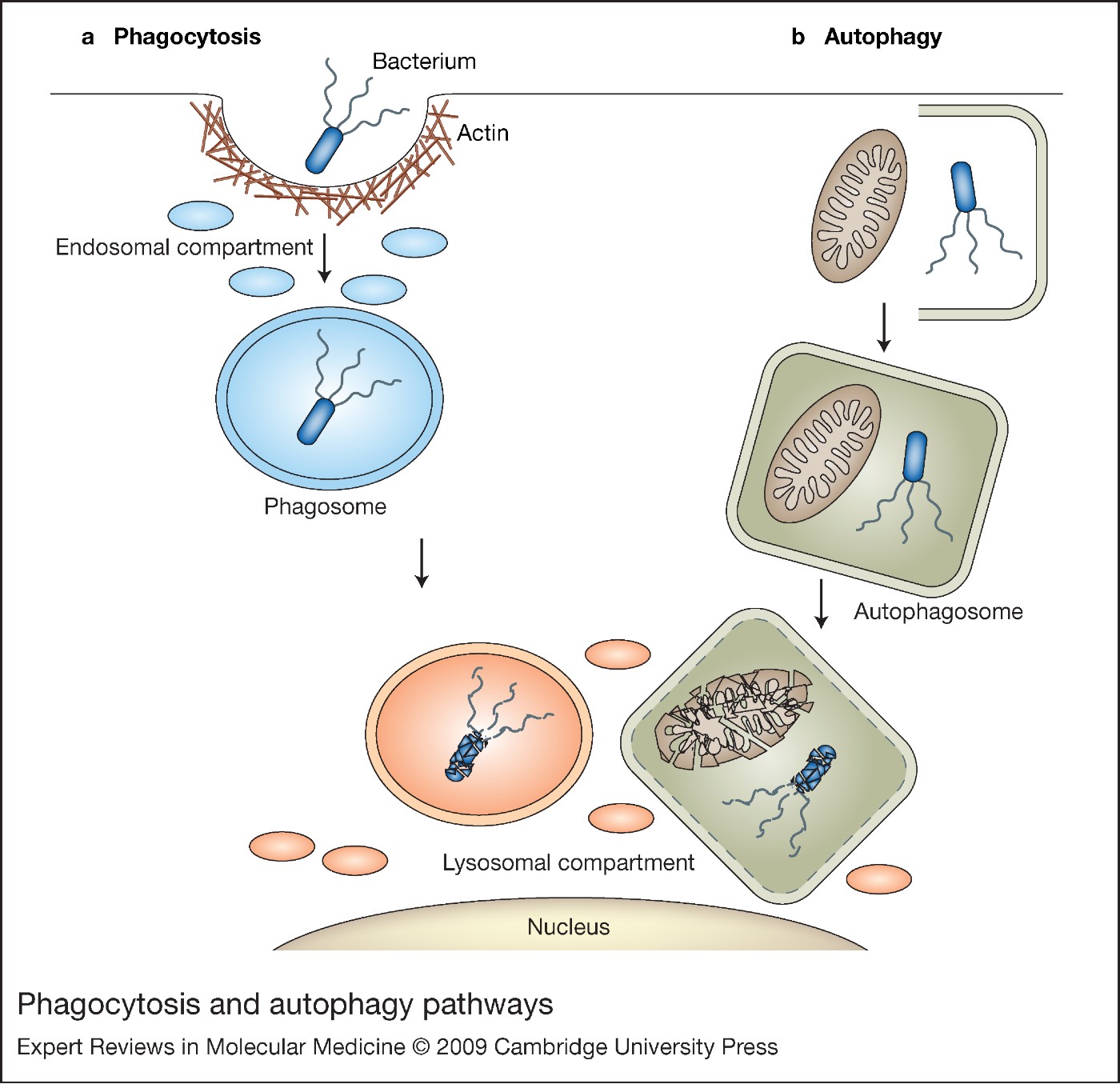
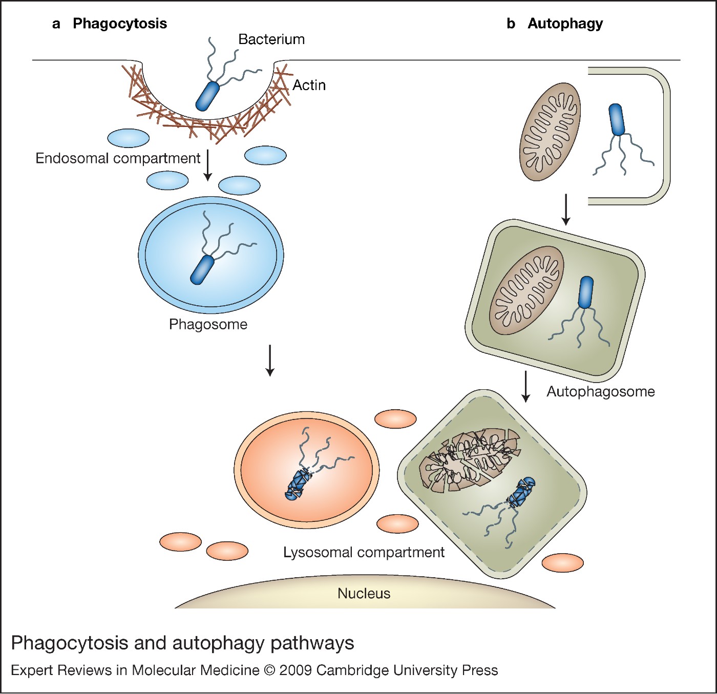
Some PRRs stimulate microbial uptake via phagocytosis (Fig. 2), a process that mediates uptake of particles larger than 0.5 µm. Upon receptor ligation, receptors cluster and mediate particle binding. Downstream signalling leads to actin-based membrane motility that shapes the membrane around the particle, forming a phagosome. Phagosomes mature by a series of fusion and fission events with endosomes and later lysosomes, creating a bacteriolytic environment in the phagolysosome (Ref. Reference Aderem and Underhill122). The variety of phagocytic receptors expressed by macrophages enables phagocytosis of a large range of particles and also provides signalling for effector responses appropriate for the ingested particle (Ref. Reference Stuart and Ezekowitz123).
Phagocytosis and autophagy pathways. (a) Phagocytosis is initiated by receptor binding, upon which the actin cytoskeleton shapes the plasma membrane around the particle. Once a phagosome is formed it matures via fusion and fission with endosomes and lysosomes, creating a degradative and microbicidal environment. (b) An autophagosome is formed by enclosure of selected cytoplasmic content by a double membrane. The autophagsome matures by fusion with lysosomes, leading to degradation of the inner membrane and autophagsosomal content.
Phagosome maturation is regulated by calcium flux and phosphoinositide signalling; it is also thought to be regulated by phagosomal pH (Ref. Reference Vieira124). During phagocytosis the pH falls, due to assembly of the ATPase complex that mediates proton translocation. This acidic environment affects pathogen growth, stimulates NADPH oxidase assembly and creates an optimal environment for hydrolytic enzyme activity. Superoxide anions produced by the NADPH oxidase complex can form hydrogen peroxide and hydroxyl radicals. Inducible NO synthase (iNOS) is another important enzyme for microbicidal activity, catalysing production of NO from arginine, oxygen and NADPH. Conversion into NO radicals leads to production of other microbicidal agents such as peroxynitrite (ONOO−), nitrogen dioxide (NO2) radicals, nitryl chloride (NO2Cl) and nitrogen trioxide (N2O3). Other killing mechanisms in phagosomes include enzymes such as proteases and lysozyme, antimicrobial peptides such as defensins, and metabolic competitors such as lactoferrin.
Macrophages from patients with inflammatory bowel disease show reduced phagocytosis and killing ability (Ref. Reference Caradonna125). By contrast, oxidative burst activity of lamina propria macrophages is increased during inflammatory bowel disease compared with macrophages in healthy mucosa (Ref. Reference Hausmann126). A polymorphism in P40phox, an adapter protein of the NADPH protein complex, has been associated with Crohn disease (Refs Reference Roberts127, Reference Rioux128). Functional effects of the polymorphism are unclear but are thought to affect microbial killing. Colitis was significantly attenuated in iNOS-deficient mice, but no link between iNOS and human inflammatory bowel diseases has been found yet (Ref. Reference Krieglstein129).
Particularly interesting is the MST-1 gene, encoding macrophage-stimulating protein (MSP; also known as hepatocyte growth-factor-like protein, HGFL), which is produced in the liver in an inactive form. It circulates in the bloodstream and is thought to be cleaved and activated at sites of inflammation by tissue proteases and macrophage membrane proteases. Active MSP binds its receptor RON, through which it influences macrophage function, including chemotaxis, spreading and phagocytosis (Ref. Reference Wang130). It also reduces macrophage responses to LPS and cytokines by downregulating IL-12, NO and cyclooxygenase (COX-2) production (Refs Reference Wang130, Reference Morrison131). From the study of RON-deficient mice, it is clear that this mechanism is important in regulating inflammatory responses (Ref. Reference Wang130). MST-1 was suggested as a gene involved in Crohn disease and recently it was shown that a SNP in MST-1, located in a region necessary for RON binding, is associated with the disease (Refs 132, Reference Fisher133). Together this suggests that MSP plays an essential role in macrophage-mediated tolerance in the intestine.
Autophagy
Autophagy (Fig. 2) is a mechanism used by all cells of the body for homeostasis and production of nutrients during starvation. It is also used for killing intracellular pathogens that have escaped phagosomal killing (Ref. Reference Kirkegaard134) and it is suggested to be a form of programmed cell death (Ref. Reference Levine and Yuan135). An autophagosome is characterised by a double membrane surrounding selected organelles or microbes. Like phagosomes, autophagosomes mature during fusion with endosomes and lysosomes, a process mediating degradation. TLR signalling has been shown to enhance autophagy and induce phagosome maturation using components of the autophagic pathway (Refs Reference Sanjuan136, Reference Xu137). ATG16L1 and IRGM, two genes involved in autophagy, have been linked with Crohn disease (Refs 132, Reference Hampe138). Little is known about IRGM (immunity-related GTPase family M protein), but human IRGM has been shown to induce autophagy and to reduce survival of Mycobacterium tuberculosis (Ref. Reference Singh139). A deletion polymorphism has also been linked with Crohn disease, resulting in altered IRGM expression and reduced autophagy of Salmonella typhimurium. It is still unclear if mutations in IRGM that have been linked with Crohn disease influence microbial killing. ATG16l1 (autophagy-related protein 16-like 1) influences S. typhimurium localisation and uptake by autophagosomes, but no role in microbial killing has been shown (Ref. Reference Kuballa140). Since autophagy increases access of the cell to antigens for MHC class II presentation, it is possible that tolerance towards the microbial flora is influenced by ATG16l1 and IRGM. Cells from ATG16l1-deficient mice show a block in autophagosome formation and subsequent IL-1β and IL-18 production (Ref. Reference Saitoh141). These mice also had more severe inflammation after colitis induced by dextran sodium sulphate (DSS), which could be due to IL-1β-induced changes in epithelial barrier function. ATG16l1 has been shown to affect Paneth cell function, and Crohn disease patients with the risk allele of ATG16l1 show similar Paneth cell abnormalities (Ref. Reference Cadwell142).
Granuloma formation
Formation of granulomata (dense structures of immune cells, particularly giant cells formed by macrophage fusion) is common in Crohn disease patients. Little is known about granulomata associated with Crohn disease but in other disorders they generally form in response to undegraded antigens (e.g. tuberculoid granulomata that form upon M. tuberculosis infection) or when antigen clearance is deficient (e.g. in patients with a defect in P91phox, part of the NADPH oxidase complex). Although likely, it is still unclear if microbial antigens cause granuloma formation in Crohn disease (Refs Reference Baksh143, Reference Ryan144). Macrophage fusion can be induced in the absence of microbial substance (Ref. Reference Helming and Gordon145) and electron microscopy has not revealed the presence of microbes within granulomata, but in vitro studies have shown that adherent-invasive Escherichia coli from Crohn disease patients can induce granuloma formation (Ref. Reference Meconi146). Underlying mechanisms of giant-cell formation remain to be unravelled; however, macrophage activation with IL-4, a Th2 cytokine, is an important factor (Ref. Reference Helming and Gordon145). Crohn disease is suggested to be Th1 driven, but it is possible that inflammation-induced tissue damage stimulates local wound healing and Th2 responses. However, one study has associated granulomata in Crohn disease with Th1 cells (Ref. Reference Kakazu147). It has been proposed that initial lack of adequate macrophage responses to intestinal flora leads to reduced neutrophil recruitment and insufficient clearance of microbes, which stimulates granuloma formation and chronic inflammation (Ref. Reference Marks and Segal148). This is supported by a recent study showing that macrophage cytokine production is normal but secretion is impaired in Crohn disease (Ref. Reference Smith149). Macrophages of these patients were shown to have a defect in vesicle trafficking leading to lysosomal degradation of inflammatory cytokines instead of secretion. These Crohn disease patients had reduced neutrophil recruitment and microbial killing, compared with a healthy control group.
Concluding remarks
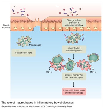
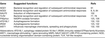
Intestinal macrophages mediate microbial recognition and killing but, unlike other tissue macrophages, do not stimulate inflammatory responses upon microbial recognition. Mechanisms for microbial recognition (NOD2), microbial killing (NADPH oxidase, ATG16L1 and IRGM) and macrophage activation (MST-1) have been shown to play essential roles in inflammatory bowel diseases, suggesting that macrophage function could be the origin of these disorders (Table 2). Interaction of macrophages with the intestinal flora steers immune responses by activating various signalling cascades after binding of specific PRRs. A change in flora or a defect in microbial clearing processes can lead to uncontrolled microbial growth in the lamina propria, which stimulates recruitment of inflammatory macrophages (Fig. 3).
The role of macrophages in inflammatory bowel diseases. (a) Macrophages in the lamina propria clear encountered microbes without inducing strong pro-inflammatory responses. This maintains the healthy balance between microbial flora and the intestinal immune system. (b) A change in flora or a defect in microbial clearing processes could lead to uncontrolled microbial growth and activation of pro-inflammatory processes. These processes mediate recruitment of inflammatory monocytes that develop into inflammatory macrophages in the lamina propria.

Genes linked to inflammatory bowel diseases that can influence macrophage function
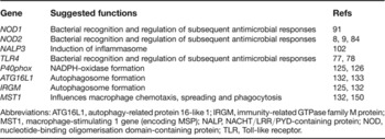
Abbreviations: ATG16L1, autophagy-related protein 16-like 1; IRGM, immunity-related GTPase family M protein; MST1, macrophage-stimulating 1 gene (encoding MSP); NALP, NACHT/LRR/PYD-containing protein; NOD, nucleotide-binding oligomerisation domain-containing protein; TLR, Toll-like receptor.
Understanding how blood monocytes develop into lamina propria macrophages may make it possible to control this process in order to dampen inflammation in patients with inflammatory bowel disease. The interactions between the intestinal flora and macrophages are also likely future targets for drugs, either in the form of probiotics or by use of receptor agonists or antagonists that stimulate intestinal immune responses to produce the required response.
In the later stages of his life, Metchnikoff became intensely interested in the gut flora and how this might influence health, disease and ageing. He advocated regular intake of lactobacilli, and although ridiculed by many in his time, current knowledge shows that there is probably more than a grain of truth in his ideas. However, there can be no doubt that macrophages and innate immunity contribute to major effector pathways in the pathogenesis of inflammatory bowel disease, providing attractive drug targets including, but not restricted to, TNF-α.
Acknowledgements and funding
The authors thank the anonymous peer reviewers for their helpful suggestions.