Device closure of secundum atrial septal defects was initially described in 1974, but it was not until the Amplatzer™ septal occluder became available in the mid-1990s that it became a routine procedure. Since then, there has been significant progress in the ability of interventionists to tackle anatomically challenging defects with high rates of procedural success.Reference Butera, Biondi-Zoccai and Sangiorgi1 Although extremely rare, tissue erosion is considered as the most feared event following atrial septal defect closure. It can occur within 72 hours of implantationReference Crawford, Brindis and Krucoff2,Reference Sauer, Ntalakoura and Haun3 or even years later requiring surgical treatment or leading to death.Reference Roberts, Parmar and Rajathurai4,Reference Mendirichaga, Smairat and Sancassani5 Early and recurrent development of pericardial effusion has been described among predictors for this complication but is it always the case?Reference Amin, Hijazi and Bass6
Case report
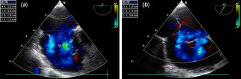
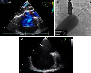
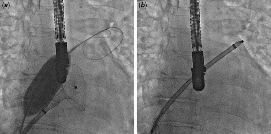
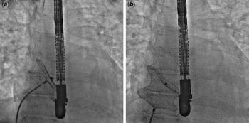
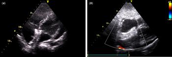
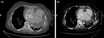
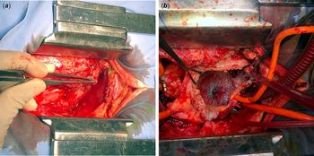
In March 2019, a 59-year-old woman with a complex atrial septal defect was referred to our centre for percutaneous closure after a 1-month history of progressively worsening dyspnoea and lower limb oedema. Trans-oesophageal echocardiography examination showed a multi-fenestrated aneurysmal type inter-atrial septum (Fig 1). All defect margins were sufficient in length except for the postero-inferior rim that hosted a 6-mm large defect. Bilateral femoral venous access points were prepared to anticipate complex sizing techniques (Fig 2). Different deployment manoeuvres were used sequentially so that the left atrial disc was delivered within the left upper pulmonary vein in an oval configuration, and with balloon assistance, a 40-mm Amplatzer™ septal occluder was positioned in place (Fig 3). Device stability was verified by the Minnesota Wiggle (Fig 4) before release and by the Valsalva manoeuvre directly after. There was no residual shunt or impingement on intra-cardiac structures but both lateral discs appeared unusually spaced. After extubation, a pericardial effusion of 7 mm was seen and was attributed to excessive manipulation (Fig 5). Daily ultrasound monitoring showed slow progression with stable serum haemoglobin levels. On the 7th post-operative day, a CT angiography urgently performed for an acute onset of chest pain and dyspnoea showed a 31-mm left postero-lateral pericardial effusion with a fine peripheral contrast enhancement without device malposition, pericardium’s thickening, or perforation (Fig 6a). Bilateral basal atelectasis with minimal left pleural effusion was seen on the pulmonary level. At this point, the patient was treated with antibiotics and diuretics. On post-operative day 11, the effusion suddenly increased in size with early echocardiographic signs of tamponade leading to surgical drainage of 700 ml serous fluid. The drain tube was removed after 2 days and the patient was discharged under colchicine and aspirin. She remained asymptomatic until 4 weeks of follow-up with no evidence of effusion. Six weeks following discharge, she presented with another episode of chest and upper back pain with severe dyspnoea with no haemodynamical instability. Urgent CT showed a pericardial effusion with loculated peripheral contrast enhancement. The device also appeared to be tenting atrial walls without cardiac perforation or pleural effusion (Fig 6b). A device-related erosion was highly suspected and it motivated an urgent surgical exploration. Surprisingly, the surgeon reported a thickened pericardium with no effusion or erosion. The occluder was splayed over the aorta and the inferior caval vein, and the inferior rim was nearly absent (Fig 7). The device was extracted and the defect was repaired. Anotomopathological examination of the pericardium showed an organised mesothelial and fibrinous reaction. The patient was discharged after 3 days and remained well until this manuscript was drafted.
TOE views showing a multi-fenestrated aneurysmal-type inter-atrial septum with a 25-mm long aneurysmal base at 0° view and two major adjacent holes on colour Doppler view with the diameters of 12 and 13 mm (a). Aneurysm base measured 32 mm at 62° view (b). TOE = Trans-oesophageal echocardiography.

Complex defect sizing technique. A 24-mm Amplatzer™ sizing balloon II unintentionally placed across a lower localised small defect, inflated and kept in place while a 34-mm Amplatzer™ sizing balloon II was passed across another adjacent hole after being introduced from the second venous access (a). ASD measured at 40 mm using the stop-flow technique during second balloon inflation (b). Note the torn thin tissue strand following deflation turning the defect into one large hole with a floppy inferior rim and deficient 3-mm-long aortic rim (c). ASD = Atrial septal defect.

Complex device positioning with balloon-assistance (a). Note the misshapen tip of the delivery sheath positioned in the left upper pulmonary vein (b).

Minnesota Wiggle manoeuvre. Note the twisted shape of the delivery cable upon pushing (a) and the anchored position of the left disc during pullback (b). These were retrospectively considered as indirect signs to space over-occupying occluder within surrounding atrial tissue.

Minimal pericardial effusion with a well-positioned device in the four chambers (a) and sub-costal (b) TTE views. TTE = Transthoracic echocardiography.

Chest MDCT images. Left postero-lateral pericardial effusion of 31 mm with a fine peripheral contrast enhancement 11 days following device closure (a). ASD closure device responsible for the tenting of the lateral left atrial wall 50 days following device closure (b). Note the pericardial thickening and dense fibrous material in the pericardial space facing the atria with only a small volume posterior effusion. MDCT = Multiple detector computed tomography; ASD = Atrial septal defect.

Surgical view. Thickened pericardium without fluid collection (a), followed by device explantation and patch closure (b).

Discussion
Although device embolisation is the most encountered complication of atrial septal defect closures, cardiac erosion looms up as the most life-threatening eventReference Moore, Hegde and El-Said7 whose causing factors and mechanisms remain controversial until this date.Reference Crawford, Brindis and Krucoff2,Reference Carroll8 Since the routine use of Amplatzer™ septal occluder, device manufacturers and interventionists have been reviewing all reported cases of confirmed and suspected erosions to identify predictors for this complication before and even after device placement.Reference Amin, Hijazi and Bass6,Reference Amin9
Post-procedure pericardial effusion was previously reported as an important risk factor for erosion but is it always device-related? As left-to-right shunt is eliminated by the closure of a large atrial defect, the right ventricle volume decreases suddenly and leads to a tiny amount of systolic separation that may be called as trace pericardial effusion.Reference Carroll8 On the other side, when the effusion accumulates during or immediately after the intervention, the mechanism is likely to be related to cardiac catheterisation or perforation.Reference Divekar, Gaamangwe and Shaikh10 Our patient had a challenging defect that required different laborious manipulations and multiple entries in the left upper pulmonary vein that might have deformed the tip of the delivery sheath (Fig 3b), leading to an iatrogenic effusion accumulation. This hypothesis was also supported by the presence of left-sided pleural effusion and by the absence of effusions over 4 weeks of follow-up.
The development of pericardial effusion in our high-risk case led to close surveillance and kept us alert to the possibility of erosion. When the patient was re-admitted for worrisome symptoms, the accumulation of effusion after a free window of time and the bulky CT appearance of the occluder, tenting excessively atrial walls, were sufficient arguments to suspect device involvement and to precipitate the surgical action. No prompt cardiac ultrasound was performed, and unfortunately, this might have been sufficient to backstop active surveillance, as no effusion would have been visualised. Basic medical imaging plays an important role in helping the clinician to better evaluate device–tissue interactions and to understand the exact mechanism of effusion.Reference Carroll8 A retrospective review of the second CT with an expert radiologist revealed that the fibrous pericardial thickening without any liquid accumulation could have been identified.
Device oversizing has been increasingly recognised by implanters as a risk factor for erosion but balloon-sizing with proper stop-flow technique in our long-term experience with these devices has limited the error of defect size overestimation. However, the lack of a decent inferior rim, the deficient aortic border, and the splaying of the device contributed in part to our rushed decision, especially when all these findings were previously reported as risk factors for erosion.Reference Roberts, Parmar and Rajathurai4
Conclusion
Among device erosion risk factors, pericardial effusion early occurrence and recurrence remain the most troubling sign. However, close monitoring and proper radiologic assessment could prevent precipitated decisions in haemodynamically stable patients. The overall rate of device erosion remains extremely low. More data are needed to better understand its incidence, true causes, warning signs, and possible preventive solutions.
Author contributions
RH collected clinical data, designed the report, and took the lead in writing the manuscript. ZS was the main operator and was assisted by RH. All of the authors have read and approved the final version of the manuscript.
Acknowledgements
None.
Financial Support
This research received no specific grant from any funding agency, commercial or not-for-profit sectors.
Conflicts of Interest
Z. Saliba is a proctor and consultant for Abbott Vascular. The other authors have no conflicts of interest to declare.
Ethical Statement and Informed Consent
The authors assert that all procedures contributing to this work comply with the ethical standards of the relevant national guidelines on human experimentation, and with the Helsinki Declaration of 1975, as revised in 2008. The patient written informed consent was obtained for the publication of this report.