Introduction
Distant metastasis of laryngeal squamous cell carcinoma (SCC) has a reported clinical incidence of between 4.3 and 30 per cent.Reference Yoskovitch, Hier, Okrainec, Black and Rochon1 The most affected sites are the lungs (66 per cent), bone (22 per cent), liver (10 per cent), mediastinum and bone marrow.Reference Licitra, Bernier, Grandi, Locati, Merlano and Gatta2 Distant skeletal muscle metastasis from laryngeal SCC is exceedingly rare.Reference Sridhar, Rao and Kunhardt3
Herein, we present a case of laryngeal SCC with distant skeletal muscle (rectus femoris) metastasis, and we review the relevant literature.
Case report
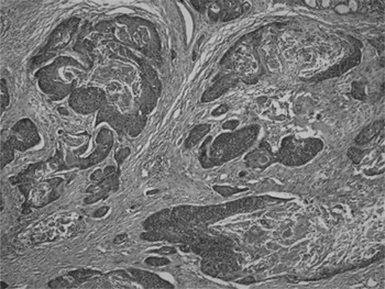
A 70-year-old man had first presented to our ENT department with an eight-month history of hoarseness and cough. Following laryngoscopy and biopsy, laryngeal SCC had been diagnosed. The ENT surgeons had performed a total laryngectomy and bilateral modified lymph node dissection. The pathological specimen had shown a poorly differentiated SCC with focal cartilage infiltration (Figure 1). Neck dissection revealed no evidence of disease. Total body computed tomography (CT) and positron emission tomography (PET) scans had been performed, and revealed no systemic spread of the disease.
Photomicrograph of primary tumour, showing squamous cell carcinoma with keratin production (H&E; ×100).

Fifteen months later, the patient re-presented with pain and a mass in the left anterior femoral region.
Physical examination revealed a tender mass in the rectus femoris muscle.
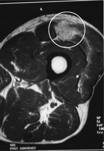
A magnetic resonance tomography (MRI) scan was performed, confirming a mass in the left rectus femoris muscle (Figure 2).
Axial magnetic resonance imaging scan used to evaluate the muscle tissue; white circle indicates a mass in the rectus femoris muscle.

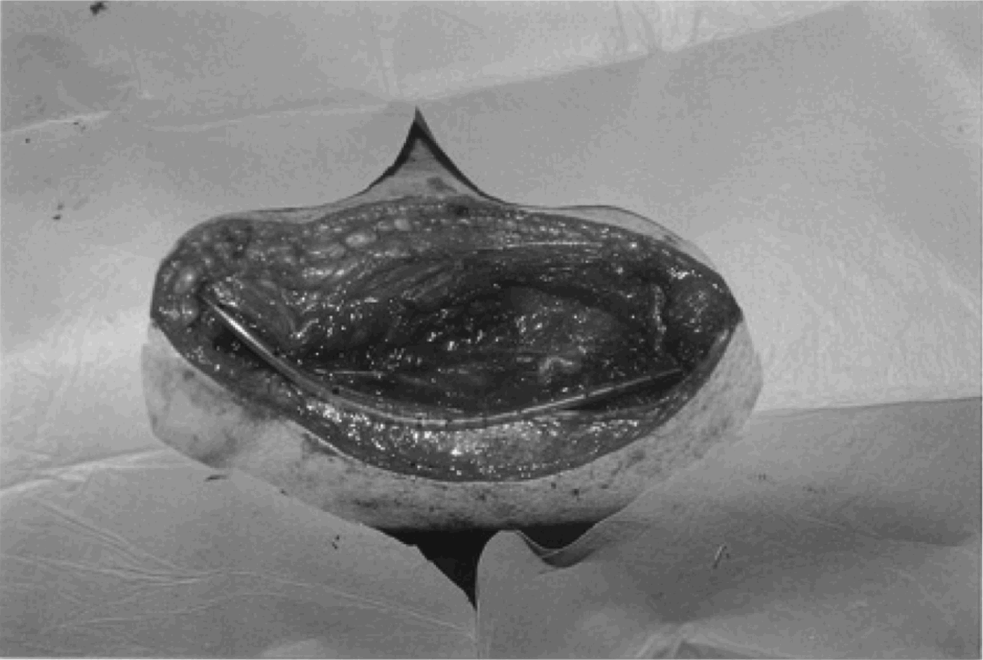
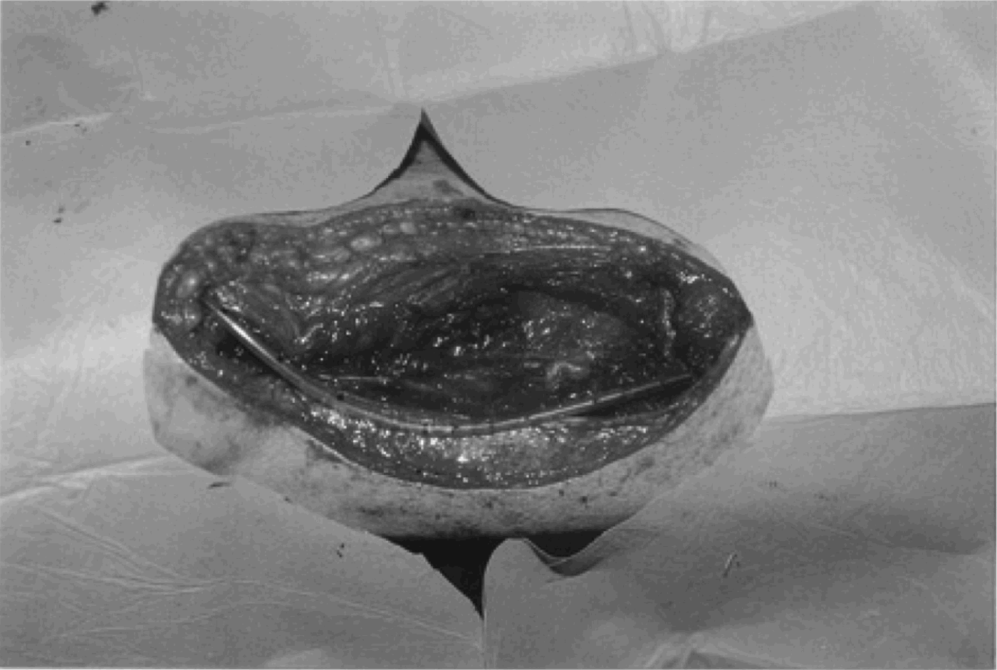
Complete surgical excision of the mass was performed (Figure 3).
Intra-operative view; the mass was completely excised.
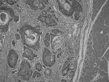
Histological study of the resected mass revealed the same morphological aspects of laryngeal SCC noted in the resected primary tumour (Figure 4).
Photomicrograph of resected metastatic tumour, adjacent to striated muscle tissue (at right of section); tumour morphology was similar to that of the primary tumour (H&E; ×100).

Whole body imaging with CT and PET did not yield any evidence of secondary lesions.
Twenty-one months after surgical excision of the metastatic mass, the patient showed no evidence of distant muscular recurrence. However, two years later, he died from brain and suprarenal metastases.
Discussion
Skeletal muscle tumours occur more commonly as primary lesions than secondary metastases. Although the skeletal muscles represent nearly 50 per cent of the total body mass and receive a large proportion of cardiac output, haematogenous metastatic spread of disease to skeletal muscle is extremely rare.Reference Sridhar, Rao and Kunhardt3 The reason for the rarity of metastatic tumour spread to skeletal muscle is unclear, but may be related to several factors, such as extremely variable and turbulent blood flow, high tissue pressure, β-adrenergic stimulation, tissue oxygen levels, metabolism (especially the effect of lactic acid on metastatic cell production) and host immune response.Reference Sridhar, Rao and Kunhardt3, Reference McKeown, Conant and Auerbach4 Distant muscular metastasis may also occur without pulmonary metastasis in cases involving pulmonary systemic circulation bypass.Reference Welsh, Welsh and Rizzo5 The pulmonary circulation may be theoretically bypassed by the azygous venous and vertebral venous systems via Batson's plexus. Disruption of the normal venous drainage pathways as a result of surgical alteration (especially that affecting the internal jugular vein) may contribute to this mode of spread.Reference Yoskovitch, Hier, Okrainec, Black and Rochon1
Most skeletal muscle metastases are of pulmonary origin. Very limited series and cases of skeletal muscle metastases have been reported from leukaemia, lymphoma and melanoma and from thyroid, genitourinary tract, pancreatic and breast malignancies.Reference Ménard and Parache6 Distant muscular metastasis from laryngeal cancer is an extremely rare occurrence.Reference Marioni, Blandamura, Calgaro, Ferraro, Stramare and Staffieri7 Marioni et al. reported the first case of haematogenous distant metastasis to the right gluteus maximus muscle from laryngeal SCC, without evidence of cervical lymph node metastases. Hitherto, this has been the only reported case describing haematogenous distant metastasis from laryngeal SCC without evidence of cervical lymph node metastases.Reference Marioni, Blandamura, Calgaro, Ferraro, Stramare and Staffieri7
There have been previously reported cases of distant muscular metastases from laryngeal SCC, but these have also had cervical lymph node metastases.Reference Ménard and Parache6, Reference Yucel, Demirel, Demiryont, Egeli and Deger8 Yucel et al. reported the first case of distant metastasis to the scapular muscles in a patient who had undergone total laryngectomy and bilateral neck dissection. However, in this case distant haematogenous spread may not have been the aetiology of metastasis, as the tumour may have reached the subscapular nodes and muscles through via lymphatic spread.Reference Yucel, Demirel, Demiryont, Egeli and Deger8 Ménard and Parache reported a case of recurrent laryngeal SCC with cervical lymph node involvement and distant skeletal muscle metastasis to the right biceps femoris muscle.Reference Ménard and Parache6
In the present report, we have described the first case of haematogenous distant metastasis to the left rectus femoris muscle from laryngeal SCC without evidence of cervical lymph node metastases. To the best of our knowledge, this is the second reported case of haematogenous distant muscular metastasis from laryngeal SCC without cervical lymph node involvement.Reference Marioni, Blandamura, Calgaro, Ferraro, Stramare and Staffieri7 In our case, whole body CT and PET scanning did not reveal systemic spread of the disease. These imaging techniques may miss small metastatic foci. It is quite difficult to explain the exact mechanism of disease spread to the rectus femoris muscle in our case of SCC of the larynx without lung or liver involvement.
Detection of skeletal muscle metastasis is not always easy. Although skeletal muscle metastases may be painful and/or palpable, as in our case, they may also be subclinical. Contrast-enhanced helical CT has been successfully used to detect such tumours, but the superior intrinsic soft tissue contrast afforded by MRI makes it the ‘gold standard’ for the imaging of muscle lesions.Reference Pretorius and Fishman9, Reference Damron and Heiner10 Therefore, we used MRI for the evaluation of our patient's rectus femoris muscle mass.
• Distant skeletal muscle metastasis from laryngeal squamous cell carcinoma is exceedingly rare
• This paper describes the first case of haematogenous distant metastasis to the left rectus femoris muscle from laryngeal squamous carcinoma without evidence of cervical lymph node metastases. This represents the second published case of such haematogenous distant muscular metastasis without cervical lymph node involvement
• Treatment options for skeletal muscle metastasis from carcinoma rarely alter the outcome; however, surgical excision may be indicated in selected cases of isolated muscular metastasis, particularly if locoregional control of the tumour is believed to have been achieved
The limited number of reported cases makes it difficult to determine the best treatment for skeletal muscle metastases. The prognosis for patients with skeletal muscle metastasis is considered to be poor, as skeletal muscle metastasis generally occurs as a feature of systemic spread.Reference Sudo, Ogihara, Shiokawa, Fujinami and Sekiguchi11 Treatment options such as radiotherapy, chemotherapy and surgical excision are believed to rarely alter the outcome.Reference Marioni, Blandamura, Calgaro, Ferraro, Stramare and Staffieri7 However, we believe that surgical excision is indicated in selected cases of isolated muscular metastasis, particularly if locoregional control of the tumour is believed to have been achieved. For this reason, we surgically excised our patient's isolated muscular metastasis. Twenty-one months after excision, the patient showed no evidence of distant muscular recurrence.
Conclusion
Distant muscular metastasis of laryngeal cancer is extremely rare; however, physicians must be cautious if they encounter a muscular mass during routine follow up of a laryngeal cancer patient. In the rare case of such a muscular metastasis, surgical excision may be a good treatment alternative if locoregional control of the tumour is believed to have been achieved. However, more reports on this subject are needed before a final conclusion is drawn.