

Introduction
A retrospective survey of the causes of death in China (Chen, Reference Chen2008) revealed that cerebrovascular disease is the most frequent cause of death in the Chinese population. Furthermore, approximately 75% of patients who survive cerebrovascular disease lose some degree of labour capacity and approximately 40% suffer from severe disability, which places heavy burdens on families and communities. The most common cerebrovascular disease is ischemic stroke, which accounts for approximately 60–80% of cerebrovascular disease cases. Currently, the prognosis of a patient with ischemic stroke can be improved significantly by treatment involving the rapid reconstruction of the blood supply toward infarcted regions. Such treatments may involve the use of recombinant tissue plasminogen activator (rt-PA); however, this agent must be applied within a strict window of time and its use is contraindicated in many scenarios, which greatly limits its clinical applications. Thrombolytic monitoring studies from developed countries have reported thrombolytic ratios of 4–10%; by contrast, this ratio is approximately 2% in China (Wang et al., Reference Wang, Liao, Zhao, Wang, Wang, Nguyen-Huynh and Wang2011). Therefore, the development of new treatment methods aimed at improving neurological function recovery in stroke patients would be expected to positively affect the quality of life of patients, reduce the economic burdens placed on families, and conserve medical resources. In recent years, in vitro and animal studies have demonstrated the abilities of selective serotonin reuptake inhibitors (SSRIs) to promote nerve regeneration after central nervous system injury (Ampuero et al., Reference Ampuero, Rubio, Falcon, Sandoval, Diaz-Veliz, Gonzalez and Wyneken2010; Guirado et al., Reference Guirado, Varea, Castillo-Gómez, Gómez-Climent, Rovira-Esteban, Blasco-Ibáñez and Nàcher2009; Homberg et al., Reference Homberg, Olivier, Blom, Arentsen, van Brunschot, Schipper and Reneman2011; Rayen, van den Hove, Prickaerts, Steinbusch, & Pawluski, Reference Rayen, van den Hove, Prickaerts, Steinbusch and Pawluski2011). In addition, SSRIs were shown to reduce the infarcted volumes and improve movement and sensory function defects in an experimental rat model of stroke (Sato, Kawamata, Kobayashi, & Okada, Reference Sato, Kawamata, Kobayashi and Okada2014). Internationally, small-scale clinical trials demonstrated that SSRIs could promote improvements in movement and cognitive function in patients with AIS through a mechanism that may be independent of the antidepressant effects of these drugs (Chollet et al., Reference Chollet, Tardy, Albucher, Thalamas, Berard, Lamy and Loubinoux2011; Jorge, Acion, Moser, Adams, & Robinson, Reference Jorge, Acion, Moser, Adams and Robinson2010; Mikami et al., Reference Mikami, Jorge, Adams, Davis, Leira, Jang and Robinson2011). Moreover, a multi-centre, double-blind, randomized controlled study demonstrated that citalopram, a SSRI, may not only exhibit neuroprotective effects but also reduce the recurrence of vascular events (Kraglund, Mortensen, Grove, Johnsen, & Andersen, Reference Kraglund, Mortensen, Grove, Johnsen and Andersen2015). As SSRIs are relatively inexpensive and their use is not restricted to within a strict time window, they could potentially become a routine treatment option for patients with ischemic stroke. To date, few Chinese clinical research studies have addressed these drugs. The present study therefore aimed to investigate the effects of paroxetine (PAR) on the recovery of motor and cognitive functions in Chinese patients with nD-AIS and to evaluate its safety in this population.
Methods
Participants
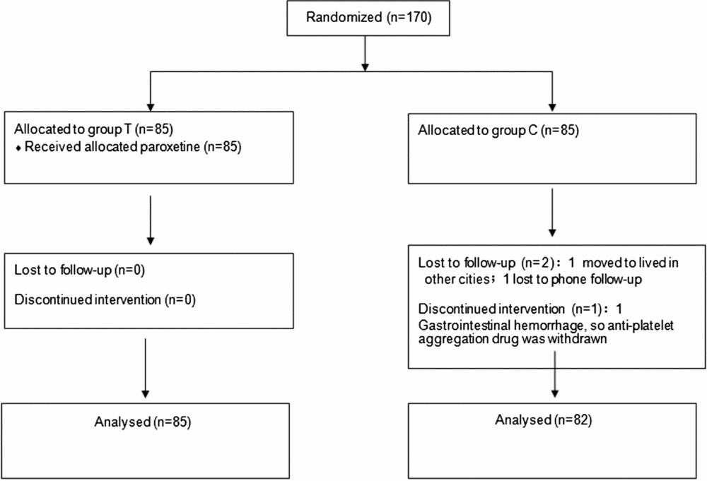
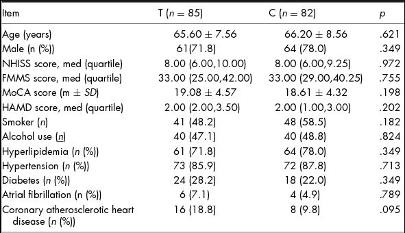
Based on similar studies (Chollet et al., Reference Chollet, Tardy, Albucher, Thalamas, Berard, Lamy and Loubinoux2011; Jorge et al., Reference Jorge, Acion, Moser, Adams and Robinson2010; Mikami et al., Reference Mikami, Jorge, Adams, Davis, Leira, Jang and Robinson2011) with a sample size of 30–60 patients in each group, we set a larger sample size of 85 patients per group in our study, due to the sufficient source of acute ischemic stroke. A total of 170 patients with acute ischemic stroke (AIS) who had been admitted to the Department of Neurology of our hospital between January 2012 and June 2014 were randomly divided into treatment (Group T) and control groups (Group C) using a random number table. Eighty-five patients were allocated to each group; after three patients in Group C were lost to follow-up, the final study included 167 enrolled patients (Group T, 85 cases; Group C, 82 cases) (Figure 1). The two study groups did not differ significantly in terms of sex, age, educational level, hypertension, diabetes, blood lipid levels, alcohol and tobacco addiction, heart disease, and National Institutes of Health Stroke Scale (NIHSS) scores (p > .05) (Table 1). The entire design of the study had been approved by the Ethics Committee of Jinhua Hospital affiliated to Zhejiang University, and all the patients or their authorized relatives had signed the informed consent.
Trial profile.

Comparison of General Information Between the Two Groups

Note: NHISS: National Institutes of Health Stroke Scale; FMMS: Fugl–Meyer Motor Scale; MoCA: Montreal Cognitive Assessment Scale; HAMD: Hamilton Depression Scale.
The following criteria were used to determine study inclusion: (1) The patient met the diagnostic criteria of acute cerebrovascular disease developed by the Fourth National Cerebrovascular Disease Conference and confirmation of AIS via magnetic resonance imaging of the head. (2) The patient exhibited clear consciousness and could participate in examinations within 1 week of stroke onset. (3) The patient had a Fugl–Meyer Motor Scale (FMMS) score of <55 points (Chollet et al., Reference Chollet, Tardy, Albucher, Thalamas, Berard, Lamy and Loubinoux2011) and/or Montreal Cognitive Assessment (MoCA) score of <26 points (Tan et al., Reference Tan, Li, Gao, Wang, Zhao, Yu and Zhou2015). (4) The patient's age was between 50 and 80 years.
The following criteria were used to determine study exclusion: (1) A NIHSS score >20 points; (2) aphasia; (3) a history of pre-stroke depression for which antidepressant and benzodiazepine drugs were used, and a first-time post-stroke Hamilton depression scale (HAMD) score >7 points; (4) receipt of thrombolytic therapy; and (5) the development of post-onset severe nosocomial infection, bed sores, or heart failure that might have affected neurological rehabilitation.
Treatments
Patients in Group C received conventional secondary cerebral infarction prevention and rehabilitation training within 48 hour of admission. Group T received the same conventional treatment provided to Group C, as well as treatment with orally administrated PAR hydrochloride tablets (Zhejiang Jianfeng Pharmaceutical Co., Ltd., Jinhua, China; State medical Approval No: H20040533) at dosages of 10 mg/d during week 1 and 20 mg/d thereafter, for a total treatment duration of 3 months.
Measures
At four designed time points (T0–3) corresponding to D0, D15, D90, and D180 after study initiation, the FMMS and MoCA scores were used to assess the patients' movement and cognitive impairment and recovery, and the HAMD score was used to evaluate the existence of depression in the patients after stroke onset. The Chinese version of MoCA-Beijing was used at T0 and T2, and the Chinese version of MoCA-Changshawas used at T1 and T3 (supplemented references). All scale evaluators were trained and tested by the main investigator and were blind to the group assignment. Scale assessments at T0 and T1 were performed during hospitalization and followed by a bi-weekly follow-up of each patient's medication. Scale assessments at T2 and T3 were completed at the 3rd and 6th month time points, respectively. Meanwhile, the types and extents of adverse reactions in the two groups were recorded.
Statistical Analysis
The baseline data and scale results of the two groups were analysed using SPSS18.0 (SPSS, Inc., Chicago, IL, USA). The normally distributed measurement data (i.e., MoCA score and age) are expressed as means ± s, and the average values between the two groups were compared using the t-test. Data with non-normal distributions (i.e., NIHSS, FMMS, and HAMD scores) are expressed as medians and quartiles, and pairwise comparison were performed using the Mann–Whitney U-test. Categorical data (sex, lifestyle factors, comorbidities, and HAMD> 8 points) are expressed as numbers and percentages and were compared using the χ2 test. A p value < .05 was considered to indicate statistical significance.
Results
Comparison of FMMS Scores
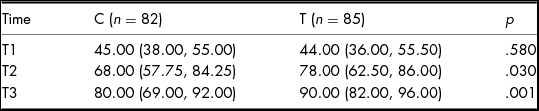
The FMMS scores differed significantly between the two groups at T2 and T3 (p <.05), but not at T1 (p > .05) (Table 2).
Comparison of Fugl–Meyer Motor Scale Scores Between the Two Groups After Treatment

Comparison of MoCA Scores
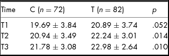
A MoCA score <26 points can be considered indicative of vascular cognitive impairment (Tan et al., Reference Tan, Li, Gao, Wang, Zhao, Yu and Zhou2015). In our study, Group C included 72 cases of cognitive impairment, whereas Group T included 82 cases. A comparison of the MoCA scores between the two groups indicated a statistically significant difference at T2 and T3 (p < .05), but not at T1 (p > .05) (Table 3).
Comparison of Montreal Cognitive Assessment Scale Scores Between the Two Groups After Treatment (x ± s)

Comparison of HAMD Scores
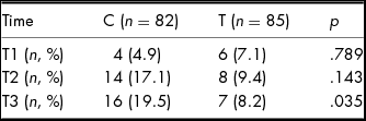
At admission, patients in both groups had HAMD scores of <8 points. However, a comparison of patients with HAMD scores ≥8 points in the two groups revealed a statistically significant difference at T3 (p <.05), but not at any other time point (p >.05) (Table 4).
Comparison of the Patients with the HAMD Score ≥8 Points in the Two Groups After Treatment

Note: HAMD: Hamilton Depression Scale.
Adverse Reactions
Group T included nine cases with adverse reactions, including five cases of nausea and anorexia, three cases of blurred vision, and one case of insomnia during the early stage of PAR administration. However, all adverse reactions were minor and tolerable. After explained by related physicians, all the patients understood the side effects; the side effects gradually relieved, so no specific medical treatment was given.
Discussion
The promotion of neurological function recovery in stroke patients has long been a hot topic in neuroscience research. Currently, recovery from central nervous system injury is thought to correlate closely with nerve regeneration and functional reorganization of the residual parts of the brain (Rossini, Calautti, Pauri, & Baron, Reference Rossini, Calautti, Pauri and Baron2003). Studies have found that after ischemic stroke, the central nervous system can self-repair and regenerate; furthermore, neural stem cells in certain areas such as the lateral subventricular zone and hippocampal dentate gyrus can migrate to the infarcted sites to induce nerve regeneration, neovascular migration, and new neuronal synapse formation (Cramer, Reference Cramer2008; Jones et al., Reference Jones, Allred, Adkins, Hsu, O'Bryant and Maldonado2009; Jones, & Adkins, Reference Jones, Adkins, Cramer and Nudo2010; Martí-Fàbregas et al., Reference Martí-Fàbregas, Romaguera-Ros, Gómez-Pinedo, Martínez-Ramírez, Jiménez-Xarrié, Marín and Garcia-Verdugo2010). However, the abovementioned processes depend on a local ‘harmonious microenvironment’, which includes brain derived neurotrophic factor (BDNF), glial cell-derived neurotrophic factor (GDNF), other neurotrophic factors, neurotransmitters, and/or neurogenic inhibitory factors. A study of a rat model of depression demonstrated that fluoxetine could promote neuronal regeneration by upregulating certain specific neurotrophic factors and increasing lactic acid levels in astrocytes, thus protecting the neurons (Allaman, Fiumelli, Magistretti, & Martin, Reference Allaman, Fiumelli, Magistretti and Martin2011). In a rat model of AIS, SSRI drugs were found to enhance the expression of neurotrophic factors, such as BDNF and GDNF, in the infarcted sites and to promote neurogenesis and angiogenesis in the infarcted area; as a result, the treatment group exhibited significantly better sensory and motor function recovery, compared with the control group (Espinera, Ogle, Gu, & Wei, Reference Espinera, Ogle, Gu and Wei2013). Among patients with vascular dementia, those treated with fluoxetine had higher serum levels of BDNF relative to the controls (Liu et al., Reference Liu, Zhang, Sun, Fan, Zhou and Fu2014), suggesting that these drugs can indeed participate in nerve regeneration and functional reorganization after a central nervous system injury, possibly by increasing the expression of neurotrophic factors and regulating the release of neurotransmitters.
SSRIs are reliable and effective antidepressants associated with good treatment compliance, and are therefore widely used to treat post-stroke depression. Researchers have found that these drugs not only improve patients’ depressive symptoms, but also appear to promote the recovery of neurological functions (Bilge, Koçer, Koçer, & TürkBörü, Reference Bilge, Koçer, Koçer and TürkBörü2008). Initially, these drugs were mainly administered because of their antidepressant effects, with the intent to improve patients’ pain and physical discomfort, enhance their confidence and motivation regarding rehabilitation, and promote the recovery of their neurological functions. However, some international small-scale clinical studies found that the beneficial effects of SSRIs on the rehabilitation of neurological function in stroke patients may be independent of the antidepressant effects (Chollet et al., Reference Chollet, Tardy, Albucher, Thalamas, Berard, Lamy and Loubinoux2011; Jorge et al., Reference Jorge, Acion, Moser, Adams and Robinson2010; Kraglund et al., Reference Kraglund, Mortensen, Grove, Johnsen and Andersen2015; Mikami et al., Reference Mikami, Jorge, Adams, Davis, Leira, Jang and Robinson2011). Although meta-analyses have shown that SSRIs have neuroprotective effects against ischemic stroke, the specific mechanisms require further investigation. In addition, SSRIs may increase the incidence of bleeding events, although this risk is extremely minor (Mortensen & Andersen, Reference Mortensen and Andersen2015; Siepmann et al., Reference Siepmann, Penzlin, Kepplinger, Illigens, Weidner, Reichmann and Barlinn2015). To date, Chinese clinical studies have focused on patients with post-stroke depression, and clinical studies regarding the use of SSRIs in patients with nD-AIS are rare. To observe whether SSRIs can promote the functional rehabilitation of patients with nD-AIS patients, the present study selected 167 patients with AIS and HAMD scores of <8 points upon admission (i.e., no accompanying depression). Whereas our analysis found no statistically significant difference in the FMMS and MoCA scores between patients treated or not with PAR at T0 and T1 (p >.05), we observed significant differences at later time points (T2 and T3, p <.05), suggesting that PAR promoted improvements in both motor and cognitive functions in patients with nD-AIS. Simultaneously, we observed that although both groups included patients with depression after treatment, this frequency was significantly lower in Group T than in Group C; in other words, PAR (or a similar drug) could prevent the occurrence of post-stroke depression, consistent with the findings of some small-scale international clinical trials. A double-blind, randomized controlled study by Chollet et al. (Reference Chollet, Tardy, Albucher, Thalamas, Berard, Lamy and Loubinoux2011) enrolled 113 non-depressed patients with ischemic stroke associated with moderate to severe hemiplegia (FMMS scores ≤55 points; 56 patients in the fluoxetine group (20 mg/day) vs. 56 patients in the placebo group). After 3 months of treatment, patients in the fluoxetine group exhibited significantly better improvements in motor function, compared with the placebo group (p <.05). Mikami et al. (Reference Mikami, Jorge, Adams, Davis, Leira, Jang and Robinson2011) and Jorge et al. (Reference Jorge, Acion, Moser, Adams and Robinson2010) respectively, studied the effects of fluoxetine and citalopram on motor function and overall cognitive function in patients with ischemic stroke, and found that these drugs enhanced both functional parameters. A multi-centre, randomized, double-blind clinical study conducted by Kraglund and colleagues revealed that in patients with AIS, citalopram not only has neuroprotective effects, but could also reduce the incidence of re-stroke (Kraglund et al., Reference Kraglund, Mortensen, Grove, Johnsen and Andersen2015). However, the present study did not have a sufficiently long follow-up time to observe re-stroke events, and additional follow-up studies are needed.
In summary, the present study showed that PAR may promote the recovery of motor and cognitive functions in patients with nD-AIS. However, our study has some limitations. First, the sample size was small, and the study featured an open, randomized controlled design rather than a double-blind design. Second, the evaluation of neurological functions was limited to motor and cognitive functions, while includes the patients’ language function, independent living ability, or other aspects. However, the positive effects of SSRI drugs on neural plasticity suggest new approaches toward the treatment of ischemic stroke, and clinical trials with larger samples will likely further direct and the applications of these drugs in patients with ischemic stroke. Accordingly, SSRI drugs are expected to underlie a new strategy for the treatment of ischemic stroke that will greatly improve the quality of life of patients and reduce the related economic burdens on families and communities.
Financial Support
This research received no specific grant from any funding agency, commercial, or not-for-profit sectors.
Conflicts of Interest
None.
Ethical Standards
The authors assert that all procedures contributing to this work comply with the ethical standards of the relevant national and institutional committees on human experimentation and with the Helsinki Declaration of 1975, as revised in 2008.