The staged Fontan procedure or total cavopulmonary connection is accepted as the palliative treatment of choice in a wide spectrum of patients with functionally univentricular hearts.Reference Gewillig 1 When studying biventricular circulations, it can be shown that cardiac output is modulated by changes in heart rate, contractility, pre-load and after-load. The ability to increase pre-load is particularly important in increasing cardiac output. In the univentricular circulation, there is no pre-pulmonary pump and therefore there is either absent or limited ability to increase preload to the systemic ventricle. In this setting, it has been established that, of the four factors listed above, pre-load has the most significant impact in altering cardiac output in these patients.Reference Gewillig, Brown and Eyskens 2 The relative chronotropic incompetence of univentricular hearts limits the impact of heart rate on cardiac output. The limited ability to adapt cardiac output to changing haemodynamic demands means that it is vital to be able to closely monitor cardiac output in this patient population. This is particularly important during transcatheter interventions that can have significant effects on pre-load and after-load.
There are several established techniques for the measurement of cardiac output. Use of the Fick principle and thermodilution are considered to be gold standards.Reference Wilkinson 3 , Reference Swan, Ganz, Forrester, Marcus, Diamond and Chonette 4 Both of these techniques are time-consuming and therefore prolong procedure time. They also provide discrete rather than continuous measurements of cardiac output. Calculation of cardiac output using the Fick method has an inherent inaccuracy because of the variability of oxygen saturation, particularly in assessing mixed venous saturations with respect to where samples are collected.Reference Rudolph 5 Several studies have looked at the accuracy of thermodilution, and standard errors of repeated measurement of cardiac output using this technique have been reported to be as high as 10%.Reference Gondos, Marjanek, Kisvarga and Halasz 6 , Reference Kadota 7
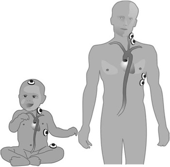
This study investigates the utility of the Icon® monitor (Osypka Medical, Berlin, Germany), which can continuously and non-invasively measure cardiac output using the concept of electrical velocimetry. During ventricular systole, erythrocytes are accelerated into the ascending aorta, which changes their alignment and produces a change in thoracic electrical bioimpedance. Using four standard electrocardiogram electrodes placed on the neck and thorax (Fig 1), Icon® measures the surface electrocardiogram and thoracic electrical bioimpedance. Changes in thoracic electrical bioimpedance during left ventricular ejection allow the rate of change in thoracic electrical bioimpedance to be derived. Using algorithms, Icon® determines indices of blood flow, contractility, and fluid status. Indices of blood flow are stroke volume and cardiac output and can be normalised to body surface area as stroke index and cardiac index. The monitor also calculates an index of contractility, as well as variations in this index. Thoracic fluid index and stroke volume variation are indicators of fluid status. Measurements can be taken at regular selectable intervals or even continuously giving instantaneous measurements of cardiac output.
Electrode placement.

Recent studies have favourably compared electrical velocimetry with thermodilutionReference Zoremba, Bickenbach, Krauss, Rossaint, Kuhlen and Schalte 8 and trans-oesophageal Doppler.Reference Schmidt, Theilmeier and Van Aken 9 Measurement of cardiac output using electrical velocimetry has also been shown to be comparable to measurements obtained using the Fick principle in children with complex congenital heart disease.Reference Norozi, Beck, Osthaus, Wille, Wessel and Bertram 10
Materials and methods
An Icon® monitor was used to assess changes in cardiac output during cardiac catheter interventions in 15 patients with cavopulmonary connections. All catheter procedures were carried out after informed consent under general anaesthesia using positive pressure ventilation. In all, 14 patients had palliation of functionally univentricular circulations with a fenestrated Fontan procedure using an extracardiac conduit. Of these 14 patients, 13 had a diagnosis of hypoplastic left heart syndrome and the 14th had tricuspid atresia with transposition of the great arteries, ventricular septal defect, and pulmonary stenosis. The remaining patient had a diagnosis of pulmonary atresia with intact ventricular septum. This patient initially had a right ventricular outflow tract patch and modified Blalock–Taussig shunt followed by a bidirectional superior cavopulmonary connection (1.5 ventricle repair). The median age of the patients was 6.1 years, with a range of 4.8–15.3 years. Median patient weight was 18.5 kg, with a range of 15–63 kg. There were 12 male patients in the study.
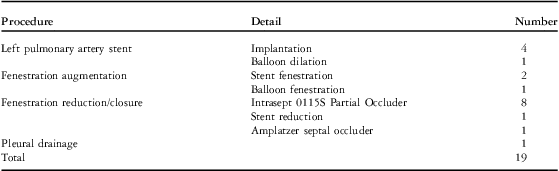
Of the 15 patients, 11 underwent a single intervention and the remaining four had two consecutive interventions under the same general anaesthetic. The procedures are detailed in Table 1. In all, four patients underwent stenting of left pulmonary artery stenoses using uncovered stents (Jostent, Abbott, Abbott Park, Illinois, United States of America) mounted on a standard angioplasty balloon (Opta, Cordis Corporation, Hialeah, Florida, United States of America) with diameters of 10–12 mm. Another had ballooning of a previously placed uncovered stent. The stent was dilated from 12 to 14 mm to match somatic growth. In all, nine patients had partial closure or reduction of their Fontan fenestrations. Of these, eight patients had partial occlusion of fenestrations using an Intrasept 0115S partial fenestration occlusion device (Cardia, Eagan, Minnesota, United States of America) and one patient had reduction of a previously placed stent fenestration using a technique described by Anderson et al.Reference Anderson, Bhole, Desai, Mehta and Stumper 11 The patient with pulmonary atresia with intact ventricular septum post bidirectional cavopulmonary shunt had complete closure of the atrial septal defect with an 8 mm Amplatzer Septal Occluder (AGA Medical, Plymouth, Minnesota, United States of America). There were two patients who had creation of new fenestrations with the placement of stent fenestrations using the Diabolo technique.Reference Stumper, Gewillig and Vettukattil 12 One of the patients who had stent fenestration had significant bilateral pleural effusions drained before his intervention under the same anaesthetic. A third simply had ballooning of a patent fenestration with a standard angioplasty balloon.
Procedure detail.

The Icon® cardiac output monitor was used from the start of each case, and indices of cardiac output were recorded at 1-minute intervals. Recordings were analysed before and immediately following interventions to assess their impact. Periods of stable heart rate were chosen so that stroke volume was directly proportional to cardiac output. Measurements were averaged over a 5-minute period. Changes in stroke volume and heart rate were compared using a Student's paired t-test.
Results
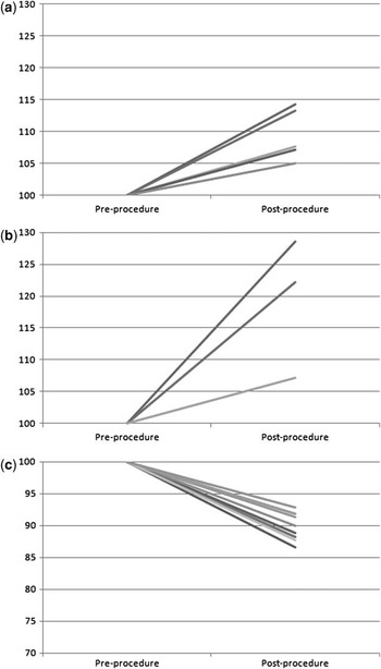
The results have been summarised in Table 2 and are graphically represented as a percentage change in stroke volume in Figure 2. All four patients who underwent stenting of left pulmonary artery stenosis, as well as the patient who had ballooning of a previously inserted stent, showed a consistent increase in their stroke volume and hence cardiac output. The median increase was 7.7% with a range of 5–14.3%. These changes were statistically significant (p = 0.007). Following stent implantation, there was a marked angiographic improvement. The minimum diameter increased from a median of 5 mm (3–9 mm) to 10 mm (10–15 mm). There was a sizeable increase in stroke volume in the patients who had augmentation of their fenestrations with a concomitant fall in saturations. Saturations pre-procedure were a median of 99% (99–100%) and fell to 85% (82–91%). The median observed change in stroke volume was an increase of 22.2%, with a range of 7.1–28.6%. The smallest increase in stroke volume was seen in the patient who only had ballooning of a fenestration as compared with the patients with creation of a de novo fenestration and stent implantation (range: 22.2–28.5%). There is a large observed change, but because there are only two patients this was found not to reach statistical significance (p = 0.094). There also was a reduction in stroke volume with complete or partial occlusion of obligate right-to-left atrial shunts, and as expected the saturations rose with reduction of right-to-left shunting. There was a median decrease in stroke volume of 10.6% (range: 7.1–13.4%; p < 0.0001). The reduction in stroke volume was again statistically significant. Changes in saturations were from a median of 83% (78–88%) to 93% (88–98%). There was one patient who had pleural drains inserted with drainage of significant pleural effusions who showed a clear increase in stroke volume and cardiac output (16%). Heart rate data were also collected at the same intervals and compared for any changes following intervention. There were no statistically significant changes in heart rate (p = 0.25).
Percent change in stroke volume pre and post intervention.

Relative changes in stroke volume for different interventions on cavopulmonary connections. ( a ) Left pulmonary artery stent/balloon; ( b ) fenestration augmentation; ( c ) fenestration reduction.

Discussion
Monitoring of cardiac output is vitally important in patients with univentricular physiology. This is particularly true during transcatheter interventions, which have considerable haemodynamic impact. Icon® is a versatile, non-invasive technique for use as a further adjunct to established monitoring in the catheter lab. The Icon® monitoring device has been shown in previous studies to be comparable to established techniques for assessment of cardiac output including thermodilutionReference Zoremba, Bickenbach, Krauss, Rossaint, Kuhlen and Schalte 8 and trans-oesophageal Doppler.Reference Schmidt, Theilmeier and Van Aken 9 It has also been validated for use in children with congenital heart disease by comparison with the Fick principle.Reference Norozi, Beck, Osthaus, Wille, Wessel and Bertram 10 This is the first such study reported in the literature of increased cardiac output associated with stenting of stenotic and hypoplastic left pulmonary arteries in a univentricular circulation. This can be explained by a reduction in the energy dissipation caused by these obstructive lesions. The effect of these lesions becomes even more important in a circulation in series. The absence of a pre-pulmonary pump means that there is a limited ability to increase the pre-load of the systemic ventricle. This limitation in provision of pre-load for the systemic ventricle means that heart rate, contractility, and after-load have reduced impact on cardiac output in the setting of the total cavopulmonary circulation. Any obstructive lesion would therefore theoretically adversely affect pre-load and ultimately cardiac output.
Hijazi et alReference Hijazi, Fahey, Kleinman, Kopf and Hellenbrand 13 have previously described a reduction in cardiac output associated with closure of fenestrations and these current results are consistent with that report. A reciprocal increase in cardiac output is also seen in this series in the patients who had augmentation of their fenestrations. Although these changes did not reach statistical significance, there is a large numerical difference observed but statistical significance is difficult to prove because of the small numbers. Finally, there was a predictable increase in cardiac output seen with drainage of pleural effusions. All of these findings provide a degree of internal validation of the reliability of the observed trends in cardiac output following interventions.
Icon® provides a non-invasive, continuous assessment of cardiac output, which is a valuable addition to current catheter lab monitoring modalities. It provides further insight into the understanding and manipulation of cardiac output in patients with complex haemodynamics and physiology. Previously established methods for the measurement of cardiac output continue to be used, but all have significant limitations. Thermodilution and the Fick principle are both time-consuming and thus prolong procedure time. They also provide discrete measurements of cardiac output and do not provide continuous, immediate assessments of cardiac output. Trans-oesophageal Doppler is an operator-dependent technique that leads to statistically significant inter-observer variability.Reference Schmidlin, Bettex and Bernard 14 The continuous, non-invasive assessment of cardiac output provided by Icon® presents an additional tool in the monitoring armamentarium for use in the catheter laboratory. Further, this non-invasive monitor has wider applications and can be useful in monitoring post-operative patients either on intensive care units or on the wards. It could also be applied to the longer term assessment of patients under outpatient follow-up as an ongoing assessment of cardiac function. In unpublished data from this unit, the Icon® monitor has been used in cardiac resynchronisation therapy using multisite pacing. The results generated from the Icon® monitor compared favourably with transthoracic echocardiographic tissue Doppler assessments.
The data presented here are initial data and are limited by the small numbers observed. It does provide further valuable insights into the complex physiology of total cavopulmonary circulations. In particular, it highlights the deleterious effect of obstructive pulmonary arterial lesions on circulations in series. Conceptually, hypoplastic and stenosed pulmonary arteries must have an adverse effect on the already compromised Fontan circulation. This study provides haemodynamic evidence to support the aggressive treatment of these lesions.
Conclusion
Icon® is a novel monitoring technique that provides non-invasive, continuous ssment of cardiac output. As such, it is ideally suited for use in the catheter laboratory and particularly during interventional procedures in patients who have compromised haemodynamics and can be inherently unstable.
Acknowledgements
None.
Financial Support
This research received no specific grant from any funding agency, commercial or not-for-profit sectors.
Conflicts of Interest
None.