Introduction
Intensive commercial poultry is reared under a heavy burden of a wide range of pathogens that range from viruses, bacteria and parasites. Innate and adaptive immune cells sense and establish a hostile environment against these pathogens. Adaptive immunity is mainly established by the B and T lymphocytes, where B cells play vital roles in the humoral immune responses whereas T cells perform functions of cell-mediated immune responses. Innate immune system consists of cells and proteins that prevent the entry, spread and subsequently activation of adaptive immune functions. Innate immune cells help in the clearance of pathogens from the body in first few hours of pathogen invasion irrespective of the nature and genetics of these pathogens. The main parts of the innate immune system include proteins, epithelial barriers, dendritic cells, phagocytes, natural killer cells, and MCs.
MCs were first identified by a German scientist Dr von Recklinghausen in 1863 from the mesentery of the frog (Paul, Reference PAUL2013). Usually the credit of discovery of these cells is given to another German scientist Dr Paul Ehrlich (Ehrlich, Reference EHRLICH1878; Crivellato et al., Reference CRIVELLATO, BELTRAMI, MALLARDI and RIBATTI2003) as he was awarded Noble Prize for his contribution to immunology in 1908. Ehrlich discovered the occurrence of MCs in different animal species including pigeons. For years, it had been considered that these are the villains of the body due to their involvement in the sneeze, wheeze and allergic reactions (Rao and Brown, Reference RAO and BROWN2008; Van Goor et al., Reference VAN GOOR, SLAWINSKA, SCHMIDT and LAMONT2016). Current scientific perceptions on MCs have been changed, and these cells are considered as important in host defence as lymphocytes and other immune cells (Marshall, Reference MARSHALL2004; Galli et al., Reference GALLI, NAKAE and TSAI2005). Scientific advancements in last decades have proved that these cells are not only involved in the allergic reactions but also perform important functions including; sentinels of the immune system, regulators of innate and adoptive immune responses, intestinal movement, mucosal secretions and wound healing (Galli et al., Reference GALLI, NAKAE and TSAI2005; Vliagoftis and Befus, Reference VLIAGOFTIS and BEFUS2005; Lu et al., Reference LU, LIND, GONDEK, BENNETT, GLEESON, PINO-LAGOS, SCOTT, COYLE, REED and VAN SNICK2006; St. John and Abraham, Reference ST. JOHN and ABRAHAM2013). Owing to several unique features of MCs such as low abundance, accommodation in tissues and difficulty in isolation, these cells are less well characterised compared to other leukocytes (Rao and Brown, Reference RAO and BROWN2008). However, recent research trends have significantly advanced our understandings on the biology of MCs, and their roles in the immune system.
In this review, recent advances in the avian mast cell (MC) biology, mechanism of activation, immune functions and possible benefits of their mitigating in vaccination are discussed. Additionally, essential roles of MCs in viral, bacterial and parasitic diseases of poultry are included.
Mast cell biology
MCs predominantly reside in the skin and mucosal surfaces. Distribution of MCs varies in different species and in different tissues of the same species. Chickens have a higher population of MCs compared to turkey, pheasants, or Japanese quail (Carlson and Hacking, Reference CARLSON and HACKING1972). Populations of MCs in oesophagus and spleen is higher than other part of digestive system, bursa, thymus, breast, and leg muscle (Carlson and Hacking, Reference CARLSON and HACKING1972). These cells are closely associated with the blood vessels, especially in the digestive system (Wight, Reference WIGHT1970).
MCs are descendants of the bone marrow precursor and pluripotent haematopoietic cells (Wernersson and Pejler, Reference WERNERSSON and PEJLER2014). These are long lived cells, without being differentiated, and can proliferate in response to specific signals (Abraham and St John, Reference ABRAHAM and ST JOHN2010). MCs progenitor are not present in blood in a mature form, however, upon their entrance to tissues, these are converted to mature MCs by the action of local microenvironment, especially stem cell factor and interleukin (IL)-3 (Wernersson and Pejler, Reference WERNERSSON and PEJLER2014). As the tissue develop, mature and proliferate, the MC population increases (Marshall, Reference MARSHALL2004; Galli et al., Reference GALLI, GRIMBALDESTON and TSAI2008). T helper 2 responses can also lead to increase in population and phenotypical changes in MCs ( Figure 1 ) (Galli et al., Reference GALLI, GRIMBALDESTON and TSAI2008).
Granules are present in the cytoplasm of these cells, which are lysosomes-like, electron dense and secretory in nature. This presence of secretory granules led to their discovery by Paul Ehrlich, who observed cells enriched with granules and named them ‘mastung’ (from the German meaning ‘well fed’). Later on, it became the criteria to identify these cells on the basis of these granules and can be easily identified when stained by the cationic dyes (Wernersson and Pejler, Reference WERNERSSON and PEJLER2014). Granular basis of MCs divided them into two distinct groups mucosal and connective tissue MCs. These can further be categorised on the basis of granule composition and response (Metcalfe et al., Reference METCALFE, BARAM and MEKORI1997), and their proliferation ability in response to different stimuli such as nature of pathogens (Abraham and St John, Reference ABRAHAM and ST JOHN2010).
MCs are distributed in the connective tissue of almost all body tissues; however, their numbers differ. Number and density of the MCs are highest in pathogen-risk tissues to provide sentinel functions (Dahlin and Hallgren, Reference DAHLIN and HALLGREN2015). Different factors such as age, stimulus and nature of pathogens determine their distribution in the tissues. In chickens, the number of MCs is highest in the bursa of Fabricius at seven days of age whereas at day 21, these cells are highest in number in the spleen and thymus (Karaca et al., Reference KARACA, YÖRÜK and USLU2006). These are mainly present in medulla of thymus, in bursa and spleen during the embryonic stages of chicken development (Jun-feng et al., Reference JUN-FENG, JIN-GUO, DENG-HUI and HONG-YAN2011). There is a positive correlation between organ-development and MCs number, and the maximum number of MCs are identified up to 18, 19, and 20 days in the spleen, bursa and thymus, respectively (Jun-feng et al., Reference JUN-FENG, JIN-GUO, DENG-HUI and HONG-YAN2011).
To fix MCs in connective tissue, Carnoy solution (Valsala et al., Reference VALSALA, JARPLID and HANSEN1986) is widely used and to fix these cells in the mucosal surfaces, isotonic formaldehyde acetic acid and basic lead acetate are best practiced (Uslu and Yörük, Reference USLU and YÖRÜK2013; Yildiz et al., 2016). Numerous MCs are present in the proventriculus, gizzard, and small intestine of fowl. These mainly reside in lamina propria (Swayne and Weisbrode, Reference SWAYNE and WEISBRODE1990) and tunica muscularis. Lower percentages of MCs can be detected in tunica submucosa and are not present in the tunica adventitia (Zhang et al., Reference ZHANG, PENG, GE, ZHONG and ZHOU2014). Number of MCs varies in different parts of the intestine (Yang et al., Reference YANG, YU, GANDAHI, BIAN, WU, LIU, ZHANG, ZHANG and CHEN2012). It has been noted that metachromatic and MC density was higher in the tunica mucosa layer of middle and distal caecum in quail (Yildiz et al., 2016).
MCs appear in lungs of chicken embryos at 15 days of age and are distributed in the mucous membranes and lamina propria in the tertiary bronchial tube; while on 16~21 days, MCs can be found in the mucous membrane, epidermis and other parts of respiratory system (Li et al., Reference LI, GAO, YAO, OU, JIANG and GUO2011). MCs are present in the stroma, hilum and theca of normal and atretic follicles in the ovaries of chicken. Age has no effect on the distribution of MCs in stroma and hilar regions of ovary (Gupta and Gilbert, Reference GUPTA and GILBERT1988; Parshad and Kathpalia, Reference PARSHAD and KATHPALIA1993). Intensity of degranulation can be observed maximum on 17 week compared to five week old birds (Parshad and Kathpalia, Reference PARSHAD and KATHPALIA1993). In different parts of oviduct, MC population is present in the following order: infundibulum > shell gland > magnum = isthmus and there is a positive correlation between the MCs and histamine secretion (Hrabia et al., Reference HRABIA, RZAĄSA, PACZOSKA-ELIASIEWICZ and SLOMCZYŃSKA2001).
MCs number increases with the increase in age of chicken (Li, Reference LI2008). The shape of MCs present in the thymus is oval, ovoid and irregular and in lungs they are circular, ellipse and anomalous shape (Li et al., Reference LI, GAO, YAO, OU, JIANG and GUO2011). In contrast, MCs of gastrointestinal tract are mainly ovoid and some are irregular in shape (Zhang et al., Reference ZHANG, PENG, GE, ZHONG and ZHOU2014).
Molecular mechanisms of MC activation
MCs are excellent sensors of different type of stimuli, and respond in rapid or selective way. To perform this function these cells have a large number of surface receptors that directly or indirectly recognise the pathogen or other signals (Rao and Brown, Reference RAO and BROWN2008). Main stimuli for the activation of MCs are pathogens ( Figure 1 ), feed, stress, or inflammatory responses (Abraham and St John, Reference ABRAHAM and ST JOHN2010).
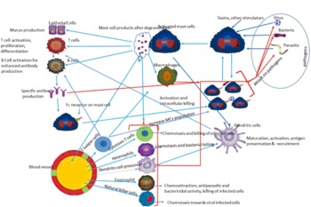
Mast cell activation by pathogens and their immune regulatory role to control infections.
Mast cells are activated by pathogens, toxins and other stress factors by receptors present on their surface. Activated mast cells produce different lipid-derived eicosanoid and cytokines and communicate with different immune cells like macrophages, dendritic cells, natural killer cells, heterophils, eosinophils, B and T cells. Communication of these cells leads to their recruitment, activation, maturation, proliferation and also facilitate their function for clearance of invading pathogens. B cells produce pathogen-specific antibodies which attach themselves to Fc receptors of mast cells and enabling them for more quick, specific and strong response to secondary or chronic challenge.

The main responsibility of MCs after the invasion of the pathogen is the recognition of pathogen associated molecular patterns (PAMPs). MCs perform this function in two distinct ways, first by recognition through toll like receptors (TLR) that are common for different classes of pathogens ( Figure 1 ) and secondly, via specific recognition through binding of antibodies which are specific to pathogens (Abraham and St John, Reference ABRAHAM and ST JOHN2010). Stimuli through different TLR effect cytokine production and degranulation of MCs (Abraham and St John, Reference ABRAHAM and ST JOHN2010). Beyond these conventional pathogen activation methods, MCs have other receptors that can be activated by bacteria including Staphylococcus aureus, Escherichia coli and Mycobacterium tuberculosis (Malaviya et al., Reference MALAVIYA, GAO, THANKAVEL, VAN DER MERWE and ABRAHAM1999; Muñoz et al., Reference MUÑOZ, HERNÁNDEZ-PANDO, ABRAHAM and ENCISO2003; Rocha-de-Souza et al., Reference ROCHA-DE-SOUZA, BERENT-MAOZ, MANKUTA, MOSES and LEVI-SCHAFFER2008). Additionally, MCs can sense danger signals from surrounding epithelial, endothelial and tissue resident immune cells (St. John and Abraham, Reference ST. JOHN and ABRAHAM2013).
Fc receptors (FcRs) also help the MCs in binding the IgE (FcεRI) and IgG (FcγRs) by which they recognise the previously encountered pathogens ( Figure 1 ). Thereafter, MCs become activated and degranulation occurs (Kawakami and Galli, Reference KAWAKAMI and GALLI2002). Simultaneous activation/stimulation by the TLR and FcεRI increase the cytokine production from the MCs by increasing the activity of mitogen-activated protein kinases (Qiao et al., Reference QIAO, ANDRADE, LISBOA, MORGAN and BEAVEN2006). MCs can be activated by different parasites that may have pathogens and in response to that degranulation occurs (Demeure et al., Reference DEMEURE, BRAHIMI, HACINI, MARCHAND, PERONET, HUERRE, ST-MEZARD, NICOLAS, BREY, DELESPESSE and MECHERI2005).
Mast cell functions
Anatomical location of MCs make them able to act as a first line of defence (St. John and Abraham, Reference ST. JOHN and ABRAHAM2013). After activation/recognition of pathogens, extensive degranulation occurs in MCs. Granules in MCs contain a large number of the active compound such as histamine, vascular endothelial growth factor, proteases (Gordon and Galli, Reference GORDON and GALLI1990; Boesiger et al., Reference BOESIGER, TSAI, MAURER, YAMAGUCHI, BROWN, CLAFFEY, DVORAK and GALLI1998), and chemokines (Abraham and St John, Reference ABRAHAM and ST JOHN2010). Most of these active components, produced by the MCs granules directly have antimicrobial activity. MCs in the intestine perform vital functions including muscle contraction, blood flow regulation, peristalsis, and acid, electrolytes, and mucus secretion by epithelial cells (Bischoff, Reference BISCHOFF2009). Moreover, these granules enter in the lymphatic vessels and trigger the recruitment of antigen presenting cells directly to the tissue (Kunder et al., Reference KUNDER, JOHN, LI, LEONG, BERWIN, STAATS and ABRAHAM2009). Histamine and other products of MCs also trigger the antigen presentation, activation of T cells, recruitment of CD8+ T and dendritic cells at the site of infection (Caron et al., Reference CARON, DELNESTE, ROELANDTS, DUEZ, HERBAULT, MAGISTRELLI, BONNEFOY, PESTEL and JEANNIN2001; Orinska et al., Reference ORINSKA, BULANOVA, BUDAGIAN, METZ, MAURER and BULFONE-PAUS2005; Shelburne et al., Reference SHELBURNE, NAKANO, ST JOHN, CHAN, MCLACHLAN, GUNN, STAATS and ABRAHAM2009). MCs perform these functions by releasing pre-formed or de novo synthesised mediators such as proteases, histamine, and heparin or lipid mediators and cytokine (Bischoff, Reference BISCHOFF2009).
These cells can also express major histocompatibility complex (MHC) class I and II. MCs cells enhance the production of mucus from epithelial cell (Bischoff, Reference BISCHOFF2009) as a defence mechanism. It has been demonstrated that stress conditions, such as heat, affect MCs. Heat stress increases the mucosal injury and oedema appearance in all parts of small intestine in broilers. There appear to be an increase in number of MCs and histamine contents in the intestine of these birds which indicate their involvement in tissue damage in heat stress (Liu et al., Reference LIU, LIU, LIU, LIN, LIU and FAN2011).
Particle size of feed affects the distribution and quantity of MCs in the intestine of chickens. Birds fed with a finely ground diet have more MCs and histamine levels in the villi of duodenum, jejunum, and ileum compared to birds fed coarsely ground diet (Liu et al., Reference LIU, PIAO, OU, CAO, HUANG and LI2006). This increase in number of MCs may be associated with better performance due to the possible involvement of the MCs in peristalsis, inflammatory and immune responses. A coarsely ground mash diet clustered the MCs in the upper part of villi in the intestine in chicken. Higher levels of histamine have appeared to be positively correlated with the MCs (Liu et al., Reference LIU, PIAO, OU, CAO, HUANG and LI2006). Deficiency in selenium can increase the number of MCs in jejunum in chickens (Hou et al., Reference HOU, LIU, LI, WANG and WANG2009). Cold stress also affects the number of MCs in the thymus and spleen. Increase in the population of MCs was observed in cold stress birds (Wang et al., Reference WANG, WANG and ZHANG2005). Taken together, MCs positively influence the activity of immune cells, similar to dendritic cells, B lymphocytes, T lymphocytes, monocytes, epithelial and endothelial cells (Galli et al., Reference GALLI, GRIMBALDESTON and TSAI2008).
Disease-specific responses of mast cells
role in viral diseases
MCs perform various functions that range from recognition and presentation of antigen, inflammation and recruitment of immune cells, cytokine production and viral clearance (Orinska et al., Reference ORINSKA, BULANOVA, BUDAGIAN, METZ, MAURER and BULFONE-PAUS2005; St John et al., Reference ST JOHN, RATHORE, YAP, NG, METCALFE, VASUDEVAN and ABRAHAM2011; Ebert et al., Reference EBERT, BECKER, LEMMERMANN, BUTTNER, MICHEL, TAUBE, PODLECH, BOHM, FREITAG, THOMAS, HOLTAPPELS, REDDEHASE and STASSEN2014). Extensive histologic lesions along with increase number of MCs in thymus, spleen, cloacal bursa, liver and kidney have been observed in very virulent infectious bursal disease virus (vvIBDV) challenged broilers at the second, third and fourth day post-challenge (Wang et al., Reference WANG, XIONG, SHE, LIU, ZHANG, LUO, LI, HU, WANG, ZHANG and SUN2008; Reference WANG, JIA, SHE and LIU2012a). These were due to activated MCs and it was confirmed by the presence of tryptases (Wang et al., Reference WANG, XIONG, SHE, LIU, ZHANG, LUO, LI, HU, WANG, ZHANG and SUN2008; Reference WANG, JIA, SHE and LIU2012a). The same group of investigators demonstrated that MCs are mainly involved in acute inflammation and the cause of bursal injury in vvIBDV-challenged birds, because inhibition of MCs degranulation lessens the bursal damage (Wang et al., Reference WANG, LIU, SHE, XU, LIU, XIONG, YANG, SUN and PENG2009a; Rehman et al., Reference REHMAN, MENG, UMAR, MUNIR and DING2016). Newcastle disease virus (NDV) infection severely damages the proventriculus, duodenum, jejunum and caecal tonsil, and increases the number of MCs and tryptase contents in these parts of digestive system (Sun et al., Reference SUN, WANG, SHE, LI, LIU, HAN, WANG and DING2008). There is a positive correlation between tissue damage and the number of MCs in these organs (Sun et al., Reference SUN, WANG, SHE, LI, LIU, HAN, WANG and DING2008).
Ketotifen is a MC membrane stabiliser agent and prevents degranulation. Research indicates that pretreatment of chicken with ketotifen reduces the tissue damage caused by the NDV (Sun et al., Reference SUN, LI, SHE, WANG, HAN, LI, DING and YUE2009), indicating its potential as immune-stimulant. It had been shown that murine MCs are involved in avian influenza virus induced immune responses and replicates in these cells (Meng et al., Reference MENG, HUO, WANG, XIAO, LIU, WEI, DONG, ZHANG, HU and SUN2016). Moreover, influenza infection accumulates MCs in the lungs (Zarnegar et al., Reference ZARNEGAR, MENDEZ-ENRIQUEZ, WESTIN, SODERBERG, DAHLIN, GRONVIK and HALLGREN2017). Use of MC inhibitors such as ketotifen significantly reduces lung injury (Hu et al., Reference HU, JIN, HAN, ZHANG, CAO, XIE, XUE, LI, MENG, FAN, SUN and WANG2012) indicating the involvement of MCs in influenza infection. However, the studies related to involvement of avian MCs in avian influenza as well as other viral diseases are not available.
role in bacterial diseases
MC contribution in bacterial infections in poultry is less understood. Following the recognition of the bacteria through TLR, MC are crucial to protect the body from bacterial infections (Marshall, Reference MARSHALL2004). MCs produce different inflammatory mediators that recruit other white blood cells along with phagocytosis of opsonised bacteria (DeBruin et al., Reference DEBRUIN, GOLD, LO, SNYDER, CAIT, LASIC, LOPEZ, MCNAGNY and HUGHES2015). Bacterial endotoxins stimulate the production of IL-1α and β that trigger the neutrophils (equal to heterophils in poultry) for killing bacteria. It has been noticed in mice that a deficiency of MC poorly recruits neutrophils, high mortality and delayed bacterial clearance (Malaviya et al., Reference MALAVIYA, IKEDA, ROSS and ABRAHAM1996). MCs play a part in preventing the distribution of bacterial pathogens to healthy tissues (Abraham and St John, Reference ABRAHAM and ST JOHN2010). Degranulation, in response to bacterial recognition, produces histamine that excites epithelial ion transport via histamine H1 receptor activation (Collins et al., Reference COLLINS, MCGRATH, BAIRD and CAMPION2007). This excitation of epithelial ion transport via histamine H1 receptor stimulates the paracellular movement of water and sodium in lumen that causes diarrhoea, which may facilitate bacterial clearance (Collins et al., Reference COLLINS, MCGRATH, BAIRD and CAMPION2007). Treatment of chicken with swine gut antimicrobial peptides increases the number of MCs in duodenum, jejunum and ileum (Wang et al., Reference WANG, MA, SHE, SUN, LIU, HU, LIU, YANG and PENG2009b). Petrone et al. (Reference PETRONE, CONSTANTINO and PRADAL-ROA2002) compared caecal submucasal MCs of chicken by challenging them with Eimeria tenella and Salmonella enteritidis. They found there was a non-significant difference of E. tenella, S. enteritidis and their combination on the caecal submucosal MCs but these were numerically higher for the combined infection group.
Antibiotics and probiotics affect the MCs population in the poultry. It has been observed that supplementation of antibiotic and Lactobacilli at the rate of 100 mg/kg significantly lessens the MCs number in duodenum of broilers. This indicates that decreasing the population of pathogenic bacteria or uses of beneficial bacteria reduce MCs in the duodenum (Wang et al., Reference WANG, ZENG, WANG, LIN, JIA and ZHANG2012b). Thus, MCs are highly skilled cells and respond differentially to pathogenic and beneficial bacteria. This is an interesting area of research that warrants future investigations.
role in parasitic diseases
The first explanations about the protective roles of MCs in infection came from studies on parasites (Woodbury et al., Reference WOODBURY, MILLER, HUNTLEY, NEWLANDS, PALLISER and WAKELIN1984; Abraham and St John, Reference ABRAHAM and ST JOHN2010; Urb and Sheppard, Reference URB and SHEPPARD2012). There was a significant increase in the MCs number in the jejunum (Darmawia et al., 2013) and significant decrease from thymic medulla of chickens infected with Ascaridia galli (Ou et al., Reference OU, GAO, WANG and XU2003). As more than 95% of the Ascaridia galli grow in the jejunum (Ferdushy et al., Reference FERDUSHY, NEJSUM, ROEPSTORFF, THAMSBORG and KYVSGAARD2012) this may be the reason for the significant change of MCs in this organ. Similarly, Luna-Olivares et al. (Reference LUNA-OLIVARES, KYVSGAARD, FERDUSHY, NEJSUM, THAMSBORG, ROEPSTORFF and IBURG2015) have found significantly higher numbers of MCs in Ascaridia galli infected chickens at 21, 28 and 42 day post infection (dpi). Significant increases in the number of MCs was found in Sphaeridiotrema globulus infected mallard duck from 4-10 dpi (Mucha and Huffman, Reference MUCHA and HUFFMAN1991).
Eimeria acervulina challenge in broilers increases the MCs count in duodenum on 3 and 4 dpi (Morris et al., Reference MORRIS, DANFORTH, CALDWELL, PIERSON and MCELROY2004). Similar findings have been observed in the caecum of chickens challenged with Eimeria tenella (Petrone et al., Reference PETRONE, CONSTANTINO and PRADAL-ROA2002; Caldwell et al., Reference CALDWELL, DANFORTH, MORRIS, AMEISS and MCELROY2004). Intestine of immunised chicken show mastocytophilia and MC hyperactivity following challenge with Eimeria sporozoite (Rose et al., Reference ROSE, OGILVIE and BRADLEY1980). This might be due to the involvement of MCs as mediators of intestinal anaphylaxis (Stenton et al., Reference STENTON, VLIAGOFTIS and BEFUS1998). Higher number of MCs in the proventriculus, gizzard, duodenum, jejunum and ileum can help with resistance to parasites (Zhang et al., Reference ZHANG, PENG, GE, ZHONG and ZHOU2014).
It has been well established that mast degranulation after pathogen exposure produces histamine, proteases and other active mediators. This increases vascular permeability, blood flow at site of infection and, most importantly, the movement of intestinal smooth muscles for the expulsion of the mucosal parasites (Maurer et al., Reference MAURER, KOSTKA, SIEBENHAAR, MOELLE, METZ, KNOP and VON STEBUT2006; Anthony et al., Reference ANTHONY, RUTITZKY, URBAN, STADECKER and GAUSE2007; Darmawia et al., 2013; Banuelos-Cabrera et al., Reference BANUELOS-CABRERA, VALLE-DORADO, ALDANA, OROZCO-SUAREZ and ROCHA2014; Ashina et al., Reference ASHINA, TSUBOSAKA, NAKAMURA, OMORI, KOBAYASHI, HORI, OZAKI and MURATA2015). These active components communicate with progenitors of MCs in bone marrow via the release of that leukotriene to increase their recruitment at the infection site (Weller et al., Reference WELLER, COLLINGTON, BROWN, MILLER, AL-KASHI, CLARK, JOSE, HARTNELL and WILLIAMS2005).
Purposeful tempering of MCs
The immunoregulatory roles of MCs provide insights for possible targeting of these cells during vaccination (McLachlan et al., Reference MCLACHLAN, SHELBURNE, HART, PIZZO, GOYAL, BROOKING-DIXON, STAATS and ABRAHAM2008). Activated MCs recruit higher antigen presenting cells to the vaccine site that may lead to increased production of antibodies. Intranasal administration of H5N2 vaccine with CpG or recombinant IL-2 increases the number of MCs, number of IgA, IgG secreting cells and intraepithelial lymphocytes and CD3+ T lymphocytes in respiratory tract (Xiaowen et al., Reference XIAOWEN, QINGHUA, XIAOFEI and QIAN2009). The increased number of immune cells in the respiratory system may inhibit the entry route of avian influenza viruses (Xiaowen et al., Reference XIAOWEN, QINGHUA, XIAOFEI and QIAN2009). Similar results showing increased MCs, IgA secretory cells, intraepithelial lymphocytes, CD3+ T lymphocytes in the intestine and higher antibody titres were found by vaccinating with mucosal immune adjuvant in chickens (Zhang et al., Reference ZHANG, ZHANG and YANG2007). These higher antibody levels caused by tempering mast and other immune cells with the use of adjuvants involve multifaceted mechanisms. They involve triggering the immune cells, signalling to lymphoid organs and the creation of a disease-like environment at the site of vaccination. Hence, MC activating adjuvants may increase the cellular, humoral and immunological memories in the poultry.
Stabilisation of MCs can reduce their contribution in the disease pathology. MCs stabilisation has shown reductions in tissue damage in IBDV and NDV challenged chickens (Sun et al., Reference SUN, LI, SHE, WANG, HAN, LI, DING and YUE2009; Wang et al., Reference WANG, LIU, SHE, XU, LIU, XIONG, YANG, SUN and PENG2009a). Furthermore, MC stabilisers are in use to treat different diseases in human (St. John and Abraham, Reference ST. JOHN and ABRAHAM2013). Similar exploitation of MCs warrants future research in avian immunology.
Conclusions
MCs are dynamic and versatile cells of immune system that operates on different levels to clear pathogens from the body. Owing to this versatility in controlling infections, they not only influence the immediate innate response but also control the adoptive immune responses. These cells enhance the host defences by triggering the cellular immune responses from blood and lymphoid organs at the site of infection. Granulation of MCs increases tissue damage in viral diseases and their deficiency leads to delayed bacterial clearance. These multifaceted roles of MCs may be targeted during vaccination to control and prevent infections in poultry.