Introduction
Autoimmune encephalitis is a newly discovered category of illness caused by antibodies against synaptic or neuronal cell-surface proteins; it can develop rapidly over several days or weeks, be associated with focal or widespread nervous system involvement, and occur at any age (Leypoldt et al., Reference Leypoldt, Armangue and Dalmau2015). At least 11 immunologically distinct varieties of autoimmune encephalitis are currently known, and this number continues to grow (Leypoldt et al., Reference Leypoldt, Armangue and Dalmau2015); of these, anti-NMDA receptor (NMDAr) encephalitis frequency is second only to acute disseminated encephalomyelitis, which primarily affects children (Granerod et al., Reference Granerod, Ambrose, Davies, Clewley, Walsh, Morgan, Cunningham, Zuckerman, Mutton, Solomon, Ward, Lunn, Irani, Vincent, Brown and Crowcroft2010). Among the autoimmune encephalitides, NMDAr encephalitis is remarkable for its ability to mimic psychiatric disorders early in the illness course. In fact, most patients with anti-NMDAr encephalitis are initially seen by a psychiatrist (Dalmau et al., Reference Dalmau, Gleichman, Hughes, Rossi, Peng, Lai, Dessain, Rosenfeld, Balice-Gordon and Lynch2008) and misdiagnosed (Lejuste et al., Reference Lejuste, Thomas, Picard, Desestret, Ducray, Rogemond, Psimaras, Antoine, Delattre, Groc, Leboyer and Honnorat2016), delaying treatment. Observed mortality rates are in the range of 4–10% (Gable et al., Reference Gable, Gavali, Radner, Tilley, Lee, Dyner, Collins, Dengel, Dalmau and Glaser2009; Dalmau et al., Reference Dalmau, Lancaster, Martinez-Hernandez, Rosenfeld and Balice-Gordon2011), and for most patients the recovery course is protracted and entails substantial disability, but a full recovery can be achieved in 50% or more of patients with prompt and effective treatment (Gable et al., Reference Gable, Gavali, Radner, Tilley, Lee, Dyner, Collins, Dengel, Dalmau and Glaser2009; Gabilondo et al., Reference Gabilondo, Saiz, Galán, González, Jadraque, Sabater, Sans, Sempere, Vela, Villalobos, Viñals, Villoslada and Graus2011; Titulaer et al., Reference Titulaer, McCracken, Gabilondo, Armangué, Glaser, Iizuka, Honig, Benseler, Kawachi, Martinez-Hernandez, Aguilar, Gresa-Arribas, Ryan-Florance, Torrents, Saiz, Rosenfeld, Balice-Gordon, Graus and Dalmau2013).
Autoimmune encephalitis can develop rapidly over days or weeks, sometimes beginning with a prodrome of headache, mild hyperthermia, or symptoms of a viral illness. All forms of autoimmune encephalitis may manifest changes in mood, behavior, and memory, but these and other psychiatric symptoms are much more common in anti-NMDAr encephalitis. In their landmark study, Dalmau et al. (Reference Dalmau, Gleichman, Hughes, Rossi, Peng, Lai, Dessain, Rosenfeld, Balice-Gordon and Lynch2008) reported that 77% of anti-NMDAr encephalitis patients were initially seen by a psychiatrist, whereas only 23% were first seen by a neurologist. Another study identified anti-NMDAr encephalitis cases through a national autoimmune encephalitis case registry (n = 111) and found that 65 patients (59%) had presented with psychiatric features; of these, 43 (66.2%) were admitted initially to a psychiatric unit, and two patients (3.1%) were transferred from a medical inpatient unit to a psychiatric unit before being correctly diagnosed (Lejuste et al., Reference Lejuste, Thomas, Picard, Desestret, Ducray, Rogemond, Psimaras, Antoine, Delattre, Groc, Leboyer and Honnorat2016).
The consequences of not treating anti-NMDAr encephalitis promptly, and its ability to mimic psychiatric disorders and thus delay correct diagnosis, place a high priority on improving its recognition by psychiatric practitioners. This study examines the frequency and temporal progression of signs and symptoms in adults with anti-NMDAr encephalitis who have prominent behavioral features suggesting a primary psychiatric disorder and are likely to be first evaluated by a psychiatrist, in order to identify clinical characteristics and patterns that should prompt active consideration of this diagnosis early in the illness course.
Method
A computerized search of the PubMed and EMBASE databases was conducted on 8 January 2017 with the aim of identifying all published reports of anti-NMDAr encephalitis that were associated with prominent behavioral or psychiatric symptoms. The syntax used for the search was: [(((((anti-nmda receptor encephalitis) OR anti-nmda encephalitis) OR anti-nmdar) OR anti-nmdar encephalitis) OR anti-nmda) OR anti-nmda receptor] AND [((((((((psychiatric disorders) OR psychiatric symptoms) OR psychiatric illness) OR psychiatric disease) OR psychiatric hospitalization) OR mental illness) OR mental disorders) OR altered mental status) OR mental state]. This search strategy was intended to identify published reports in which the clinical presentation was likely to have resulted in a psychiatric evaluation, rather than cases with more obvious neurological impairments such as delirium. The search yielded 354 PubMed citations and 78 EMBASE citations. Manual review of citations retrieved by the initial search disclosed 29 overlapping citations, and four citations that were duplicated within EMBASE. Thus, 354 potentially unique PubMed and 45 potentially unique EMBASE citations were identified. Manual search of the bibliographies of the initial citation list identified an additional 20 potentially unique and relevant citations. After a thorough initial manual review of these citations to identify pseudo-unique citations (e.g. non-English titles and/or surnames were sometimes indexed differently by the databases), 385 distinct citations were considered for study inclusion.
Unique citations were evaluated for study inclusion criteria: age 19 years or older; behavioral disturbance or reported symptoms suggesting a psychiatric disorder, or for which a psychiatrist was likely to be consulted; a clinical diagnosis of anti-NMDAr encephalitis was made and supported by the presence of anti-NMDAr antibodies; and information was provided regarding the temporal sequence in which features were observed. Clinical features considered relevant to case selection included mood symptoms, delusions or bizarre thought content, sensory disturbances, self-injurious behaviors, and behaviors that were odd or uncharacteristic for the individual. There were a few cases in which the only reported behavioral symptom was ‘agitation’; these were included in the analysis because a psychiatrist might be consulted in a case of unexplained agitation. Patients described as part of a case series were included if age, sex and behavioral features were presented individually. Cases presented in more than one publication were counted only once, but the information provided in the overlapping reports was aggregated to create the most complete picture possible. A total of 230 unique cases (185 female, 45 male) met these inclusion criteria. These cases were described in at least 144 unique reports (see online Supplement 1).
Each report was scored for the presence of clinical features including seizures, autonomic dysfunction (abnormal vital signs, urinary incontinence, diaphoresis, sialorrhea, pallor/flushing), cognition (memory, language, attention), arousal, motor dysfunction (dyskinesia, ataxia, rigidity, dysarthria, nystagmus, myclonus, weakness), catatonic features (stereotypy, mutism, staring, echolalia, echopraxia), insomnia, and psychiatric features (more complex behavioral signs and symptoms typically associated with psychiatric disorders, e.g. aggressive or disruptive behaviors, mood symptoms, delusions, and hallucinations). If the anatomical location of a dyskinesia was not specified, it was scored as ‘other’. An effort was made to distinguish primary central hypoventilation from instances in which patients were sedated and intubated due to intractable seizures. If unambiguous scoring was not possible, hypoventilation was scored as absent when this intervention occurred in the context of intractable seizures without mention of respiratory insufficiency; and as present when seizures were absent or adequately controlled, or when respiratory concerns preceded them. Staring was scored as a catatonic sign when there was no clinically overt seizure disorder because it is likely that a psychiatrist would make the same assumption under those circumstances. When the likelihood that an abnormality had been observed was considered to be 51% or greater, it was scored as present, otherwise it was scored absent. For example, a patient exhibiting only brief staring spells with negative electroencephalograms (EEGs) would not be scored as having seizures.
The temporal sequence in which each feature was initially observed or reported was recorded numerically as ranks 1, 2, 3, etc. (1 = first, 2 = second, etc.). Features that were initially observed simultaneously or nearly simultaneously (i.e. on the same day) were assigned the same temporal rank, depending on when in the course of the episode they were noted. When the order in which a feature had been first observed could not be determined (i.e. it was not explicitly stated and could not be reasonably inferred from the narrative report), it was scored simply as ‘present’; in these cases, the feature was included in frequency analyses, but not in analyses of chronology.
Results
A prodrome was reported in 70 (30.4%) patients. Fifteen (8.1%) patients were pregnant, at least 68 (29.6%) had a teratoma [an additional 3 (1.3%) patients probably had a teratoma, but the report was not sufficiently detailed to be certain], and 12 (5.2%) patients had other types of malignant tumors.
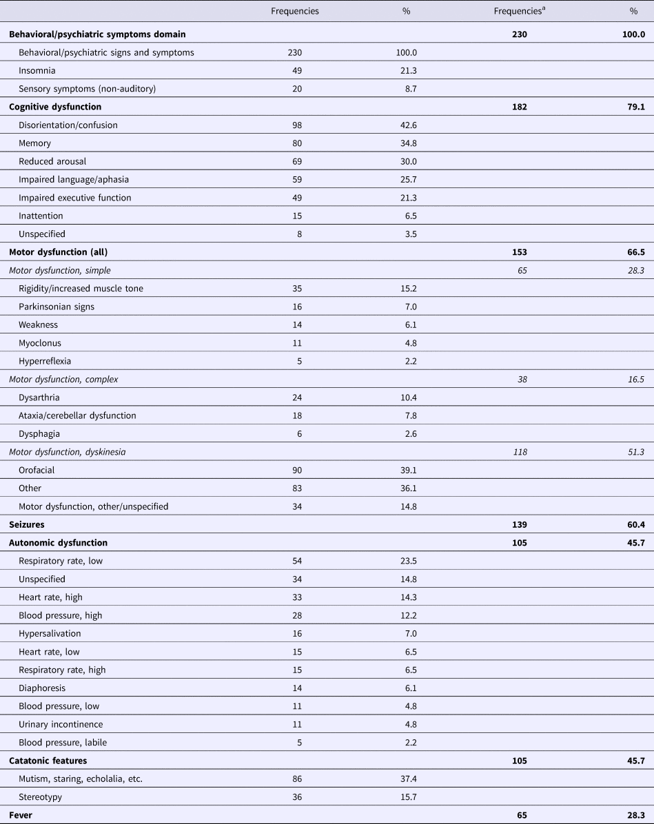
For purposes of analysis, symptoms were aggregated into seven symptom domains: behavioral/psychiatric, cognitive, motor, catatonic, seizures, autonomic, and fever. The behavioral/psychiatric domain included features such as shouting, crying, laughing, odd repetitive behaviors, pacing/agitation, responding to internal stimuli or reports of perceptual disturbances, verbalizing delusional or bizarre ideation, suicidal ideation or self-injurious behaviors, insomnia, and refusing to eat. Table 1 provides the frequencies with which individual features were reported, and cumulative frequencies within symptom domains. After behavioral/psychiatric symptoms, which were universally present (100%) because they were an inclusion criterion for the study, the most commonly observed features were seizures (60.4%), disorientation/confusion (42.6%), orofacial dyskinesias (39.1%), mutism or staring (37.4%), dyskinesias involving other body parts (36.1%), memory disturbance (34.8%), reduced arousal (30.0%), fever (28.3%), and impaired language/aphasia (25.7%). Notably, auditory hallucinations were common but often atypical for psychiatric disorders (e.g. several patients reported hearing musical sounds).
Clinical features in psychiatric presentations of anti-NMDAr encephalitis

a The domain (boldface) frequency is less than the sum of individual frequencies within that domain because many patients had more than one symptom within a domain. Frequencies <1% not reported.
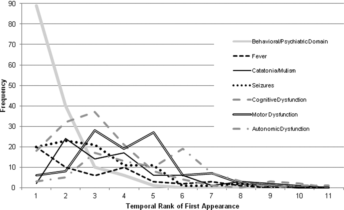
Information about the temporal order in which individual clinical features were initially observed was available in the majority of cases across all elements, with availability ranging from 55.6% (cerebellar signs/ataxia) to 100% (several elements). The sole exception was unspecified/other movement disorder, for which temporal information was available in only 47.1% of cases in which it was observed. For each patient, the temporal rank for each of the seven symptom domains was represented by the median temporal rank across elements within that domain for that patient. A temporal rank could be assigned to a symptom domain in 63.0–83.1% of cases in which at least one element from that domain was present. For each patient, the median temporal rank for each symptom domain was plotted on a frequency graph (Fig. 1), providing a visual representation of when in the illness episode initial observations of features from each symptom domain tended to peak.
Temporal sequence of anti-NMDAr encephalitis clinical features grouped into seven domains. The x-axis represents the temporal rank for each clinical domain (i.e. when it was first observed relative to other domains for a patient); the y-axis represents the frequency with which each rank was assigned to a domain, across patients. The temporal rank for each clinical domain represents the median value for all elements within that domain. The range of the x-axis depicts the highest temporal rank assigned to any domain.

Figure 1 illustrates that in this series of adult anti-NMDAr encephalitis patients whose illness episode was associated with clinical features suggesting a psychiatric disorder, those features dominated the earliest phase of the illness. Seizures, fever, and cognitive dysfunction were often among the earliest features to emerge, but psychiatric signs and symptoms were overwhelmingly prominent early in the course. Catatonic features and seizures tended to appear soon after psychiatric features, followed by cognitive and motor dysfunction. Autonomic dysfunction demonstrated a secondary peak for initial observation coinciding with those for cognitive and motor dysfunction, but its principal peak for initial observation occurred after other features had appeared and followed a secondary peak of initial observation for motor dysfunction. Fever was the least common domain, and when present it tended to be first observed in the earliest phase of the illness. Autonomic dysfunction was the last symptom domain to emerge.
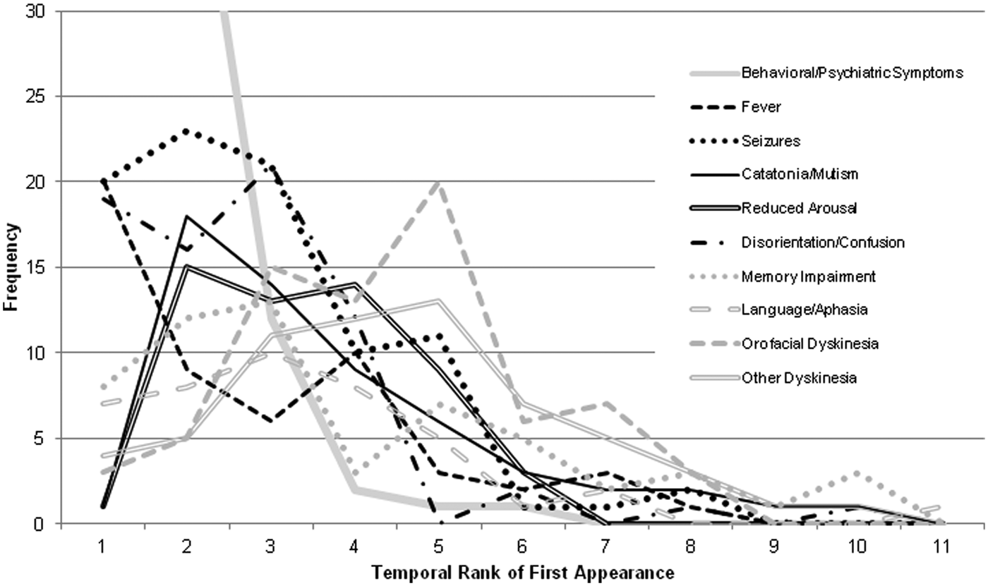
In order to develop a clearer picture of how an episode of anti-NMDAr encephalitis evolves, the chronology of the most common clinical features (i.e. those with frequencies >25%) was examined more closely. Psychiatric symptoms were present universally because they were a prerequisite for study inclusion, and nine other features were present in 25.7–60.4% of cases (Table 1). Each feature could be temporally ranked in the majority of cases when it was present, ranging from 64.0% (89 out of 139 cases) for seizures to 83.1% (54 out of 65 cases) for fever. On average, these nine most common clinical features could be temporally ranked 74.0% of the time when present. Their temporal progression patterns are presented in Fig. 2. Fever was often the first of these nine features to emerge, coinciding with the peak frequency for initial observation of psychiatric signs and symptoms, followed by seizures, catatonia, and hypoarousal. The third frequency peak included initial observations of disorientation/confusion, impaired language/aphasia and impaired memory. Dyskinesias (orofacial and other) were the last to appear, with a peak frequency immediately preceding that for initial observation of autonomic dysfunction (Fig. 1).
Temporal sequence of individual clinical features in anti-NMDAr encephalitis. The x-axis represents the temporal rank for each clinical feature (i.e. when it was first observed relative to other clinical features for a patient); the y-axis represents the frequency with which each rank was assigned to a feature, across patients. Therefore, the range of the x-axis depicts the highest temporal rank assigned to any of these individual features. Note that the line for ‘behavioral/psychiatric symptoms’ has been truncated to make the graph more readable for other features; actual frequencies for ranks 1 and 2 are 84 and 44, respectively.

Since early diagnosis of anti-NMDAr encephalitis in the context of a psychiatric assessment hinges on recognizing patterns of clinical features that are atypical for psychiatric disorders, the frequency with which these nine most prevalent features co-occurred was tabulated. At least two of these nine features (seizures, disorientation/confusion, orofacial dyskinesias, mutism/staring, dyskinesias involving other body parts, memory disturbance, reduced arousal, fever, and impaired language/aphasia) were present in 196 (84.3%) patients, at least three were present in 153 (66.5%) patients, and 109 (47.4%) patients demonstrated four or more of these nine features.
Discussion
Data from the present study demonstrate that patients with anti-NMDAr encephalitis who manifest signs and symptoms suggesting primary psychiatric disorders typically do so early in the course of illness, before more distinctive diagnostic clues (e.g. orofacial dyskinesias) are evident. Other features that are most likely to occur in the earliest stage of illness, i.e. seizures, fever and, disorientation/confusion, do so much less frequently (<10% of the time), and the latter two are so non-specific they may not prompt most clinicians to consider a diagnosis of anti-NMDAr encephalitis. Catatonic features occur relatively early, but these are also non-specific and may lead clinicians prematurely to a psychiatric diagnosis. NMDAr encephalitis-induced orofacial dyskinesias can be dramatic (e.g. there are multiple reports of self-biting), but they tend to occur later in the acute illness course, limiting their usefulness as signals for considering this diagnosis early. The same is true for stereotypies, which can be somewhat distinctive for this disorder (e.g. there are several reports of ‘bicycle pedaling’ or ‘piano playing’) but tend to occur even later.
The temporal sequence of clinical features described in this study is generally consistent with previous observations (Irani et al., Reference Irani, Bera, Waters, Zuliani, Maxwell, Zandi, Friese, Galea, Kullmann, Beeson, Lang, Bien and Vincent2010), but the present sample is much larger and the methodology employed provides greater resolution of clinical features and their relative timing. The frequencies of the most common clinical features in the present sample are also broadly consistent with those reported in another large series of adult patients (Titulaer et al., Reference Titulaer, McCracken, Gabilondo, Armangué, Glaser, Iizuka, Honig, Benseler, Kawachi, Martinez-Hernandez, Aguilar, Gresa-Arribas, Ryan-Florance, Torrents, Saiz, Rosenfeld, Balice-Gordon, Graus and Dalmau2013). A typical illness course has been described as consisting of four well-demarcated stages that progress from prodrome to psychiatric symptoms accompanied by speech abnormalities and seizures, followed by neurologic complications including movement disorders and dysautonomia, and finally a prolonged period of residual deficits (Kayser and Dalmau, Reference Kayser and Dalmau2016). However, while there may be a general pattern in the way that NMDAr encephalitis unfolds, the present data suggest very considerable inter-individual variation exists with respect to which features appear and when they are most likely to be observed.
The clinical morphology of anti-NMDAr encephalitis reflects its affinity for hippocampal neurons and NMDA receptors, which produces effects closely resembling those of NMDAr antagonists such as ketamine and phencyclidine (Lancaster et al., Reference Lancaster, Martinez-Hernandez and Dalmau2011; Dalmau, Reference Dalmau2016). Unlike those of paraneoplastic syndromes, the antibodies of autoimmune encephalitis target extracellular components of cell surface or synaptic proteins, alter the structure or function of these target antigens, can develop in the absence of a tumor, and respond to immunotherapy in the majority of cases (Leypoldt et al., Reference Leypoldt, Armangue and Dalmau2015). Anti-NMDAr encephalitis is caused specifically by IgG antibodies binding to the GluN1 subunit of the NMDAr; antibody binding causes capping and cross-linking of receptors on excitatory and inhibitory neurons, inducing their disappearance from the cell surface through reversible internalization, without affecting the number of synapses, dendrite structure or cell survival (Kayser and Dalmau, Reference Kayser and Dalmau2016). Cerebrospinal fluid (CSF) antibody titers correlate better than serum titers with clinical outcome and relapses (Kayser and Dalmau, Reference Kayser and Dalmau2016), and most clinical features of anti-NMDAr encephalitis are consistent with the hypothesis that NMDAr hypofunction is central to its pathophysiology (Maneta and Garcia, Reference Maneta and Garcia2014). Based on evidence from studies of NMDA antagonists, Dalmau (Reference Dalmau2016) has suggested that the diversity of anti-NMDAr encephalitis clinical presentations and outcomes reflects varying degrees of NMDA receptor impingement, with milder forms causing perceptual disturbances, ideas of reference, and paranoia together with impaired performance of executive function tasks. These milder forms would account for the clinical findings observed at illness onset or during recovery, whereas more extensive involvement could produce the psychotic symptoms, agitation, memory disturbance, and stereotyped or repetitive motor behaviors typically seen after several weeks of illness. In cases where NMDA receptor impingement is most severe, profound unresponsiveness and coma may ensue (Dalmau, Reference Dalmau2016), with central hypoventilation requiring assisted ventilation.
Patients often present with symptoms typical of common psychiatric disorders, and sometimes with seemingly pertinent family history, so clinicians may be challenged to make the correct diagnosis early in the clinical course. For example, one of the reports included in the present study describes a 47-year-old man who presented with anxiety, derealization, and atypical auditory hallucinations (music) following a viral prodrome. He did not respond to antiviral therapy and developed suicidal ideation for which electroconvulsive therapy (ECT) was administered. Following a course of bilateral ECT treatments, he recovered sufficiently to be discharged, and when the diagnosis was made retrospectively 2 years later he was doing well (Braakman et al., Reference Braakman, Moers-Hornikx, Arts, Hupperts and Nicolai2010). Other reports included in the present analysis describe patients who probably had relapsing anti-NMDAr encephalitis over years that were misdiagnosed as recurring psychotic episodes (Hopkins et al., Reference Hopkins, Moodley and Chan2013; Heekin et al., Reference Heekin, Catalano, Frontera and Catalano2015; Huang et al., Reference Huang, Kang, Zhang, Li, Qiu, Liu, Ren, Yang, Liu, Li and Guo2015; Yoshimura et al., Reference Yoshimura, Yada, Horigome and Kishi2015). In one of these cases (Heekin et al., Reference Heekin, Catalano, Frontera and Catalano2015), a 24-year-old woman presented with symptoms consistent with psychotic mania preceded for 6 months by complaints of upper extremity weakness and parasthesias. Although she was found to have significant word-finding difficulty and short-term memory impairment during her initial psychiatric evaluation, she was treated for this and subsequent episodes with antipsychotic medications; the diagnosis of anti-NMDArE was not made until 14 years later, after she suffered seizures during a hospitalization. In another of these cases (Yoshimura et al., Reference Yoshimura, Yada, Horigome and Kishi2015), a 48-year-old woman presented with confusion, disorientation, visual hallucinations, bizarre behavior, and a past history of recurrent delirious mania that had been treated with ECT, antipsychotic medications, and lithium. The only unambiguous neurocognitive symptom was retrograde amnesia for the index episode; the diagnosis of anti-NMDArE was made during a subsequent hospitalization 7 months later (Yoshimura et al., Reference Yoshimura, Yada, Horigome and Kishi2015). Kayser et al. (Reference Kayser, Titulaer, Gresa-Arribas and Dalmau2013) found that 23 of 571 (4%) patients with anti-NMDAr encephalitis exhibited isolated psychiatric syndromes in which delusional thinking, hallucinations, mood disturbances and aggression predominated without any neurological abnormalities. Eighteen (78.3%) of these patients were experiencing a relapse, but five (27.1%) patients presented with isolated psychiatric syndromes during their initial anti-NMDArE episode; of these five initial episode patients, family members recalled observing ‘excessive blinking’ in one patient, and hypersalivation in another (Kayser et al., Reference Kayser, Titulaer, Gresa-Arribas and Dalmau2013). Lejuste et al. (Reference Lejuste, Thomas, Picard, Desestret, Ducray, Rogemond, Psimaras, Antoine, Delattre, Groc, Leboyer and Honnorat2016) observed that in a series of anti-NMDAr encephalitis patients admitted to psychiatric units, six (13%) had prior psychiatric episodes that were probably caused by undiagnosed anti-NMDAr encephalitis.
Psychiatric symptoms are among recently proposed international consensus diagnostic criteria (Graus et al., Reference Graus, Titulaer, Balu, Benseler, Bien, Cellucci, Cortese, Dale, Gelfand, Geschwind, Glaser, Honnorat, Höftberger, Iizuka, Irani, Lancaster, Leypoldt, Prüss, Rae-Grant, Reindl, Rosenfeld, Rostásy, Saiz, Venkatesan, Vincent, Wandinger, Waters and Dalmau2016), which were published after most of the cases included in this study had been clinically diagnosed. These criteria indicate that a definite diagnosis of anti-NMDAr encephalitis can be made in the presence of IgG anti-GluN1 antibodies and at least one of the following major groups of symptoms when onset has been rapid (<3 months), after reasonable exclusion of other disorders: cognitive dysfunction or abnormal behavior suggesting a psychiatric disorder, speech dysfunction including mutism, seizures, movement disorder, decreased level of consciousness, or autonomic dysfunction (includes central hypoventilation). Antibody testing should include CSF to avoid false negative and false positive results; if only serum is available, confirmatory tests should be used. A probable diagnosis can be made if antibody testing is not available when at least three of the above major symptom groups are present and the EEG is abnormal or CSF reveals pleocytosis or oligoclonal bands; or a teratoma is present (Graus et al., Reference Graus, Titulaer, Balu, Benseler, Bien, Cellucci, Cortese, Dale, Gelfand, Geschwind, Glaser, Honnorat, Höftberger, Iizuka, Irani, Lancaster, Leypoldt, Prüss, Rae-Grant, Reindl, Rosenfeld, Rostásy, Saiz, Venkatesan, Vincent, Wandinger, Waters and Dalmau2016). CSF pleocytosis is present in up to 91% of cases (Dalmau et al., Reference Dalmau, Lancaster, Martinez-Hernandez, Rosenfeld and Balice-Gordon2011) and may precede detectable CSF-specific oligoclonal bands by as much as 2 weeks (Irani et al., Reference Irani, Bera, Waters, Zuliani, Maxwell, Zandi, Friese, Galea, Kullmann, Beeson, Lang, Bien and Vincent2010). New onset of seizures should prompt clinicians to consider a diagnosis of anti-NMDAr encephalitis, and there is an approximately 30% likelihood that an EEG will reveal a very distinctive pattern of activity termed ‘extreme delta brush’ (Schmitt et al., Reference Schmitt, Pargeon, Frechette, Hirsch, Dalmau and Friedman2012). Neuroimaging studies generally are of limited value in anti-NMDAr encephalitis (Irani et al., Reference Irani, Bera, Waters, Zuliani, Maxwell, Zandi, Friese, Galea, Kullmann, Beeson, Lang, Bien and Vincent2010; Dalmau et al., Reference Dalmau, Lancaster, Martinez-Hernandez, Rosenfeld and Balice-Gordon2011; Lim et al., Reference Lim, Lee, Jung, Kim, Shin, Moon, Byun, Kim, Shin, Lee, Kim, Park, Lee and Chu2014), but resting-state functional magnetic resonance imaging measurement of large-scale brain network connectivity shows some promise as a diagnostic aid (Peer et al., Reference Peer, Prüss, Ben-Dayan, Paul, Arzy and Finke2017).
A related diagnostic challenge arises when clinical features of anti-NMDAr encephalitis overlap with those of neuroleptic malignant syndrome (NMS). Several reports included in the present study (Punja et al., Reference Punja, Pomerleau, Devlin, Morgan, Schier and Schwartz2013; Howard et al., Reference Howard, Kass, Bandi and Guntupalli2014; Koksal et al., Reference Koksal, Baybas, Mutluay, Altunkaynak and Keskek2015), and a pediatric case (Slettedal et al., Reference Slettedal, Dahl, Sandvig, Dalmau and Strømme2012), attest to the difficulty distinguishing these disorders in the context of a severely ill patient who has been treated with antipsychotic medications for behavioral symptoms consistent with a psychiatric disorder. In the series of 45 anti-NMDAr encephalitis patients erroneously admitted to a psychiatric unit, NMS was suspected strongly enough in 21 patients (46.6%) that they were transferred to a medical service (Lejuste et al., Reference Lejuste, Thomas, Picard, Desestret, Ducray, Rogemond, Psimaras, Antoine, Delattre, Groc, Leboyer and Honnorat2016). In addition to sharing features of hyperthermia, extrapyramidal dysfunction, altered mental status and autonomic dysregulation, 7% of anti-NMDAr encephalitis patients have elevated serum creatine kinase (Dalmau et al., Reference Dalmau, Gleichman, Hughes, Rossi, Peng, Lai, Dessain, Rosenfeld, Balice-Gordon and Lynch2008). Hyperthermia is three times more frequent in anti-NMDAr encephalitis patients treated with antipsychotic medications than in those who are not, suggesting heightened neuroleptic intolerance (Lejuste et al., Reference Lejuste, Thomas, Picard, Desestret, Ducray, Rogemond, Psimaras, Antoine, Delattre, Groc, Leboyer and Honnorat2016), and NMS risk may also be increased (Caroff and Campbell, Reference Caroff and Campbell2015). The possibility of precipitating NMS by administering antipsychotic medications to a patient with anti-NMDArE who has been misdiagnosed with a psychiatric disorder is another reason clinicians need to be vigilant for forms of encephalitis that mimic mental illness. The best chance of avoiding a scenario in which complications accumulate while effective and appropriate treatment is delayed rests with making the correct diagnosis as soon as possible.
This study is limited by its reliance on a review of published case material, which may not be representative of the patient population. Relevant information may have been omitted inadvertently, and statements or inferences regarding the temporal sequence of features may not be consistently reliable across reports. It can be difficult to distinguish certain behavioral abnormalities from one another (e.g. catatonic mutism v. partial complex seizure v. profound aphasia), and any ambiguities in the published reports are likely to have amplified coding imprecision. Another limitation is the use of a single reviewer to select and score the cases included in this study; adjudication and scoring of outcomes might have been different in some cases if conducted by additional or other case reviewers. This study is not a systematic review and is not meant to report or assess the risk of bias in individual case reports, or to synthesize the data in anything more than a descriptive manner. These limitations are offset to some extent by a large study size, which mitigates contributions from random error related to inadvertent omissions, incomplete information, and possible author bias in the published reports that were included in the analyses. This study focused on patients who exhibited clinical features likely to prompt referral to a psychiatrist, so any findings reported here should not be extrapolated to anti-NMDAr patients without such features.
In conclusion, although anti-NMDAr encephalitis is uncommon, it is likely that every psychiatrist will encounter these patients in the course of his/her practice. The clinical outcome with prompt and effective treatment is often good, but without appropriate treatment, patients are likely to suffer a protracted course with significant long-term disability or death. The best strategy for recognizing this disorder is to have a high index of suspicion when an individual develops new psychiatric symptoms in the context of a recent viral prodrome (malaise, headache, loss of appetite), when accompanied by seizures or unexplained fever, or when the quality of the psychiatric symptoms is unusual (e.g. non-verbal auditory hallucinations). This study affirms that the concurrence of new or atypical psychiatric symptoms and non-psychiatric features such as seizures or fever should prompt active consideration of a diagnosis of anti-NMDArE. The presentation and course of anti-NMDArE varies considerably from one individual to another, so it is important to be vigilant for any of these features and not to predicate diagnosis on any single feature. Although the abrupt onset of orofacial dyskinesias is somewhat distinctive for this disorder, this feature often emerges relatively late, so relying on its presence to initiate treatment may lead to unnecessary delays.
Supplementary material
The supplementary material for this article can be found at https://doi.org/10.1017/S0033291718003665.
Author ORCIDs
Ronald J. Gurrera 0000-0001-6648-4704.
Acknowledgments
This material is the result of work supported with resources and the use of facilities at the VA Boston Healthcare System. The contents do not represent the views of the US Department of Veterans Affairs or the United States Government.
Financial support
This research received no specific grant from any funding agency, commercial or not-for-profit sectors.
Conflict of interest
None.