Case report
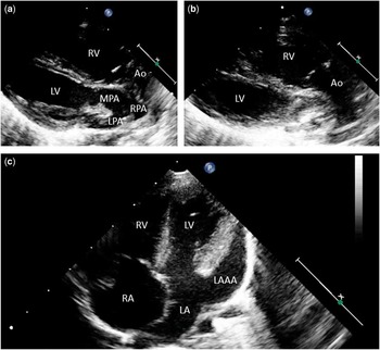
A 35-day-old boy weighing 4.5 kg was referred to our department from a general paediatric hospital due to low oxygen saturation. His transcutaneous oxygen saturation was 75%. Transthoracic echocardiography showed discordant ventriculo-arterial connections, a non-restrictive atrial septal defect, and patent ductus arteriosus, in addition to a 23 mm×10-mm left atrial appendage aneurysm (Fig 1). The patient underwent an arterial switch operation, patent ductus arteriosus ligation, and downsizing of the atrial septal defect. As the patient’s left atrial appendage aneurysm was not huge in size and in order not to increase the complexity of the operation or lead to co-morbid pathologies, the surgeon decided that no surgical intervention targeted at the left atrial appendage was warranted. The patient was extubated on the second post-operative day and no problems were observed.
Two-dimensional echocardiography demonstrates in the ventricular long-axis view ( a and b ) discordant ventriculo-arterial connections, whereas the apical four-chamber view ( c ) shows concordant atrioventricular connections and the left atrial appendage aneurysm. Ao=aorta; LA=left atrium; LAAA=left atrial appendage aneurysm; LPA=left pulmonary artery; LV=left ventricle; MPA=main pulmonary artery; RA= right atrium; RPA=right pulmonary artery; RV=right ventricle.

Congenital left atrial appendage aneurysms are caused by congenital dysplasia of the atrial muscle.Reference Victor and Nayak 1 These aneurysms probably increase in size as the patient ages.Reference Bhattarai, Padalino and Stellin 2 Despite its congenital cause, symptoms usually do not arise until the second or third decade of life.Reference Victor and Nayak 1 , Reference Foale, Gibson and Guyer 3 Once the aneurysms reach a larger size, they predispose the patient to supra-ventricular arrhythmias, cardiac dysfunction, atypical chest pain episodes due to compression of the left coronary artery or any of its divisions, an increased risk of intra-atrial thrombus formation, and systemic embolisation.Reference Victor and Nayak 1 , Reference Foale, Gibson and Guyer 3 – Reference Zhao, Ge, Yan, Pan and Liao 5 Diagnosis is established by transthoracic echocardiography and, very rarely, by cardiac catheterisation if additional information is likely to be gained. In particular, the colour Doppler echocardiogram was very helpful in this diagnosis, showing the exchange of blood between the left atrium and the left atrial appendage. This entity is different from acquired enlargement of the left atrium secondary to rheumatic heart disease or severe mitral regurgitation.Reference Terada, Tanaka, Kashima, Sannou and Arima 6 , Reference Chen, Chang, Guo and Chang 7 Congenital left atrial appendage aneurysms are extremely rare. In reported cases, the body of the atrium is not usually enlarged, and such aneurysms have not been found to be associated with other pathologies.Reference Tanoue, Kado, Shiokawa and Sagawa 4 In contrast, in our case, a congenital left atrial appendage aneurysm was found in the presence of transposition of the great arteries and an intact ventricular septum. As far as we are aware, this association has not been previously reported in the literature.
Acknowledgements
We would like to express our endless thanks to Cuneyt Aslan and Gulsah Ilter Candan for their devoted work in the administration of echocardiography.
Financial Support
This research received no specific grant from any funding agency, commercial or not-for-profit sectors.
Conflicts of Interest
None.