When Howard Carter discovered Tutankhamun's tomb in 1922, he found a box with two coffins of 20- and 32-week female foetuses whose paternity was later attributed to the Pharaoh.Reference Hawass, Gad and Ismail 1 Indeed, both blood group data and deoxyribonucleic acid analysis have demonstrated the royal affiliation, thus invalidating the hypothesis of a placement in the tomb for a symbolic purpose of allowing the king to live as a newborn in the afterlife.Reference Hawass, Gad and Ismail 1 , Reference Farag and Iskandar 2 Confirming the special attention accorded to the foetus in ancient Egypt, we report the case of a foetus placed in a sort of shrine. Allowing the diagnosis of dextrocardia, the computed tomography scan tells us a little more about what happened more than 24 centuries ago.
Case report
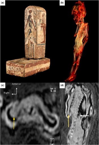
A wooden casket of the Louvre's collection, whose form evokes a small chapel, was brought in our radiology unit for computed tomography scan analysis on the occasion of a temporary exhibition.Reference Pierrat-Bonnefois 3 This shrine presented ornamentation (Fig 1a) that allowed its approximate dating to the very late period of Egyptian pharaonic civilisation – about 4th to 3rd century B.C. This curious object, unique as much as egyptologists can say, was formerly part of the collection of Dr Clot, a French doctor who lived in Egypt during the 19th century. He never opened the case to investigate its content, for this would have damaged the painted gesso over it. About 30 years ago, an X-ray examination revealed that it contains the remains of a foetus.Reference Hours 4
Computed tomography scan analysis of a foetal mummy placed in a shrine from the Louvre's collection. ( a ) Photograph of the shrine. Ornamentation of the wooden casket allowed its approximate dating at about 4th to 3rd century B.C. 2009 (Musée du Louvre/Georges Poncet). ( b ) Three-dimensional computed tomography reconstruction of the foetus. The right hand was positioned on the left side of the chest. The foetal measurements allowed an estimation of its term at 15 weeks of gestation. ( c ) Computed tomography axial image showing a teardrop-shaped mass (yellow arrow) in the right hemithorax. Owing to the absence of similar image in the left hemithorax, we assumed that it was a dextrocardia, a rare condition that is often associated with congenital heart disease. ( d ) Computed tomography coronal image showing the supposed cardiac structure (yellow arrow) in the right hemithorax and the left-side single spleen (white arrow).

To improve this first view, a computed tomography scan was performed in 2009 using a Dual-Source CT machine (Siemens, Flash Definition, Erlangen, Germany) with 80 kilovoltage peaks and 260-millisecond settings. Slices of 0.6 millimetre were reconstructed every 0.3 millimetre, providing high-resolution images in every plane. Thus, three-dimensional reconstructions depicted accurately a mummified foetus. As shown by the swaddled feet, the body was probably wrapped. The skull was crushed in the frontal plane, suggesting an extraction with forceps. The rest of the body had no external deformation. The femur length was 18 millimetres – measured from the greater trochanter to the lateral condyle – and the tibia length was 16.5 millimetres. Measurement of the biparietal diameter was not feasible because the head was flattened. The crown rump length was 100 millimetres. Although the left arm was placed along the body, the right hand was positioned next to the supposed location of the heart, that is to say, on the left side of the chest (Fig 1b). No aspect of cardiac structure was visualised in the left hemithorax. In contrast, a teardrop-shaped mass, whose axis was pointed to the right, was clearly depicted in superposition of the right lung (Fig 1c). No image of stomach was seen in the chest of the mummy, suggesting primary dextrocardia. The spleen was on the left side of the body (Fig 1d) and the skeleton was normal. The computed tomography scan also revealed that this foetus was fitted by bundles of textile in the cavity carved inside the wooden log.
Discussion
First necropsies performed on the mummies began in the 18th and 19th centuries. They often required their unwrapping and their dismemberment. Modern technologies, using non-destructive methods, have significantly improved paleopathological studies both for the integrity of specimens and for the diagnosis of diseases that could have affected them.Reference David 5 Indeed, medical techniques such as radiographs, computed tomography scan, magnetic resonance imaging, and deoxyribonucleic acid analysis are now widely used in this context.Reference Gray 6 – Reference Pääbo 10 Recently, a congenital malformation of the neural tube was diagnosed in an infant mummy using computed tomography scan.Reference Boano, Fulcheri and Martina 11 In ancient Egypt, consanguinity, a well-known cause of congenital malformations, was common, especially in the royal family, as evidenced by the Tutankhamun pedigree.Reference Hawass, Gad and Ismail 1 However, owing to the embalming process that required a complete evisceration, no congenital heart disease has been reported so far to our knowledge. Owing to the right position of the heart in the chest and its axial orientation – apex pointing to the right – we assumed that it was a primary dextrocardia rather than a simple displacement of the heart.Reference Garson and Edin 12 As proved by a foetal echocardiographic study, when dextrocardia is the consequence of a disease that causes pushing of the heart towards right, also named secondary dextrocardia, a diaphragmatic hernia is almost always found.Reference Walmsley, Hishitani and Sandor 13 Primary dextrocardia has a foetal incidence of approximately 0.22% and nearly half of the foetuses with primary dextrocardia have visceral situs solitus.Reference Garg, Agarwal, Modi, Radhakrishnan and Sinha 14 , Reference Bernasconi, Azancot, Simpson, Jones and Sharland 15 The left-side single spleen of our mummy seems to invalidate the hypothesis of a heterotaxy syndrome, frequently found in case of situs inversus.Reference Ticho, Goldstein and Van Praagh 16 Regardless of the situs status, dextrocardia is highly associated with intracardiac anomalies, and the incidence of foetal demise secondary to congenital heart disease is around 7%, significantly higher than that observed in live-born infants.Reference Garg, Agarwal, Modi, Radhakrishnan and Sinha 14 , Reference Bernasconi, Azancot, Simpson, Jones and Sharland 15 , Reference Hoffman 17 However, we cannot state categorically that a congenital heart defect was the cause of this foetal death. Indeed, as previously noted, a right-sided heart does not indicate that the heart itself was congenitally malformed. Furthermore, the most frequent lesion found with dextrocardia in presence of situs solitus is congenitally corrected transposition of the great arteries, which is not automatically lethal for the foetus.Reference Garg, Agarwal, Modi, Radhakrishnan and Sinha 14 Another relatively common aetiology of dextrocardia is Kartagener's syndrome or primary ciliary dyskinesia. This rare genetic disorder is responsible for dysfunction of embryological nodal cilia and may lead to laterality defects in 50% of patients.Reference Leigh, Pittman and Carson 18 This hypothesis is strengthened by an observation of an increased incidence of primary ciliary dyskinesia in consanguineous populations.Reference O'Callaghan, Chetcuti and Moya 19 Finally, we assume that the foetal death was related to this dextrocardia without being able to provide more precise diagnosis. According to the mummy's measurements and to normal foetal echographic values, the term was estimated at 15 weeks of gestation.Reference Brons, van Geijn, Bezemer, Nauta and Arts 20 , Reference Larsen, Petersen, Greisen and Larsen 21 We potentially underestimated this term because of the desiccation.
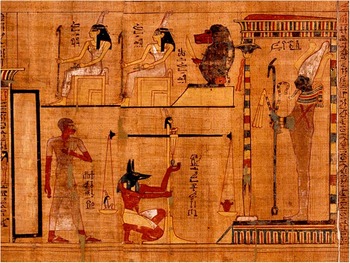
As the Egyptians believed that the “soul” was eternal, a special preparation of the body was necessary in order to reach the kingdom of the dead. The body, one component of the individuality, ideally required to be kept to host the other parts of the “soul”. Depending on the social class, the embalming procedure was not the same for everyone. Poor people were simply buried in the desert, whereas upper and middle classes were embalmed in two stages: evisceration and treatment of the tissues.Reference David 5 , Reference Garson and Edin 12 Owing to the practice of evisceration, ancient Egyptians had a good knowledge of human anatomy. They knew the normal location of the heart and its vital role. Moreover, in the Papyrus of Neferoubenef (The Louvre Museum), which is similar to the papyrus of Ani (British Museum) also called “book of the dead”, the weighing of the heart was a crucial step for the dead to access the kingdom of Osiris (Fig 2). Indeed, in Egyptian mythology, the heart, which was believed to contain the “soul”, was weighed to determine whether the dead lived correctly under the laws of divine order. Thus, one could imagine that the position given to this foetus – right hand on the left hemithorax – had a special significance, although no Egyptian text confirms that suggestion. Despite the fact that our foetus was probably from a rich family as suggested by the shrine, the evisceration process was not achieved because, presumably, of the small body size. Thus, we were able to find this highly probable dextrocardia.
Funerary papyrus representing the weighing of the heart. Papyrus of Neferoubenef (Department of Egyptian Antiquities, N 3092, The Louvre Museum/Georges Poncet). Around 1400 B.C. The deceased was judged by weighing his heart in the presence of the god Osiris. The god Anubis (jackal head) was in charge of this proceeding.

Conclusion
Computed tomography scan is increasingly used for paleopathological examinations. This modern medical tool was very helpful in depicting a highly probable dextrocardia in a mummy of an Egyptian foetus. It is an incomparable research tool both for comprehension of the hidden parts of the ancient customs of humanity and for investigation of its diseases. Although the heart was not located, ironically, at the right place, this case seems to show the interest that was given to the foetuses in ancient Egypt.
Acknowledgement
The authors thank Sylvain Ordureau (Useful progress, Paris) for his technical assistance.