INTRODUCTION
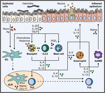
Helminths are complex macroparasites with large genomes and a highly evolved capacity to infect mammalian hosts, sometimes persisting in a single host for decades. Parasitic helminths infect over two billion of the world's population, many of whom suffer significant levels of morbidity and disability as a result. Parasitic helminths are also important pathogens of domestic livestock, inflicting significant economic cost on the agricultural industry. Type 2 immune activation is the primary protective immune response against these multicellular macroparasites. This response can lead to expulsion of the parasite and resolution of infection, but this is not always the case. In many situations, Type 2 immunity functions essentially as a wound repair mechanism that protects the host from worm-induced damage. Both helminth expulsion and wound repair mechanisms require host production of the Type 2 cytokines IL-4, IL-5, IL-9 and IL-13 (Allen and Maizels, Reference Allen and Maizels2011; Gause et al. Reference Gause, Wynn and Allen2013). Up until recently the overarching view was that the primary mediator of this immune response was the adaptive T helper 2 (Th2) CD4+ T cell population, which is a rich source of Type 2 cytokines following helminth-induced activation (Van Dyken et al. Reference Van Dyken, Nussbaum, Lee, Molofsky, Liang, Pollack, Gate, Haliburton, Ye, Marson, Erle and Locksley2016). However, more recently we have come to appreciate the complex interplay of the innate and adaptive immune systems during helminth infection. Work in the last decade has revealed how cells of the innate immune system contribute to Type 2 immune responses, both as early instigators of Type 2 immune activation – as key sources of Type 2 cytokines – and as important downstream effectors that orchestrate modulation of the tissue microenvironment (Fig. 1).
Innate immune cells participate in a network that facilitates anti-helminth Type 2 immunity. Worms cause significant damage to the epithelium as they migrate and complete their lifecycle. This causes release of damage signals such as IL-25, IL-33 and TSLP that promote the activation of innate cells present in the tissue. Activated DCs sense Ag and migrate to the dLN, where they release pro-Th2 mediators such as IFN-I, CCL17 and Relmα and induce polarization of naïve T cells to a Th2 cell fate. Tissue-released damage signals also activate basophils, ILC2s and MCs that play distinct roles in the tissue. Basophils produce various inflammatory mediators such as chemokines and histamine and may interact with ILC2s. ILC2s are rich sources of Type 2 cytokines and wound repair-related molecules (not shown). Activated MCs release inflammatory mediators including PGD2, which activates and recruits ILC2s. Critically, activated basophils, ILC2s and MCs produce significant amounts of IL-4, IL-5 and IL-13 that can contribute to the polarization of Th2 cells in the dLN and may potentiate the activity of Th2 cells within the tissue. Finally, Type 2 cytokines from these innate cells and activated Th2 cells in the tissue recruit eosinophils, induce the AAMΦs that produce anti-helminth and pro-wound healing mediators such as Relmα, and activate anti-helminth responses from the epithelium, such as goblet cell hyperplasia, mucus production, tuft cell activation and epithelial cell turnover. AAMΦ, alternatively-activated macrophage; Ag, antigen; DC, dendritic cell; dLN, draining lymph node; IL, interleukin; IFN-I, Type I interferon; ILC2, group 2 innate lymphoid cell; Th2, T helper 2 cell; TSLP, thymic stromal lymphopoietin; PGD2, prostaglandin D2.

The innate immune system comprises many different cell types including some that are found in relative abundance during inflammation such as macrophages, monocytes, eosinophils and neutrophils. Other innate immune cell types are much more rare, making up less than 5% of the circulating leukocyte population. These include group 2 innate lymphoid cells (ILC2s), basophils, mast cells (MCs) and dendritic cells (DCs). Many other reviews have focused on the importance of the more abundant cell types, such as macrophages and eosinophils (Murray and Wynn, Reference Murray and Wynn2011; Gause et al. Reference Gause, Wynn and Allen2013; Huang and Appleton, Reference Huang and Appleton2016). Thus, here we will focus on the importance of rare innate immune cell populations, which make up for what they lack in abundance with a potent capacity to modulate the immune microenvironment. The main focus of this review will be recent important findings highlighting the role of ILC2s, basophils, MCs and DCs in Type 2 inflammation against helminths.
Rare innate immune cells – right time, right place?
ILC2s, basophils, MCs and DCs are highly specialized for their specific roles, be that activation in response to IgE or antigen, or a capacity to rapidly release large quantities of immune chemical messengers such as cytokines, chemokines, histamine and prostaglandins. Thus, while these innate immune cells may be rare, they are poised to perform their specific role robustly. Further, while these cells can be found throughout the body in lymphoid and mucosal tissues, they are particularly enriched in sites that are subject to invasion by helminth parasites and other pathogens (Rivera et al. Reference Rivera, Siracusa, Yap and Gause2016) such as the skin, lung and gut. Since helminth infections often affect more than one mucosal or barrier tissue, the conserved presence of these cells at multiple sites ensures a coordinated response on many fronts. The importance of these cells at mucosal surfaces is clear, but their influence on immune responses in lymphoid tissues should not be underestimated. We know that DC migration to the lymph node is essential for DC function in priming naïve T cells. ILC2s and granulocytes can also be found in lymphoid tissues and may influence the formation of adaptive immune activation by their potent production of inflammatory mediators (Pulendran and Artis, Reference Pulendran and Artis2012). In addition, these rare innate immune cells are uniquely specialized to instigate immune activation promptly. ILC2s have been shown to employ genomic adaptations that maintain important genes in open states that allow rapid transcription of effector molecules in response to stimulation (Shih et al. Reference Shih, Sciumè, Mikami, Guo, Sun, Brooks, Urban, Davis, Kanno and O'Shea2016), and future studies will address if this is also the case in other innate cell types. Basophils and MCs also maintain stores of premade mediators, such as cytokines, eicosanoids and histamine, and are ready to rapidly release high concentrations of these chemicals at sites of inflammation (Voehringer, Reference Voehringer2013). Finally, rare innate cell function is highly tunable by effector molecules found in the microenvironment, and these cells are responsive to multiple pathways that control molecular mechanisms of activation and gene expression, such as worm-induced damage signals from the epithelium, Notch signalling and eicasonoids (Rivera et al. Reference Rivera, Siracusa, Yap and Gause2016; Zhang et al. Reference Zhang, Xu, Asghar Pasha, Siebel, Costello, Haczku, MacNamara, Liang, Zhu, Bhandoola, Maillard and Yang2017). Hence, innate cell function can be rapidly modulated by tissue factors, making rare innate cells highly responsive to changes in the immune microenvironment and key players in Type 2 immune activation.
ILC2s – unique cytokine power houses of the innate immune system
ILC2s are the most recently described rare innate immune cell that we will discuss. The existence of a novel innate cell population that could influence Type 2 immunity was first reported in the early 2000s (Fallon et al. Reference Fallon, Ballantyne, Mangan, Barlow, Dasvarma, Hewett, McIlgorm, Jolin and McKenzie2006). ILC2s were fully described in 2010 by three independent laboratories as a non-B, non-T cell population that drove Type 2 immunity in mice (Moro et al. Reference Moro, Yamada, Tanabe, Takeuchi, Ikawa, Kawamoto, Furusawa, Ohtani, Fujii and Koyasu2010; Neill et al. Reference Neill, Wong, Bellosi, Flynn, Daly, Langford, Bucks, Kane, Fallon, Pannell, Jolin and McKenzie2010; Price et al. Reference Price, Liang, Sullivan, Reinhardt, Eisley, Erle and Locksley2010). They are characterized by their lack of expression of markers for common cell lineages and their expression of c-kit, CD90 (Thy1) and CD127 (IL-7R) (Moro et al. Reference Moro, Yamada, Tanabe, Takeuchi, Ikawa, Kawamoto, Furusawa, Ohtani, Fujii and Koyasu2010; Neill et al. Reference Neill, Wong, Bellosi, Flynn, Daly, Langford, Bucks, Kane, Fallon, Pannell, Jolin and McKenzie2010; Price et al. Reference Price, Liang, Sullivan, Reinhardt, Eisley, Erle and Locksley2010). ILC2s arise from a common lymphoid progenitor found in the bone marrow. Their development and function is dependent on a range of transcription factors (TFs), including RORα, Id2, GATA-3 and ETS1 (Cherrier et al. Reference Cherrier, Sawa and Eberl2012; Hoyler et al. Reference Hoyler, Klose, Souabni, Turqueti-Neves, Pfeifer, Rawlins, Voehringer, Busslinger and Diefenbach2012; Klose and Artis, Reference Klose and Artis2016; Zook et al. Reference Zook, Ramirez, Guo, van der Voort, Sigvardsson, Svensson, Fu and Kee2016).
ILC2s are found in the skin (Kim et al. Reference Kim, Siracusa, Saenz, Noti, Monticelli, Sonnenberg, Hepworth, Van Voorhees, Comeau and Artis2013) and at mucosal surfaces, as well as other distal sites and lymphoid tissues. At these sites, they are poised to respond to a range of signals, including epithelial-derived damage signals elicited by worm feeding or migration, through their expression of receptors for the cytokines IL-25 (IL-17RB), IL-33 (ST2) and thymic stromal lymphopoietin (TSLP) (Tait Wojno and Artis, Reference Tait Wojno and Artis2016). In response to IL-25, IL-33 and TSLP, ILC2s potently produce large quantities of IL-5 and IL-13 (Moro et al. Reference Moro, Yamada, Tanabe, Takeuchi, Ikawa, Kawamoto, Furusawa, Ohtani, Fujii and Koyasu2010; Neill et al. Reference Neill, Wong, Bellosi, Flynn, Daly, Langford, Bucks, Kane, Fallon, Pannell, Jolin and McKenzie2010; Price et al. Reference Price, Liang, Sullivan, Reinhardt, Eisley, Erle and Locksley2010), which is likely important for inducing eosinophil recruitment, epithelial cell hyperplasia, tuft cell differentiation and goblet cell activity – all mechanisms involved in worm expulsion (Cliffe et al. Reference Cliffe, Humphreys, Lane, Potten, Booth and Grencis2005; Hammad and Lambrecht, Reference Hammad and Lambrecht2015; Gerbe et al. Reference Gerbe, Sidot, Smyth, Ohmoto, Matsumoto, Dardalhon, Cesses, Garnier, Pouzolles, Brulin, Bruschi, Harcus, Zimmermann, Taylor, Maizels and Jay2016; von Moltke et al. Reference von Moltke, Ji, Liang and Locksley2016). In addition, epithelial cell-derived IL-33 and TSLP induce IL-9 release from ILC2s that acts in an autocrine manner to potentiate production of IL-5, IL-13 and the growth factor amphiregulin during infection with Nippostrongylus brasiliensis (Turner et al. Reference Turner, Morrison, Wilhelm, Wilson, Ahlfors, Renauld, Panzer, Helmby and Stockinger2013; Mohapatra et al. Reference Mohapatra, Van Dyken, Schneider, Nussbaum, Liang and Locksley2015), a mouse-adapted helminth used as a model for intestinal hookworm infection (Camberis et al. Reference Camberis, Le Gros and Urban2003). Recent studies have identified a key role for tuft cells, a population of specialized epithelial cells that expand upon worm infection and respond to IL-4 by producing large amounts of IL-25, for promoting IL-13 production by ILC2s (Gerbe et al. Reference Gerbe, Sidot, Smyth, Ohmoto, Matsumoto, Dardalhon, Cesses, Garnier, Pouzolles, Brulin, Bruschi, Harcus, Zimmermann, Taylor, Maizels and Jay2016; Howitt et al. Reference Howitt, Lavoie, Michaud, Blum, Tran, Weinstock, Gallini, Redding, Margolskee, Osborne, Artis and Garrett2016; von Moltke et al. Reference von Moltke, Ji, Liang and Locksley2016). Thus, ILC2s are particularly tuned to the state of the epithelium and respond to damage signals with rapid and robust production of Type 2 cytokines.
Notably, the ability of ILC2s to respond to signals from the damaged epithelium also promotes their interactions with other rare innate immune cell types to coordinate Type 2 immunity. ILC2s respond to IL-33 that has been cleaved by proteases released by MCs, leading to the potent expansion of the ILC2 population, demonstrating cooperation between these two cell types that enhances the function of ILC2s (Lefrançais et al. Reference Lefrançais, Duval, Mirey, Roga, Espinosa, Cayrol and Girard2014). ILC2s have also been shown to act on DCs, with ILC2-derived IL-13 inducing migration of DCs into the draining lymph node (dLN), where DCs prime Th2 cells. ILC2-derived IL-13 is also required for CCL17 production by DCs, which recruits activated Th2 cells into the tissue during Type 2 inflammation (Halim et al. Reference Halim, Steer, Mathä, Gold, Martinez-Gonzalez, McNagny, McKenzie and Takei2014, Reference Halim, Hwang, Scanlon, Zaghouani, Garbi, Fallon and McKenzie2015), further demonstrating how innate cells work in a complex network to drive Type 2 inflammatory responses.
In addition to their production of IL-5 and IL-13, ILC2s promote the development of Th2 activation by interacting with adaptive CD4+ T cells. In this context, ILC2s are an essential source of IL-4 for the effective development of lamina propria (LP) Th2 responses against the gastrointestinal helminth Heligmosomoides polygyrus (Pelly et al. Reference Pelly, Kannan, Coomes, Entwistle, ckerl, Seddon, MacDonald, McKenzie and Wilson2016), which causes a persistent natural rodent infection in which larvae burrow into the small intestine submucosa and emerge as adult worms into the lumen (Reynolds et al. Reference Reynolds, Filbey and Maizels2012). While this study did not address where the interaction between ILC2s and T cells may occur – in the tissue site (LP) or in the dLN – the authors showed that ILC2s could drive the differentiation of Th2 cells without direct contact (Pelly et al. Reference Pelly, Kannan, Coomes, Entwistle, ckerl, Seddon, MacDonald, McKenzie and Wilson2016). In addition, IL-13-producing ILC2s can also cooperate with CD4+ T cells during N. brasiliensis infection to induce larval killing following secondary infection (Bouchery et al. Reference Bouchery, Kyle, Camberis, Shepherd, Filbey, Smith, Harvie, Painter, Johnston, Ferguson, Jain, Roediger, Delahunt, Weninger, Forbes-Blom and Le Gros2015) as the larvae transit through the lung before becoming resident in the small intestine (Camberis et al. Reference Camberis, Le Gros and Urban2003), and these IL-13+ ILC2s are dependent on epithelial cell production of Surfactant Protein D (Thawer et al. Reference Thawer, Auret, Schnoeller, Chetty, Smith, Darby, Roberts, Mackay, Whitwell, Timms, Madsen, Selkirk, Brombacher, Clark and Horsnell2016). Notably, whether ILC2s can initiate priming of Th2 cells via direct contact with naïve T cells is somewhat contentious. Though a number of studies have suggested that ILC2s can directly prime Th2 responses via expression of MHC II (Mirchandani et al. Reference Mirchandani, Besnard, Yip, Scott, Bain, Cerovic, Salmond and Liew2014; Oliphant et al. Reference Oliphant, Hwang, Walker, Salimi, Wong, Brewer, Englezakis, Barlow, Hams, Scanlon, Ogg, Fallon and McKenzie2014), other work suggests that these cells do not act as antigen presenting cells (Maizels and Withers, Reference Maizels and Withers2014).
While early studies suggested that ILC2s were a relatively homogenous population, recent work has identified that not all ILC2s are created equal, at least in the mouse. Paul and colleagues showed that there are ‘natural’ ILC2s resident in the lung tissue that express high levels of ST2, while upon helminth-induced inflammation, there is an influx of ST2lo KLRG1hi inflammatory ILC2s that potently respond to IL-25 (Huang et al. Reference Huang, Guo, Qiu, Chen, Hu-Li, Siebenlist, Williamson, Urban and Paul2014). Exactly what the function of KLRG1 is in this context is not currently known, though it can be inhibitory in other settings. These inflammatory ILC2s appear to differentiate into natural ILC2s and are important in the expulsion of N. brasiliensis from the intestine (Huang et al. Reference Huang, Guo, Qiu, Chen, Hu-Li, Siebenlist, Williamson, Urban and Paul2014). Inflammatory ILC2s were also active in IL-17-associated infection with Candida albicans (Huang et al. Reference Huang, Guo, Qiu, Chen, Hu-Li, Siebenlist, Williamson, Urban and Paul2014), with the Notch pathway acting as a key regulator of these cells' ability to demonstrate functional plasticity (Zhang et al. Reference Zhang, Xu, Asghar Pasha, Siebel, Costello, Haczku, MacNamara, Liang, Zhu, Bhandoola, Maillard and Yang2017).
In addition, another subset of ILC2s has recently been identified, a long-lived persistent population that was maintained after amelioration of Type 2 antigen exposure in mouse models (Martinez-Gonzalez et al. Reference Martinez-Gonzalez, Mathä, Steer, Ghaedi, Poon and Takei2016). How these ‘memory-like’ ILC2s function in secondary worm infection has not yet been addressed, but IL-13-producing ILC2s and IL-13+ T cells must be recruited to the lung to activate alternatively-activated macrophages to kill larvae in N. brasiliensis reinfection (Bouchery et al. Reference Bouchery, Kyle, Camberis, Shepherd, Filbey, Smith, Harvie, Painter, Johnston, Ferguson, Jain, Roediger, Delahunt, Weninger, Forbes-Blom and Le Gros2015), suggesting one possible functional pathway. Notably, these long-lived memory-like ILC2s appear to resemble memory CD8+ T cells genetically (Martinez-Gonzalez et al. Reference Martinez-Gonzalez, Mathä, Steer, Ghaedi, Poon and Takei2016), suggesting that they may share some characteristics with adaptive cells in this context.
Complementing these data, recent studies have shown that some subsets of ILC2s can be key mediators of tissue repair in mouse models through their production of Type 2 cytokines, including IL-9 (Turner et al. Reference Turner, Morrison, Wilhelm, Wilson, Ahlfors, Renauld, Panzer, Helmby and Stockinger2013). In addition, ILC2s produce amphiregulin, a growth factor that can facilitate wound repair after pathogen insult (Monticelli et al. Reference Monticelli, Sonnenberg, Abt, Alenghat, Ziegler, Doering, Angelosanto, Laidlaw, Yang, Sathaliyawala, Kubota, Turner, Diamond, Goldrath, Farber, Collman, Wherry and Artis2011, Reference Monticelli, Osborne, Noti, Tran, Zaiss and Artis2015). However, IL-25 activation of IL-13+ ILC2s can also induce a pathological wound healing response (Hams et al. Reference Hams, Armstrong, Barlow, Saunders, Schwartz, Cooke, Fahy, Crotty, Hirani, Flynn, Voehringer, McKenzie, Donnelly and Fallon2014). This pathology is characterized by collagen deposition and fibrosis in the murine lung in response to eggs of the parasitic trematode, Schistosoma mansoni, a major human parasite that lives in the hepatic blood system, depositing eggs that induce a striking Type 2 response (Pearce and MacDonald, Reference Pearce and MacDonald2002). Thus, ILC2s can act as important regulators of inflammation and wound repair, and in some cases, their primary role may be to promote wound healing, but this response can become pathological if left unchecked.
These studies show that there is a level of sophistication to the form and function of ILC2s that is only now becoming appreciable. Recent work has also identified numerous novel molecular and genetic checkpoints that control the recruitment and diverse functions of these cells. For instance, recruitment of ILC2s into the murine lung during N. brasiliensis infection is partially dependent on ILC2 expression of the prostaglandin D2 receptor CRTH2, and this pathway mediated persistence of Type 2 inflammation in the lung after worm clearance (Tait Wojno et al. Reference Tait Wojno, Monticelli, Tran, Alenghat, Osborne, Thome, Willis, Budelsky, Farber and Artis2015 ). Further, expression of the enzyme Arginase 1 is required for optimal proliferative capacity and cytokine production by these cells (Monticelli et al. Reference Monticelli, Buck, Flamar, Saenz, Wojno, Yudanin, Osborne, Hepworth, Tran, Rodewald, Shah, Cross, Diamond, Cantu, Christie, Pearce and Artis2016) and metabolically, ILC2s depend on fatty acid oxidation during helminth infection (Wilhelm et al. Reference Wilhelm, Harrison, Schmitt, Pelletier, Spencer, Urban, Ploch, Ramalingam, Siegel and Belkaid2016). In addition, a key danger signal released by damaged epithelium, adenosine, induces IL-33 through the A2B receptor to drive ILC2 activation (Patel et al. Reference Patel, Wu, Mishra, Chen, Millman, Csóka, Koscsó, Eltzschig, Haskó and Gause2014). Finally, a key metabolic enzyme, AMPKα1, must be expressed by myeloid cells in the lung to ensure IL-13+ ILC2s are recruited to this tissue (Nieves et al. Reference Nieves, Hung, Oniskey, Boon, Foretz, Viollet and Herbert2016). Together, these studies show that a variety of pathways regulate the myriad functions of ILC2s and key interactions between innate cell types.
Alongside these functional studies, with the development of new genomic tools, we are now able to appreciate how ILC2s are genetically adapted for their function. Recent work has demonstrated that even at the chromatin level, naïve ILC2s are programmed differently than their adaptive counterparts, naïve T cells. In the absence of Type 2 inflammation, naïve ILC2s maintain key effector genes, such as Il13, in an open state, allowing these genes to be readily transcribed upon pathogen insult (Shih et al. Reference Shih, Sciumè, Mikami, Guo, Sun, Brooks, Urban, Davis, Kanno and O'Shea2016; Van Dyken et al. Reference Van Dyken, Nussbaum, Lee, Molofsky, Liang, Pollack, Gate, Haliburton, Ye, Marson, Erle and Locksley2016). In contrast, T cells only show this level of differentiation following priming towards a Th2 cell fate. However, ILC2s and Th2 cells are surprisingly similar in their genetic makeup, both at the chromatin level and in their gene expression (Shih et al. Reference Shih, Sciumè, Mikami, Guo, Sun, Brooks, Urban, Davis, Kanno and O'Shea2016; Van Dyken et al. Reference Van Dyken, Nussbaum, Lee, Molofsky, Liang, Pollack, Gate, Haliburton, Ye, Marson, Erle and Locksley2016). Thus, it seems fair to assume that ILC2s act as an innate counterpart to their adaptive cousins, perhaps responding readily and robustly in the tissue site upon pathogen challenge, prior to the development of the adaptive response. Notably, these studies were performed in mouse models of disease, so it remains to be determined whether this finding holds true for human ILC2s.
Most of the studies discussed here have highlighted the importance of ILC2s to the development of Type 2 inflammation in mouse models (Moro et al. Reference Moro, Yamada, Tanabe, Takeuchi, Ikawa, Kawamoto, Furusawa, Ohtani, Fujii and Koyasu2010; Neill et al. Reference Neill, Wong, Bellosi, Flynn, Daly, Langford, Bucks, Kane, Fallon, Pannell, Jolin and McKenzie2010; Price et al. Reference Price, Liang, Sullivan, Reinhardt, Eisley, Erle and Locksley2010), but some studies have been performed evaluating these cells' function in humans. Surprisingly, recent work has suggested that ILCs are not required for the proper functioning of the immune system in humans (Vély et al. Reference Vély, Barlogis, Vallentin, Neven, Piperoglou, Ebbo, Perchet, Petit, Yessaad, Touzot, Bruneau, Mahlaoui, Zucchini, Farnarier, Michel, Moshous, Blanche, Dujardin, Spits, Distler, Ramming, Picard, Golub, Fischer and Vivier2016). This work demonstrated that humans suffering from severe combined immunodeficiency that underwent a hematopoietic stem cell transplant did not efficiently reconstitute ILC populations, yet did not suffer any ill effects from the absence of ILC2s, as long as their T and B cell compartment was intact (Vély et al. Reference Vély, Barlogis, Vallentin, Neven, Piperoglou, Ebbo, Perchet, Petit, Yessaad, Touzot, Bruneau, Mahlaoui, Zucchini, Farnarier, Michel, Moshous, Blanche, Dujardin, Spits, Distler, Ramming, Picard, Golub, Fischer and Vivier2016). However, there are clear constraints to this study, specifically given that it did not investigate the incidence of Type 2 inflammation in these patients, and given that this work was done in the Western world, where there is little incidence of Type 2-inducing parasites. In studies performed in regions where humans are commonly affected by helminth parasites, children endemically infected with the trematode fluke S. haematobium had reduced ILC2 numbers in the peripheral blood, perhaps suggesting that peripheral ILC2s have been recruited into the tissue, but more work is required to verify this (Nausch et al. Reference Nausch, Appleby, Sparks, Midzi, Mduluza and Mutapi2015). In contrast, in human filarial infection, IL-13-producing ILCs were expanded in the blood of filarial-infected individuals compared with uninfected subjects, and these ILCs were found to express MHC II and a range of cytokine receptors, rendering them responsive to antigen and damage signals alike (Boyd et al. Reference Boyd, Ribeiro and Nutman2014). Ultimately, while mouse models of helminth infection have advantages, allowing for the manipulation and fine dissection of pathways and cell types in various tissues, these models do not accurately represent the complexities of human infection, which features varied and repeated dosing and high incidence of coinfection. Thus, further studies in healthy humans and in patients that selectively lack ILCs in natural Type 2 immune settings will be required to clarify the importance of ILC2s to human immunity, and to verify findings from the mouse.
BASOPHILS – rare but essential for parasite clearance
Basophils are perhaps the most rare of the innate immune cells that we will discuss here, making up less than 1% of circulating leukocytes. The rarity of these cells can make them difficult to study, particularly in tissues where purifying cells is difficult, such as the gastrointestinal tract. However, the development of mouse models such as basophil-deficient mice in which the mast cell protease 8 promoter drives expression of the high-affinity diphtheria toxin receptor, allowing for toxin-mediated basophil deletion, and a basophil-specific cre-lox system have made these studies easier to perform (Voehringer, Reference Voehringer2013). Basophils are known perhaps most significantly for their capacity to respond to IgE cross-linking due to their expression of the high-affinity IgE receptor, FcεR1 (Mitre et al. Reference Mitre, Taylor, Kubofcik and Nutman2004; Schramm et al. Reference Schramm, Gronow, Knobloch, Wippersteg, Grevelding, Galle, Fuller, Stanley, Chiodini, Haas and Doenhoff2006). However, basophils also respond to cytokines, such as IL-3, IL-18, IL-33 and TSLP (Yoshimoto et al. Reference Yoshimoto, Tsutsui, Tominaga, Hoshino, Okamura, Akira, Paul and Nakanishi1999; Shen et al. Reference Shen, Kim, Do, Wang, Lantz, Urban, Le Gros and Min2008; Kroeger et al. Reference Kroeger, Sullivan and Locksley2009; Pecaric-Petkovic et al. Reference Pecaric-Petkovic, Didichenko, Kaempfer, Spiegl and Dahinden2009; Siracusa et al. Reference Siracusa, Saenz, Hill, Kim, Headley, Doering, Wherry, Jessup, Siegel, Kambayashi, Dudek, Kubo, Cianferoni, Spergel, Ziegler, Comeau and Artis2011; Schwartz et al. Reference Schwartz, Turqueti-Neves, Hartmann, Yu, Nimmerjahn and Voehringer2014b ), as well as helminth-derived products (Phillips et al. Reference Phillips, Coward, Pritchard and Hewitt2003; Tawill et al. Reference Tawill, Le Goff, Ali, Blaxter and Allen2004; Reese et al. Reference Reese, Liang, Tager, Luster, van Rooijen, Voehringer and Locksley2007).
Basophils are potent producers of IL-4 following stimulation (Min et al. Reference Min, Prout, Hu-Li, Zhu, Jankovic, Morgan, Urban, Dvorak, Finkelman, LeGros and Paul2004; Gessner et al. Reference Gessner, Mohrs and Mohrs2005; van Panhuys et al. Reference van Panhuys, Prout, Forbes, Min, Paul and Le Gros2011), a response that has been shown to be dependent on the TF GATA-1 (Nei et al. Reference Nei, Obata-Ninomiya, Tsutsui, Ishiwata, Miyasaka, Matsumoto, Nakae, Kanuka, Inase and Karasuyama2013). They also degranulate in response to activation, releasing large quantities of pre-stored mediators, including histamine, chemokines and IL-6. Perhaps surprisingly, mouse basophils release much more IL-6 than IL-4 during Type 2 immune inflammation, but the role of this cytokine in this setting is largely under-appreciated. Previous studies have tested whether basophil-derived IL-6 may drive worm-specific Th17 responses, but it does not seem to play a role in this facet of immune activation (Sharma et al. Reference Sharma, Zheng, Deshmukh, Jarjour, Sung, Fu and Ju2007, Reference Sharma, Stephen-Victor, Poncet, Kaveri and Bayry2015). Finally, studies in mice show that one of the key mechanisms of action of basophils is their capacity to recruit other cell types to the site of inflammation, interacting with and attracting innate immune effectors (Perrigoue et al. Reference Perrigoue, Saenz, Siracusa, Allenspach, Taylor, Giacomin, Nair, Du, Zaph, van Rooijen, Comeau, Pearce, Laufer and Artis2009). This capacity has primarily been demonstrated in allergic models, where basophils recruit ILC2s, alternatively-activated macrophages and eosinophils through production of chemokines and cytokines (Egawa et al. Reference Egawa, Mukai, Yoshikawa, Iki, Mukaida, Kawano, Minegishi and Karasuyama2013; Noti et al. Reference Noti, Wojno, Kim, Siracusa, Giacomin, Nair, Benitez, Ruymann, Muir, Hill, Chikwava, Moghaddam, Sattentau, Alex, Zhou, Yearley, Menard-Katcher, Kubo, Obata-Ninomiya, Karasuyama, Comeau, Brown-Whitehorn, de Waal Malefyt, Sleiman, Hakonarson, Cianferoni, Falk, Wang, Spergel and Artis2013; Kim et al. Reference Kim, Wang, Siracusa, Saenz, Brestoff, Monticelli, Noti, Tait Wojno, Fung, Kubo and Artis2014; Schwartz et al. Reference Schwartz, Turqueti-Neves, Hartmann, Yu, Nimmerjahn and Voehringer2014b ; Cheng et al. Reference Cheng, Sullivan, Retana, Allen, Liang and Locksley2015). It seems likely that this process is also active in helminth infection, though this has yet to be shown.
In addition, some work suggests that basophils are players in the mobilization of adaptive Th2 responses. Basophils may act as the essential IL-4 producers that instigate Type 2 inflammation by activating naïve T cells towards a Th2 cell fate (Perrigoue et al. Reference Perrigoue, Saenz, Siracusa, Allenspach, Taylor, Giacomin, Nair, Du, Zaph, van Rooijen, Comeau, Pearce, Laufer and Artis2009). However, basophils are found at very low levels in the dLN, though at greater numbers in the spleen, and thus it is unclear whether they can truly act as the essential source of IL-4 for Th2 priming in the dLN. Indeed, they are dispensable for the onset of primary immunity against some helminth species, including H. polygyrus and N. brasiliensis (Ohnmacht et al. Reference Ohnmacht, Schwartz, Panzer, Schiedewitz, Naumann and Voehringer2010; Smith et al. Reference Smith, Harcus, Garbi, Hämmerling, MacDonald and Maizels2012), suggesting that their role in Th2 priming in primary infection is limited in some cases. Previous studies have also suggested that basophils may act directly as an antigen-presenting cell (APC) population in mice, as they express some level of MHC II and can prime IL-4 production from T cells and Ag-specific T cell proliferation in vitro (Perrigoue et al. Reference Perrigoue, Saenz, Siracusa, Allenspach, Taylor, Giacomin, Nair, Du, Zaph, van Rooijen, Comeau, Pearce, Laufer and Artis2009). One study also suggests that basophils have some brief interaction with T cells in the dLN (Sokol et al. Reference Sokol, Chu, Yu, Nish, Laufer and Medzhitov2009). However, basophils lack the specific machinery to take up and process antigen (Eckl-Dorna et al. Reference Eckl-Dorna, Ellinger, Blatt, Ghanim, Steiner, Pavelka, Valent, Valenta and Niederberger2012) and they are not required for Th2 priming against egg products of the helminth parasite S. mansoni and during infection with H. polygyrus and N. brasiliensis (Phythian-Adams et al. Reference Phythian-Adams, Cook, Lundie, Jones, Smith, Barr, Hochweller, Anderton, Hammerling, Maizels and MacDonald2010; Smith et al. Reference Smith, Harcus, Garbi, Hämmerling, MacDonald and Maizels2012). In fact, two photon microscopy studies have demonstrated that T cell–basophil interactions in the dLN are rare. However, basophils do participate in prolonged interactions with activated T cells in the tissue (Sullivan et al. Reference Sullivan, Liang, Bando, Wu, Cheng, McKerrow, Allen and Locksley2011), suggesting that they interact primarily with activated effector T cells, presenting antigen to this population in order to maintain T cell cytokine production.
Despite the ambiguous nature of basophil–T cell interactions, basophils expand and can act as an important early source of IL-4 in some infection settings, including against the gastrointestinal helminths N. brasiliensis, Trichuris muris, Trichinella spiralis and H. polygyrus in murine models (Siracusa et al. Reference Siracusa, Saenz, Hill, Kim, Headley, Doering, Wherry, Jessup, Siegel, Kambayashi, Dudek, Kubo, Cianferoni, Spergel, Ziegler, Comeau and Artis2011; Giacomin et al. Reference Giacomin, Siracusa, Walsh, Grencis, Kubo, Comeau and Artis2012); (Herbst et al. Reference Herbst, Esser, Prati, Kulagin, Stettler, Zaiss, Hewitson, Merky, Verbeek, Bourquin, Camberis, Prout, Maizels, Le Gros and Harris2012). During N. brasiliensis infection, a non-T cell, non-eosinophil source of IL-4 was found to be important at tissue sites (Voehringer et al. Reference Voehringer, Reese, Huang, Shinkai and Locksley2006). During infection with T. muris, a murine whipworm that infects mice orally and resides in the epithelium of the cecum, basophils expand significantly from naïve levels, which promotes the effective clearance of the parasite (Siracusa et al. Reference Siracusa, Saenz, Hill, Kim, Headley, Doering, Wherry, Jessup, Siegel, Kambayashi, Dudek, Kubo, Cianferoni, Spergel, Ziegler, Comeau and Artis2011). In this setting, expansion of basophils is dependent on the cytokine TSLP (Siracusa et al. Reference Siracusa, Saenz, Hill, Kim, Headley, Doering, Wherry, Jessup, Siegel, Kambayashi, Dudek, Kubo, Cianferoni, Spergel, Ziegler, Comeau and Artis2011), which also drives extramedullary hematopoiesis in the spleen and has both basophils and MCs as its product (Siracusa et al. Reference Siracusa, Saenz, Wojno, Kim, Osborne, Ziegler, Benitez, Ruymann, Farber, Sleiman, Hakonarson, Cianferoni, Wang, Spergel, Comeau and Artis2013a ). Further, basophil development in response to murine T. spiralis infection, an infection in which adult worms in the small intestine produce live larvae that burrow through the intestine and encyst in muscle, depends on TSLP, and these TSLP-induced basophils were required for optimal Type 2 cytokine responses during infection (Giacomin et al. Reference Giacomin, Siracusa, Walsh, Grencis, Kubo, Comeau and Artis2012). Early during H. polygyrus infection, IL-3 production by effector T cells or direct antibody binding induced basophil expansion in the bone marrow and spleen (Herbst et al. Reference Herbst, Esser, Prati, Kulagin, Stettler, Zaiss, Hewitson, Merky, Verbeek, Bourquin, Camberis, Prout, Maizels, Le Gros and Harris2012), though basophils were not required for primary clearance of this parasite (Smith et al. Reference Smith, Harcus, Garbi, Hämmerling, MacDonald and Maizels2012) or for Type 2 inflammation and infection outcome in S. mansoni-infected mice (Schwartz et al. Reference Schwartz, Oeser, Prazeres da Costa, Layland and Voehringer2014a ). During S. mansoni infection, basophils were also dispensable for granuloma formation and protection from fatal disease (Schwartz et al. Reference Schwartz, Oeser, Prazeres da Costa, Layland and Voehringer2014a ), suggesting that basophils are not required for wound healing responses in this setting. Further studies will be required to determine how early basophil expansion and cytokine production during primary infection affects the course of disease and wound healing responses in multiple models of murine helminth infection.
In addition to their roles during early primary infection, basophils are also an essential component of the cellular milieu that facilitates clearance of H. polygyrus during secondary infection in mice, responsible for IgE-elicited IL-4 production (Schwartz et al. Reference Schwartz, Turqueti-Neves, Hartmann, Yu, Nimmerjahn and Voehringer2014b ). They are also required for protection in secondary infection with N. brasiliensis (Ohnmacht and Voehringer, Reference Ohnmacht and Voehringer2010; Ohnmacht et al. Reference Ohnmacht, Schwartz, Panzer, Schiedewitz, Naumann and Voehringer2010; Schwartz et al. Reference Schwartz, Turqueti-Neves, Hartmann, Yu, Nimmerjahn and Voehringer2014b ), where they are thought to co-operate with eosinophils to trap larvae in the skin (Obata-Ninomiya et al. Reference Obata-Ninomiya, Ishiwata, Tsutsui, Nei, Yoshikawa, Kawano, Minegishi, Ohta, Watanabe, Kanuka and Karasuyama2013). Any parasites that make it past the onslaught of alternatively-activated macrophages in the lung will be confronted by a further increased number of basophils compared to primary infection, where again IgE-mediated activation results in IL-4 and IL-13 production that confers expulsion of worms (Schwartz et al. Reference Schwartz, Turqueti-Neves, Hartmann, Yu, Nimmerjahn and Voehringer2014b ). However, basophils are not required for vaccine-induced clearance of H. polygyrus following injection of excretory/secretory products (Hewitson et al. Reference Hewitson, Filbey, Esser-von Bieren, Camberis, Schwartz, Murray, Reynolds, Blair, Robertson, Harcus, Boon, Huang, Yang, Tu, Miller, Voehringer, Le Gros, Harris and Maizels2015), suggesting that while some helminth-associated products can directly activate basophils, this is not the case in all settings.
As concerns human helminth infection, basophils act as an important source of IL-4 in filarial infection (Min et al. Reference Min, Prout, Hu-Li, Zhu, Jankovic, Morgan, Urban, Dvorak, Finkelman, LeGros and Paul2004). In an experimental infection of human subjects with a gastrointestinal parasite, basophilia was a reliable biomarker of early infection prior to the upregulation of IgE (Falcone et al. Reference Falcone, Telford, Hooi, Brown, Seabra, Feary, Venn, Britton and Pritchard2009). In contrast, basophil responsiveness was abrogated in chronic human hookworm infection, suggesting that the parasite may modulate basophil responses to maintain chronicity (Larson et al. Reference Larson, Cooper, Hübner, Reyes, Vaca, Chico, Kong and Mitre2012a ; Pinot de Moira et al. Reference Pinot de Moira, Fitzsimmons, Jones, Wilson, Cahen, Tukahebwa, Mpairwe, Mwatha, Bethony, Skov, Kabatereine and Dunne2014). Work in mice suggests that downregulation of basophil responsiveness to IgE stimulation may be due to chronic production of IL-10, which could be induced by secretory/excretory products from Litomosoides sigmodontis, a rodent parasite used as a model of lymphatic filariasis (Larson et al. Reference Larson, Hubner, Torrero, Morris, Brankin, Swierczewski, Davies, Vonakis and Mitre2012b ). However, this murine model does not recapitulate all aspects of human filarial infection and it remains unclear whether this mechanism is at play in humans during infection with filarial parasites or hookworms. Thus, while basophils are key mediators of Type 2 inflammation during helminth infection, acting as part of a complex network of innate immune cells within the tissue site, further studies in mouse models and patients will be needed to tease out the role of these cells in worm expulsion in primary and secondary infection with various helminths in humans.
MCs – long-lived producers of inflammatory mediators
Like other rare innate cells, MCs are found at barrier sites such as the skin and mucosal tissues and will accumulate at inflamed sites (Hepworth et al. Reference Hepworth, Maurer and Hartmann2012b ). Unlike basophils, which have a lifespan of just a matter of hours, MCs are long-lived, surviving for weeks or months, at least in mice, producing cytokines, chemokines and releasing pre-stored mediators (Voehringer, Reference Voehringer2013). While the developmental pathways that govern MC differentiation and accumulation in tissues during inflammation are not fully described, recent work from Siracusa and colleagues shows that MC development depends on the expression of carbonic anhydrase enzymes in MC progenitors (Henry et al. Reference Henry, Sy, Inclan-Rico, Espinosa, Ghanny, Dwyer, Soteropoulos, Rivera and Siracusa2016). Expression of these enzymes was required for infection-induced mastocytosis during T. spiralis infection, and absence of these enzymes led to a reduction in MC-induced inflammation in the host during murine infection. The cytokines TSLP and IL-33 are also associated with MC population expansion (Hepworth et al. Reference Hepworth, Daniłowicz-Luebert, Rausch, Metz, Klotz, Maurer and Hartmann2012a ), with TSLP inducing MC hematopoiesis in the spleen (Siracusa et al. Reference Siracusa, Saenz, Wojno, Kim, Osborne, Ziegler, Benitez, Ruymann, Farber, Sleiman, Hakonarson, Cianferoni, Wang, Spergel, Comeau and Artis2013b ). The Notch pathway has also been implicated in MC development and distribution during infection with the gastrointestinal murine parasite Strongyloides venezuelensis (Sakata-Yanagimoto et al. Reference Sakata-Yanagimoto, Nakagami-Yamaguchi, Saito, Kumano, Yasutomo, Ogawa, Kurokawa and Chiba2008, Reference Sakata-Yanagimoto, Sakai, Miyake, Saito, Maruyama, Morishita, Nakagami-Yamaguchi, Kumano, Yagita, Fukayama, Ogawa, Kurokawa, Yasutomo and Chiba2011). MC accumulation in the skin during L. sigmodontis infection was controlled by CCL17 production by DCs, preventing larval entry by reducing MC-dependent vascular permeability (Specht et al. Reference Specht, Frank, Alferink, Dubben, Layland, Denece, Bain, Förster, Kirschning, Martin and Hoerauf2011).
A number of studies have suggested that MCs are necessary for helminth expulsion in mice, employing Kit-deficient mouse models that lack MCs. While these studies should be viewed with caution as these mice have other defects, including defective intestinal peristalsis (Voehringer, Reference Voehringer2013), this work suggests that immune clearance of worms during N. brasiliensis, T. muris and T. spiralis infection are all delayed in the absence of MCs (Voehringer, Reference Voehringer2013). During S. mansoni infection, mice deficient in IgE responses have smaller granulomas and have a higher worm burden, demonstrating that IgE-mediated activation of MCs and basophils contributes to host immunity and potentially wound healing mechanisms during infection with this parasite (King et al. Reference King, Xianli, Malhotra, Liu, Mahmoud and Oettgen1997). These antibody-mediated responses have also been shown to be protective in humans suffering from S. haematobium infection and infection with the filarial parasite Brugia malayi (Lobos et al. Reference Lobos, Nutman, Hothersall and Moncada2003; Mitchell et al. Reference Mitchell, Mutapi, Savill and Woolhouse2012). The mechanism of action in this context is unclear, although it is likely that IgE induces immune attack of the larvae by MCs and other Fc receptor-bearing cells, thus inhibiting establishment of infection.
Whether MCs directly contribute to worm expulsion or not, during Type 2 inflammation they rapidly release large quantities of a multitude of inflammatory mediators, including histamine, proteases and cytokines. For instance, during murine infection with T. spiralis, MCs contribute to an increase in intestinal permeability via their production of proteases, particularly MCP1 (Lawrence et al. Reference Lawrence, Paterson, Wright, Knight and Miller2004), which can in this case contribute to enhanced expulsion of the parasite (McDermott et al. Reference McDermott, Bartram, Knight, Miller, Garrod and Grencis2003). In the later stages of worm infection, MCs respond to helminth-specific IgE crosslinking of the surface receptor FcεR1 by degranulating and releasing large quantities of inflammatory mediators, including IL-4 and IL-6 (Gessner et al. Reference Gessner, Mohrs and Mohrs2005; Liu et al. Reference Liu, Dwyer, Jones, Bankova, Shen, Katz, Austen and Gurish2013). Another MC-derived mediator is prostaglandin D2 (Nakamura et al. Reference Nakamura, Maeda, Horiguchi, Maehara, Aritake, Choi, Iwakura, Urade and Murata2015), an eicosanoid that recruits ILC2s to the site of Type 2 inflammation via their expression of the CRTH2 receptor (Xue et al. Reference Xue, Salimi, Panse, Mjösberg, McKenzie, Spits, Klenerman and Ogg2014; Tait Wojno et al. Reference Tait Wojno, Monticelli, Tran, Alenghat, Osborne, Thome, Willis, Budelsky, Farber and Artis2015). Thus, it is likely that MCs are an important source of this molecule to mediate further innate cell recruitment during helminth infection. Finally, MC cytokine production is critical in the Type 2 inflammatory response in tissues, as mice deficient in MC IL-4 or TNFα responses displayed delayed clearance of T. spiralis, suggesting that MC production of these cytokines is required for anti-helminthic responses (Ierna et al. Reference Ierna, Scales, Saunders and Lawrence2008).
An important role for MCs is to recruit and support the activation of other innate cell types. In addition to their ability to produce prostaglandins that recruit innate immune cells (Nakamura et al. Reference Nakamura, Maeda, Horiguchi, Maehara, Aritake, Choi, Iwakura, Urade and Murata2015), MCs express Tslp, a mediator of innate and adaptive cell recruitment (Dewas et al. Reference Dewas, Chen, Honda, Junttila, Linton, Udey, Porcella, Sturdevant, Feigenbaum, Koo, Williams and Paul2015). MCs are also active at early stages of helminth infection in mice, inducing production of protective tissue factors such as IL-25, IL-33 and TSLP from the epithelium that activate and recruit other innate immune cell types in response to H. polygyrus and T. muris (Hepworth et al. Reference Hepworth, Daniłowicz-Luebert, Rausch, Metz, Klotz, Maurer and Hartmann2012a ). MC-derived proteases may elicit production of these tissue factors by activating protease-activated receptor 2, which is required for optimal Type 2 immunity against T. spiralis infection (Park et al. Reference Park, Cho, Kang, Park, Kim, Kim, Ahn, Kim and Yu2011). Further, proteases secreted by MCs are actively involved in the cleavage of IL-33, enhancing its potency and leading to the activation of ILC2s (Lefrançais et al. Reference Lefrançais, Duval, Mirey, Roga, Espinosa, Cayrol and Girard2014). It is also thought that MC degranulation and histamine release may have adjuvant activity during allergic inflammation, enhancing adaptive Th2 responses by inducing emigration of DCs to the dLN (Dudeck et al. Reference Dudeck, Dudeck, Scholten, Petzold, Surianarayanan, Köhler, Peschke, Vöhringer, Waskow, Krieg, Müller, Waisman, Hartmann, Gunzer and Roers2011), but it is unknown if this mechanism is at play during helminth infection. Myeloid-derived suppressor cells induce optimal secretion of cytokines from MCs and are thought to help mediate clearance of N. brasiliensis and T. spiralis in this regard (Morales et al. Reference Morales, Saleem, Martin, Saunders, Barnstein, Faber, Pullen, Kolawole, Brooks, Norton, Sturgill, Graham, Bear, Urban, Lantz, Conrad and Ryan2014), highlighting that MCs may interact with APCs and myeloid cells in numerous contexts. Finally, ILC2s in the skin have also been shown to recruit and interact with activated MCs in contact dermatitis models in mice, though whether this interaction is active in helminth infection has not yet been addressed (Roediger et al. Reference Roediger, Kyle, Yip, Sumaria, Guy, Kim, Mitchell, Tay, Jain, Forbes-Blom, Chen, Tong, Bolton, Artis, Paul, de St Groth, Grimbaldeston, Le Gros and Weninger2013). These results underscore the importance of MCs' ability to communicate with a wide variety of cell types in the tissue.
Together, these data show that MCs are key mediators of immune activation at the early stages of infection and also function at later stages, promoting worm clearance and participating in protective immune responses in secondary infection. Conversely, one recent study has suggested that MCs may play a role in immune hyporesponsiveness in the tissue site following repeated infection with S. mansoni in a rodent model, as deletion of MCs was associated with a reduction in IL-10 and an increase in MHC II+ infiltrating cells (Prendergast et al. Reference Prendergast, Sanin and Mountford2016). Further studies will be needed to dissect the molecular mechanisms by which MCs promote and limit Type 2 inflammation and whether these cells mediate wound healing mechanisms, both in mouse models and particularly in helminth-infected humans. However, MCs seemingly have a multifaceted role to play in helminth infection, with a wide array of functions that differ based on the tissue location and stimulation.
DCs – an essential bridge between innate and adaptive immunity
DCs come in many flavours, but their essential role in priming adaptive Type 2 immune activation is a feature in a variety of infection settings (Phythian-Adams et al. Reference Phythian-Adams, Cook, Lundie, Jones, Smith, Barr, Hochweller, Anderton, Hammerling, Maizels and MacDonald2010; Smith et al. Reference Smith, Harcus, Garbi, Hämmerling, MacDonald and Maizels2012; Connor et al. Reference Connor, Tang, Camberis, Le Gros and Ronchese2014). DC subsets can be defined, broadly, by the TFs they express. Conventional DCs (cDCs) are dependent on the TF Zbtb46 (Meredith et al. Reference Meredith, Liu, Darrasse-Jeze, Kamphorst, Schreiber, Guermonprez, Idoyaga, Cheong, Yao, Niec and Nussenzweig2012; Satpathy et al. Reference Satpathy, KC, Albring, Edelson, Kretzer, Bhattacharya, Murphy and Murphy2012a ), and cDC subsets can be further subdivided by the TFs they depend on (Satpathy et al. Reference Satpathy, Wu, Albring and Murphy2012b ), while plasmacytoid DCs (pDCs) depend on E2-2 (Cisse et al. Reference Cisse, Caton, Lehner, Maeda, Scheu, Locksley, Holmberg, Zweier, den Hollander, Kant, Holter, Rauch, Zhuang and Reizis2008). Recently it has become clear that these various DC subsets have different roles in Type 2 immunity against helminths. Specifically, IRF4-dependent CD11b+ DCs (cDC2s) are required for Th2 immune activation (Tussiwand et al. Reference Tussiwand, Everts, Grajales-Reyes, Kretzer, Iwata, Bagaitkar, Wu, Wong, Anderson, Murphy, Pearce and Murphy2015), while IRF8-dependent CD103+/CD8+ DCs (cDC1s) instigate Th1 immune activation during worm infection (Everts et al. Reference Everts, Tussiwand, Dreesen, Fairfax, Huang, Smith, O'Neill, Lam, Edelson, Urban, Murphy and Pearce2016). While the role of pDCs in Type 2 immunity has been contentious, with some suggestion that they modulate Type 2 activation (de Heer et al. Reference de Heer, Hammad, Soullié, Hijdra, Vos, Willart, Hoogsteden and Lambrecht2004), it now seems that they are not essential for Th2 responses in the tissue site (Lundie et al. Reference Lundie, Webb, Marley, Phythian-Adams, Cook, Jackson-Jones, Brown, Maizels, Boon, O'Keeffe and MacDonald2016). However, they can elicit Type 2 cytokine production from activated T cells (Lundie et al. Reference Lundie, Webb, Marley, Phythian-Adams, Cook, Jackson-Jones, Brown, Maizels, Boon, O'Keeffe and MacDonald2016) and may have a role to play in promoting Th2 responses the dLN (unpublished findings).
Very recent studies have revealed that DCs can perform other functions that support their direct induction of Th2 responses during helminth infection in mice, dependent upon the subset and the immune microenvironment. In this context, DCs adopt a Type 1 interferon (IFN-I) immune signature in response to stimulation with Type 2-associated antigen from S. mansoni and N. brasiliensis in murine models (Connor et al. Reference Connor, Tang, Cognard, Ochiai, Hilligan, Old, Pellefigues, White, Patel, Smith, Eccles, Lamiable, McConnell and Ronchese2016; unpublished findings). This immune signature was previously described in DCs following stimulation with S. mansoni egg products (Trottein et al. Reference Trottein, Pavelka, Vizzardelli, Angeli, Zouain, Pelizzola, Capozzoli, Urbano, Capron, Belardelli, Granucci and Ricciardi-Castagnoli2004), but it has now been shown to emerge in response to cytokine release from activated DCs (unpublished findings). Notably, this IFN-I activation and responsiveness was required for DCs to prime Th2 immune activation effectively (Connor et al. Reference Connor, Tang, Cognard, Ochiai, Hilligan, Old, Pellefigues, White, Patel, Smith, Eccles, Lamiable, McConnell and Ronchese2016). Thus, for the first time, a specific cytokine signature has been identified that typifies DC phenotype and function following exposure to Type 2 inflammation-associated antigens.
This is significant, as a specific suite of surface molecules expressed by murine DCs that initiate a Th2 program from naïve T cells has not been identified to date. Th2-inducing DCs express CD301b and PDL2 (Gao et al. Reference Gao, Nish, Jiang, Hou, Licona-Limón, Weinstein, Zhao and Medzhitov2013; Kumamoto et al. Reference Kumamoto, Linehan, Weinstein, Laidlaw, Craft and Iwasaki2013) and the Notch ligand Jagged 1 (Okamoto et al. Reference Okamoto, Matsuda, Joetham, Lucas, Domenico, Yasutomo, Takeda and Gelfand2009). However, these markers are not unique to DCs that will induce polarization of Th2 and only Th2 cells. Thus, researchers have looked further afield to characterize Type 2-specific signals from DCs. Specifically, gene expression changes specific for Th2-inducing DCs have been identified (Connor et al. Reference Connor, Tang, Cognard, Ochiai, Hilligan, Old, Pellefigues, White, Patel, Smith, Eccles, Lamiable, McConnell and Ronchese2016). Further, epigenetic control of a Th2-inducing profile has been described, with methyl-CpG-binding protein 2 (Mbd2) isolated as an epigenetic regulator of a range of genes, including Irf4, Cd40, Cd80, Cd86 and Retnla (Cook et al. Reference Cook, Owen, Deaton, Borger, Brown, Clouaire, Jones, Jones, Lundie, Marley, Morrison, Phythian-Adams, Wachter, Webb, Sutherland, Thomas, Grainger, Selfridge, McKenzie, Allen, Fagerholm, Maizels, Ivens, Bird and MacDonald2015), all molecules associated with Type 2 activation by DCs (MacDonald and Maizels, Reference MacDonald and Maizels2008; Cook et al. Reference Cook, Jones, Jenkins, Wynn, Allen and MacDonald2012; Bouchery et al. Reference Bouchery, Kyle, Ronchese and Le Gros2014). In addition, DC responsiveness to Type 2-associated antigen may depend on the TF STAT5, at least in TSLP-induced responses (Bell et al. Reference Bell, Kitajima, Larson, Stoklasek, Dang, Sakamoto, Wagner, Reizis, Hennighausen and Ziegler2013). Finally, specific patterns of chemokine responsiveness may be a hallmark of Th2-inducing DCs. For instance, expression of CCR7 was critical for the migration of DCs in a Type 2 environment (unpublished findings), and Th2-inducing DCs are directed by their expression of CXCR5 to locate at the fringe of B cell follicles (León et al. Reference León, Ballesteros-Tato, Browning, Dunn, Randall and Lund2012), a site which may favour the development of T follicular helper cells, key adaptive producers of IL-4 in the dLN (King and Mohrs, Reference King and Mohrs2009; Fairfax et al. Reference Fairfax, Everts, Amiel, Smith, Schramm, Haas, Randolph, Taylor and Pearce2015). However, the search continues for a set of cell surface markers that typify all Th2-inducing DCs and could be used to identify and characterize these cells ex vivo in mice and humans.
Regardless, significant evidence exists that DC function in guiding Type 2 inflammatory responses is directly dependent on environmental signals that occur in the context of helminth infection. DCs, similar to other innate effectors, respond to innate cytokines produced by damaged epithelium, including TSLP (Ito et al. Reference Ito, Wang, Duramad, Hori, Delespesse, Watanabe, Qin, Yao, Cao and Liu2005; Wang et al. Reference Wang, Ito, Wang, Homey, Watanabe, Martin, Barnes, McIntyre, Gilliet, Kumar, Yao and Liu2006). In the absence of TSLP, Th2 immune activation is reduced in T. muris infection, with a consequent increase in IL-12, IFNγ and IL-17 (Taylor et al. Reference Taylor, Zaph, Troy, Du, Guild, Comeau and Artis2009), suggesting that TSLP is an important polarizing signal for Th2 induction in mice. The epithelium- and macrophage-derived factor IL-33 may also shape DC responses, as IL-33 can activate DCs (Besnard et al. Reference Besnard, Togbe, Guillou, Erard, Quesniaux and Ryffel2011; de Kleer et al. Reference de Kleer, Kool, de Bruijn, Willart, van Moorleghem, Schuijs, Plantinga, Beyaert, Hams, Fallon, Hammad, Hendriks and Lambrecht2016) and can induce polarization of Th2 immunity from naïve T cells (Besnard et al. Reference Besnard, Togbe, Guillou, Erard, Quesniaux and Ryffel2011), though whether IL-33 acts on DCs directly in helminth infection has not been investigated. Further, it has been suggested that DCs themselves must produce IL-33 to polarize Th2 immune activation (Williams et al. Reference Williams, Tjota, Clay, Vander Lugt, Bandukwala, Hrusch, Decker, Blaine, Fixsen, Singh, Sciammas and Sperling2013), and inflammatory DCs and respiratory DCs may upregulate IL-33 in certain circumstances (Wills-Karp et al. Reference Wills-Karp, Rani, Dienger, Lewkowich, Fox, Perkins, Lewis, Finkelman, Smith, Bryce, Kurt-Jones, Wang, Sivaprasad, Hershey and Herbert2012; Tjota et al. Reference Tjota, Williams, Lu, Clay, Byrd, Hrusch, Decker, de Araujo, Bryce and Sperling2013), though IL-33 is not essential for Th2 induction (unpublished findings).
In addition to their Th2 priming capabilities, DCs also influence Type 2 inflammation by interacting with adaptive and innate cells in other ways. DCs are important recruiters of Th2 cells, via their production of CCL17, dependent on ILC2-derived IL-13 (Halim et al. Reference Halim, Hwang, Scanlon, Zaghouani, Garbi, Fallon and McKenzie2015). Further, migration of DCs to the dLN during Type 2 inflammation may well also depend on eosinophil activity (Chu et al. Reference Chu, Jimenez-Saiz, Verschoor, Walker, Goncharova, Llop-Guevara, Shen, Gordon, Barra, Bassett, Kong, Fattouh, McCoy, Bowdish, Erjefalt, Pabst, Humbles, Kolbeck, Waserman and Jordana2014), another example of an innate–innate cell interaction that is key to Type 2 immunity. As previously discussed, it is possible that basophils act as an early source of IL-4 for polarizing Th2 cells in the presence of activating signals from DCs, while it is also possible that ILC2s are an important early source of IL-4 in the dLN (Pelly et al. Reference Pelly, Kannan, Coomes, Entwistle, ckerl, Seddon, MacDonald, McKenzie and Wilson2016). IL-4 has also been shown to act directly on DCs to induce Relmα expression, which was required for the induction of optimal IL-10 and IL-13 production from T cells (Cook et al. Reference Cook, Jones, Jenkins, Wynn, Allen and MacDonald2012). Importantly, these data suggest that DCs play a critical role in wound healing responses during helminth infection, since Relmα and other factors, including Relmβ and Arginase1, are actively involved in wound healing mechanisms and can also limit Type 2 inflammation, perhaps through the induction of T cell IL-10 (Nair et al. Reference Nair, Du, Perrigoue, Zaph, Taylor, Goldschmidt, Swain, Yancopoulos, Valenzuela, Murphy, Karow, Stevens, Pearce and Artis2009; Pesce et al. Reference Pesce, Ramalingam, Mentink-Kane, Wilson, El Kasmi, Smith, Thompson, Cheever, Murray and Wynn2009a , Reference Pesce, Ramalingam, Wilson, Mentink-Kane, Thompson, Cheever, Urban and Wynn b ; Cook et al. Reference Cook, Jones, Jenkins, Wynn, Allen and MacDonald2012; Esser-von Bieren et al. Reference Esser-von Bieren, Mosconi, Guiet, Piersgilli, Volpe, Chen, Gause, Seitz, Verbeek and Harris2013; Knipper et al. Reference Knipper, Willenborg, Brinckmann, Bloch, Maaß, Wagener, Krieg, Sutherland, Munitz, Rothenberg, Niehoff, Richardson, Hammerschmidt, Allen and Eming2015).
Taken together, these studies demonstrate that DCs exist within a complex immune microenvironment that includes many players, both innate and adaptive. Clearly, more work is required to address how DCs from humans reflect the complex interplay that has been identified in mice (de Jong et al. Reference de Jong, Vieira, Kalinski, Schuitemaker, Tanaka, Wierenga, Yazdanbakhsh and Kapsenberg2002). One study suggests that a mechanism of immune hyporesponsiveness during chronic helminth infection may be due to the functional impairment of DCs in humans chronically infected with S. haematobium (Everts et al. Reference Everts, Adegnika, Kruize, Smits, Kremsner and Yazdanbakhsh2010), but human DC subsets that act in infected tissues and prime human Th2 responses remain poorly understood. Thus, while DCs act as an essential bridge between innate and adaptive immune states, controlling the greater immune state of the host, future work will be needed to fully dissect the form and function of these cells during helminth infection in both murine models and in humans.
Concluding remarks
Innate immune activation – a collaborative effort
One over-arching message from our discussion of rare innate immune cell types during helminth infection is that Type 2 immune activation is a collaborative effort that requires a host of cells (Fig. 1). For example, here we have described basophils recruiting ILC2s and eosinophils, while ILC2s are responsible for effective recruitment of DCs, and MCs produce mediators that recruit ILC2s. Clearly, rare innate cells, and more numerous alternatively-activated macrophages and eosinophils, act in a complex interconnected network that mediates immune activation against helminth parasites. Importantly, these cells are uniquely adapted to their function, employing a variety of cellular and molecular mechanisms to perform the right task at the right time and place. While we do not yet fully understand these mechanisms, ongoing and future efforts will unravel novel pathways that control these highly regulated and powerful cell types.
It is worth emphasizing that different cell types are required for protective immunity and wound repair mechanisms against different species of helminths – there is not one unified response that can be described as protective for all helminth infections. Likely, the route of infection, the tissues affected, the parasite lifecycle, and the parasite's arsenal of excretory/secretory products dictate which cell types are active against a particular species of helminth. Many helminth pathogens are highly immunomodulatory and may act to inhibit or exacerbate certain responses, which could also shape the host immune response and influence the function of rare innate immune cell types. Thus, innate cells and helminths participate in a complex interplay that is unique to every parasite. In light of this, conclusions from studies in murine models of infection must be evaluated in studies of human helminth infection to reveal the nature of the interplay between rare innate immune cells and helminth parasites in humans.
Importantly, the studies discussed here and such future work will also shed light on potential novel pathways and cell types that could be targeted to enhance anti-helminth immune responses. Given that anti-helminth therapies rarely confer lasting protection and that resistance to current chematherapeutics is now becoming a reality, it is important to focus efforts towards the development of therapies that can induce protective Type 2 immunity and effective wound repair pathways. Further, the increasing scourge of allergic Type 2 immune activation in the Western world means that mechanisms to inhibit Type 2 immunity are also required. With a greater understanding of the role of rare innate immune cells we can move one step closer to tailored therapies that could alleviate the suffering of millions of people around the world. However, we must take into account the fact that these cells do not function in isolation, rather acting in a complex, highly adapted network that orchestrates Type 2 immunity.
ACKNOWLEDGEMENTS
We thank members of the Tait Wojno laboratory for assistance in preparing the manuscript.
FINANCIAL SUPPORT
Research in the Tait Wojno laboratory is supported by Cornell University, the Cornell University College of Veterinary Medicine Research in Animal Health Award (to E.D.T.W.) and the National Institutes of Health/National Institute of Allergy and Infectious Diseases (K22 AI116729 to E.D.T.W.).