INTRODUCTION
The Trypanosomatidae family is well known for comprising protozoa that cause diseases in humans, such as Trypanosoma cruzi, the aetiological agent of Chagas disease, Leishmania species, which promote different clinical forms of leishmaniasis, and Trypanosoma brucei gambiense and Trypanosoma brucei rhodesiense, which cause sleeping sickness in sub-Saharan Africa (WHO, 2007). However, most protozoa within this family are not pathogenic to humans and usually inhabit insects throughout the entirety of their life cycle. Such trypanosomatids are named monoxenics, and six species, including Angomonas deanei, harbour a symbiotic bacterium within the cytoplasm that has coevolved with the host in a mutualistic relationship. Thus, symbiont-containing trypanosomatids constitute an excellent model to study the origin of organelles and the evolution of the eukaryotic cell (Motta, Reference Motta2010).
The symbiotic bacteria found in trypanosomatids are classified as Gram-negative prokaryotes of the β-Proteobacteria group and are phylogenetically related to the Alcaligenaceae family (Du et al. Reference Du, McLaughlin and Chang1994; Alves et al. Reference Alves, Voegtly, Matveyev, Lara, Silva, Serrano, Buck, Teixeira and Camargo2011; Teixeira et al. Reference Teixeira, Borghesan, Ferreira, Santos, Takata, Campaner, Nunes, Wilder, De Souza and Camargo2011; Motta et al. Reference Motta, Martins, de Souza, Catta-Preta, Silva, Klein, de Almeida, de Lima Cunha, Ciapina, Brocchi, Colabardini, de Araujo Lima, Machado, de Almeida Soares, Probst, de Menezes, Thompson, Bartholomeu, Gradia, Pavoni, Grisard, Fantinatti-Garboggini, Marchini, Rodrigues-Luiz, Wagner, Goldman, Fietto, Elias, Goldman, Sagot, Pereira, Stoco, de Mendonça-Neto, Teixeira, Maciel, de Oliveira Mendes, Ürményi, de Souza, Schenkman and de Vasconcelos2013), clustered as a sister group of the Taylorella genus (Alves et al. Reference Alves, Klein, da Silva, Costa-Martins, Serrano, Buck, Vasconcelos, Sagot, Teixeira, Motta and Camargo2013a , Reference Alves, Serrano, Maia da Silva, Voegtly, Matveyev, Teixeira, Camargo and Buck b ). Previous studies have shown that intensive metabolic interactions occur between the bacterium and the protozoan. The treatment of A. deanei with chloramphenicol generated an aposymbiotic strain, thereby providing an important tool in the investigation of the bacterium's influence on the host protozoan's metabolism (Mundim et al. Reference Mundim, Roitman, Hermans and Kitajima1974). Previous research has revealed that symbiont-bearing trypanosomatids present an enhanced growth rate (Frossard et al. Reference Frossard, Seabra, DaMatta, de Souza, de Mello and Motta2006) and lower nutritional requirements when compared with aposymbiotic strains (Chang et al. Reference Chang, Chang and Sassa1975; Mundim and Roitman, Reference Mundim and Roitman1977; Fiorini et al. Reference Fiorini, Faria e Silva, Soares and Brasil1989). This is related to the fact that the symbiont completes essential metabolic routes of the host trypanosomatid, such as those involving the production of amino acids, vitamins and heme (Alves et al. Reference Alves, Voegtly, Matveyev, Lara, Silva, Serrano, Buck, Teixeira and Camargo2011, Reference Alves, Klein, da Silva, Costa-Martins, Serrano, Buck, Vasconcelos, Sagot, Teixeira, Motta and Camargo2013a , Reference Alves, Serrano, Maia da Silva, Voegtly, Matveyev, Teixeira, Camargo and Buck b ; Klein et al. Reference Klein, Alves, Serrano, Buck, Vasconcelos, Sagot, Teixeira, Camargo and Motta2013). Furthermore, the presence of a symbiotic bacterium promotes alterations in the trypanosomatid ultrastructure (Freymuller and Camargo, Reference Freymuller and Camargo1981), in its surface charge (Oda et al. Reference Oda, Alviano, Costa e Silva Filho, Angluster, Roitman and De Souza1984) and in its carbohydrate plasma membrane composition (Esteves et al. Reference Esteves, Andrade, Angluster, de Souza, Mundim, Roitman and Pereira1982). In return, the host protozoan protects the symbiont from osmotic lysis because the envelope of the bacterium is composed of two membrane units and a reduced cell wall (Motta et al. Reference Motta, Monteiro-Leal, de Souza, Almeida and Ferreira1997a ). Furthermore, some evidence indicates that the endosymbiont exploits the trypanosomatid's energy metabolism by utilizing the ATP generated by host glycosomes (Motta et al. Reference Motta, Soares, Attias, Morgado, Lemos, Saad-Nehme, Meyer-Fernandes and De Souza1997b ).
The mitochondrion is the centre of energy production in eukaryotic cells. Through oxidative phosphorylation, an electron circuit is coupled to ATP production and consumes O2, the electron's final acceptor. Four complexes that form the electron transport system (ETS) are located in the mitochondrial inner membrane (MIM). Donated by NADH to NADH:ubiquinone oxidoreductase (complex I) or by succinate and FADH2 to succinate:fumarate dehydrogenase (complex II), electrons enter the ETS and promote proton transport from the mitochondrial matrix to the positive site (P side) of the MIM. With the exception of complex II, the other complexes (complex I, cytochrome bc1- complex III- and cytochrome c oxidase- complex IV [COX]) carry out proton transport and build an electrochemical H+ gradient. This proton-motive force causes the protons to cross the MIM back into the mitochondrial matrix, which occurs through FoF1-ATP synthase (complex V) while ATP is synthesized from ADP and Pi. In addition to oxidative phosphorylation, the mitochondrion of trypanosomatids receives metabolites from the cytosol and produces precursors for essential biosynthetic pathways. This organelle is considered the main source of reactive oxygen species (ROS) as a by-product of respiration (Guénebaut et al. Reference Guénebaut, Schlitt, Weiss, Leonard and Friedrich1998; Grigorieff, Reference Grigorieff1999; Schägger, Reference Schägger2001; Brandt, Reference Brandt2006; Belevich and Verkhovsky, Reference Belevich and Verkhovsky2008; Strauss et al. Reference Strauss, Hofhaus, Schöder and Kühlbrandt2008; Lenaz and Genova, Reference Lenaz and Genova2009).
Compounds that specifically inhibit the activity of each complex of the ETS have been used as important tools for evaluating the extent of mitochondrial metabolism in eukaryotic cells (Lenaz and Genova, Reference Lenaz and Genova2009). Complex I can be inhibited by rotenone, but a rotenone non-sensitive NADH: ubiquinone dehydrogenase has been described in some species, including T. brucei (Opperdoes and Michels, Reference Opperdoes and Michels2008). Cyanide is a potent inhibitor of complex IV but cannot completely block oxygen consumption in some organisms. In this case, an alternative oxidase (AOX) to COX is responsible for the generation of H2O molecules from O2 but has no capacity to pump protons in the ETS (Kronick and Hill, Reference Kronick and Hill1974; Edwards and Chance, Reference Edwards and Chance1982).
In the present work, we investigated the influence of the symbiotic bacterium on host O2 consumption by comparing the respiratory chain functioning of wild-type (Wt) and aposymbiotic strains of A. deanei. Moreover, we identified the respiratory chain genes of this protozoan and its endosymbiont and compared these sequences with available ones from other trypanosomatids and from β-Proteobacteria, which are considered the symbiont's ancestors.
MATERIALS AND METHODS
Cell growth
Wild-type (Wt) and aposymbiotic strains of A. deanei were grown for 24 h at 28 °C in Warren's culture medium (Warren, Reference Warren1960) supplemented with 10% foetal calf serum.
Endosymbiont fractions
Cells were grown to log phase for 24 h in 800 mL of Warren's medium. The endosymbiont fraction was obtained using the procedure described by Azevedo-Martins et al. (2007).
Oxygen uptake measurements
Oxygen consumption rates were measured using high-resolution respirometry (OROBOROS Oxygraph-O2K). The electrode was calibrated between 0 and 100% saturation with atmospheric oxygen at 28 °C. Oxygen consumption was measured using 107 cells mL−1 incubated with 2 mL of Krebs–Ringer Buffer Solution containing 119 mm NaCl, 2·5 mm KC1, 1 mm NaH2PO4, 2·5 mm CaCl2(2H2O), 1·3 mm MgCl2(6H2O), 20 mm HEPES and 11 mm D-glucose. In the case of isolated symbionts, the fraction was incubated in a Krebs–Ringer Buffer solution containing a higher D-glucose concentration (0·2 m) to avoid bacterial lysis.
Different inhibitors were used to evaluate the activity of the mitochondrial respiratory chain: 1·0–12·0 μ m rotenone for complex I; 1·0–6·5 mm thenoyltrifluoroacetone (TTFA) for complex II; 0·25–5·50 μ m antimycin A for complex III; 4·5–80·0 μ m potassium cyanide (KCN) for complex IV; 0·25–8·0 μg mL−1 oligomycin and 10·0–300·0 μ m carboxyatractyloside (CAT) for FoF1-ATP synthase. Oligomycin binds the Fo portion of complex V, whereas CAT inhibits the ATP-ADP translocator of the same complex. P-trifluoromethoxy carbonyl cyanide phenylhydrazone (FCCP) was used at concentrations of 0·5–10·0 μ m to obtain cells with fully uncoupled mitochondrial respiration. Each compound was added to the Krebs–Ringer Buffer solution after the addition of 107 cells mL−1. The points on the plots show the mean values and standard deviation (±SD). The number of experimental replications (n) is indicated in the figure captions.
All compounds used in this work were obtained from Sigma-Aldrich (USA).
Statistical analyses
The results were expressed as the mean±standard error (±SE), and the statistical analyses involved the use of a two-way ANOVA for comparisons between the wild-type and aposymbiotic strains, followed by a post-hoc Tukey's test for comparisons between compounds concentrations. Data were considered statistically significant when P<0·05. All experiments with compounds utilized a concentration of 107 cells mL−1. In the statistical analyses, the cell concentration was normalized to 106 cells mL−1, as presented in the results.
Genomic analysis
For the genomic analysis, data for A. deanei and its respective symbiotic bacteria were obtained from our previous publication in which DNA extraction and sequencing followed by gene calling and functional annotation were described (Motta et al. Reference Motta, Martins, de Souza, Catta-Preta, Silva, Klein, de Almeida, de Lima Cunha, Ciapina, Brocchi, Colabardini, de Araujo Lima, Machado, de Almeida Soares, Probst, de Menezes, Thompson, Bartholomeu, Gradia, Pavoni, Grisard, Fantinatti-Garboggini, Marchini, Rodrigues-Luiz, Wagner, Goldman, Fietto, Elias, Goldman, Sagot, Pereira, Stoco, de Mendonça-Neto, Teixeira, Maciel, de Oliveira Mendes, Ürményi, de Souza, Schenkman and de Vasconcelos2013). All open reading frames (ORFs) identified were deposited at NCBI GenBank within the accession number ID ATMG00000000. Therefore, in this work, the relevant sequences are indicated according to the respective identification number. The reference sequences used in the present genomic analyses were selected from phylogenetically related organisms, as follows: Leishmania major, Leishmania infantum, T. cruzi and Trypanosoma brucei from TryTripDB, release 3·3, (Aslett et al. Reference Aslett, Aurrecoechea, Berriman, Brestelli, Brunk, Carrington, Depledge, Fisher, Gajria, Gao, Gardner, Gingle, Grant, Harb, Heiges, Hertz-Fowler, Houston, Innamorato, Iodice, Kissinger, Kraemer, Li, Logan, Miller, Mitra, Myler, Nayak, Pennington, Phan, Pinney, Ramasamy, Rogers, Roos, Ross, Sivam, Smith, Srinivasamoorthy, Stoeckert, Subramanian, Thibodeau, Tivey, Tratman, Velarde and Wang2010); Crithidia fasciculata from GenBank (Speijer et al. Reference Speijer, Breek, Muijsers, Groenevelt, Dekker, De Haan and Benne1996); Escherichia coli, Bordetella parapertussis and Allochromatium vinosum from METACYC (release 15·1 – http://metacyc.org/).
RESULTS
Oxygen consumption analyses of A. deanei strains and isolated symbionts using inhibitors for ETS complexes
A comparison of the oxygen consumption rates between the wild-type (Wt) and aposymbiotic cells of A. deanei revealed that the symbiont-containing protozoan consumes 29·4% more (P<0·0001) O2 than the aposymbiotic strain (Fig. 1A). The Wt cells (106 cells) consumed 4·4 pmol O2* s−1 (±0·20), whereas the aposymbiotic strain (also 106 cells) consumed 3·4 pmol O2* s−1 (±0·47).
High-resolution respirometry of A. deanei and the uncoupling effect. In (A), the Wt strain (black bar) demonstrates a high oxygen-consumption rate compared with the aposymbiotic strain (white bar) (n = 7). In (B), the titration of FCCP increased oxygen consumption in both cells, though the stimulation was different. Wt cells (closed circle) showed increased oxygen consumption by 60·5%, whereas that in aposymbiotic cells (open circles) was increased by 44·6% (n = , P<0·0001).

To characterize the functioning of the A. deanei respiratory chain, we analysed the effects of classical inhibitors such as oligomycin, rotenone, TTFA, antimycin A and KCN (each one specifically targets a protein complex of ETS) and a respiration uncoupler (FCCP) on the oxygen consumption of non-permeabilized protozoan cells.
FCCP titration was used to reach the maximal respiration rate, which represents full ETS capacity. Both strains reached the maximal respiration rate in the range of 9–15 μ m. The data showed that the aposymbiotic strain reached 4·9 pmol O2* s−1(±0·47) (Fig. 1B), whereas the wild-type cells reached 7·1 pmol O2* s−1(±0·34) (Fig. 1B). Despite the augmented values, the aposymbiotic strain was less stimulated by FCCP (P<0·0001), presenting an increase in oxygen consumption of 44·1%, whereas an enhancement of 61·4% was observed in Wt (Fig. 1B).
Protozoa of both strains were not affected by the complex I inhibitor rotenone up to 45 μ m. The small inhibitions observed in Fig. 2A and B (P<0·0001) are due to the ethanol that was used as a solvent in the preparation of the rotenone stock solution (data not shown).
High-resolution respirometry of Wt and aposymbiotic strains of A. deanei. The titration of rotenone is represented in (A) and normalized values of oxygen consumption (0–1) in (B) (n = 6). Rotenone did not affect oxygen consumption in either strain; the slight inhibition is due to the ethanol in the stock solution. The titration of TTFA (C) and normalized values of oxygen consumption (0–1) in (D) reveal strongly inhibited oxygen consumption in both strains; the aposymbiotic strain (open circles) was more resistant at a low concentration (1 mm), whereas Wt (closed circles) respiration was reduced by half (n = 3, P<0·0001). The titration of antimycin A inhibited oxygen consumption of both strains (E); normalized values of oxygen consumption (0–1) are shown in (F). The aposymbiotic strain (open circles) was more sensitive to the inhibitor but became resistant at the concentration of 0·8 μ m; the Wt strain (closed circles) was more resistant but was ultimately more strongly inhibited, with a decrease in oxygen consumption by approximately 90% (n = 5, P<0·0101). Similarly, KCN strongly inhibited both strains (G); normalized values of oxygen consumption (0–1) are shown in (H). For KCN titration, we observed varying slopes (illustrative lines) in both strains due to differing affinity for KCN between the strains. The aposymbiotic strain (open circles) presented two slopes (a and b) on the titration curve; in contrast, the Wt strain (closed circles) presented three slopes (a’, b’, and c’) on the titration curve. The second slope in the Wt strain, b’, may be explained by the presence of the symbiotic bacterium increasing the resistance of the host to inhibition by KCN (n = 5, P<0·0001).

To evaluate the contribution of complex II to protozoan respiration, cells were titrated with TTFA, an inhibitor of succinate dehydrogenase activity (Fig. 2C). Our results revealed that the aposymbiotic strain was more resistant to TTFA (P<0·0001), with an inhibition of approximately 20% that was detectable only when the cells were exposed to concentrations above 1 mm (Fig. 2D, open circles). In contrast, the symbiont-containing strain was less resistant and was almost 50% inhibited when exposed to 1 mm TTFA (Fig. 2D, closed circles). Both strains were completely inhibited with 6·5 mm TTFA treatment.
The titration curve of antimycin A, an inhibitor of complex III, revealed different sensitivities of the A. deanei strains (P<0·0101). At a low concentration range (up to 0·4 μ m), the aposymbiotic strain showed a greater sensitivity to antimycin A (almost 70% inhibition) compared with that of the symbiont-bearing cells (50% of inhibition) (Fig. 2E–F). However, at concentrations above 0·6 μ m, the aposymbiotic strain exhibited respiration that was resistant to antimycin A (70–75%), whereas the Wt strain was almost completely inhibited (90%).
The participation of COX activity in trypanosomatid respiration was investigated using KCN. The data showed that the concentration required to inhibit 50% of respiration was different for the two strains (P<0·0001). In the aposymbiotic cells, approximately 8–10 μ m KCN provided 50% inhibition, whereas for the symbiont-containing strain, this same value of inhibition was observed with approximately 25–35 μ m KCN (Fig. 2G–H). Moreover, the general profile of the KCN inhibition between the two strains was different, suggesting an influence of the symbiont in the global respiration of A. deanei, which can be noted in the shape of the curves. The aposymbiotic strain presented two different trends due to different sites of interaction with KCN (Fig. 2H, open circles), whereas the Wt strain curve shows three different trends due to multisite interaction with KCN (Fig. 2H closed circle). This difference is clear after comparing the distinctive slopes obtained for the wild-type strain and the aposymbiotic cells. These slopes were obtained by drawing a line over the inclination of the curve to emphasize each trend.
We also tested the effects of KCN on the symbiont fraction. The same methodology utilized with the cell experiment was adopted for the isolated bacteria. In this case, we fractionated the host protozoa using a pre-established methodology (Azevedo-Martins et al. Reference Azevedo-Martins, Frossard, de Souza, Einicker-Lamas and Motta2007) and incubated the symbionts in Krebs–Ringer buffer solution with 0·2 m D-glucose. The strongest inhibition of symbiont respiration was observed with 20 mm KCN (Fig. 3).
High-resolution respirometry of A. deanei, the symbiont fraction and KCN inhibition. Symbiotic bacteria from the host protozoan can be obtained by cell fractionation. Titration with KCN strongly inhibited oxygen consumption by the symbionts. The graphic is normalized by mg/protein of the symbiotic fraction and shows the Wt strain as closed circles (as Fig. 3D) and the symbiotic fraction as opened triangles. Oxygen consumption by the symbiotic fraction is inhibited by 50% at concentrations of approximately 0·1 mm but is only fully inhibited at concentrations of 20 mm.

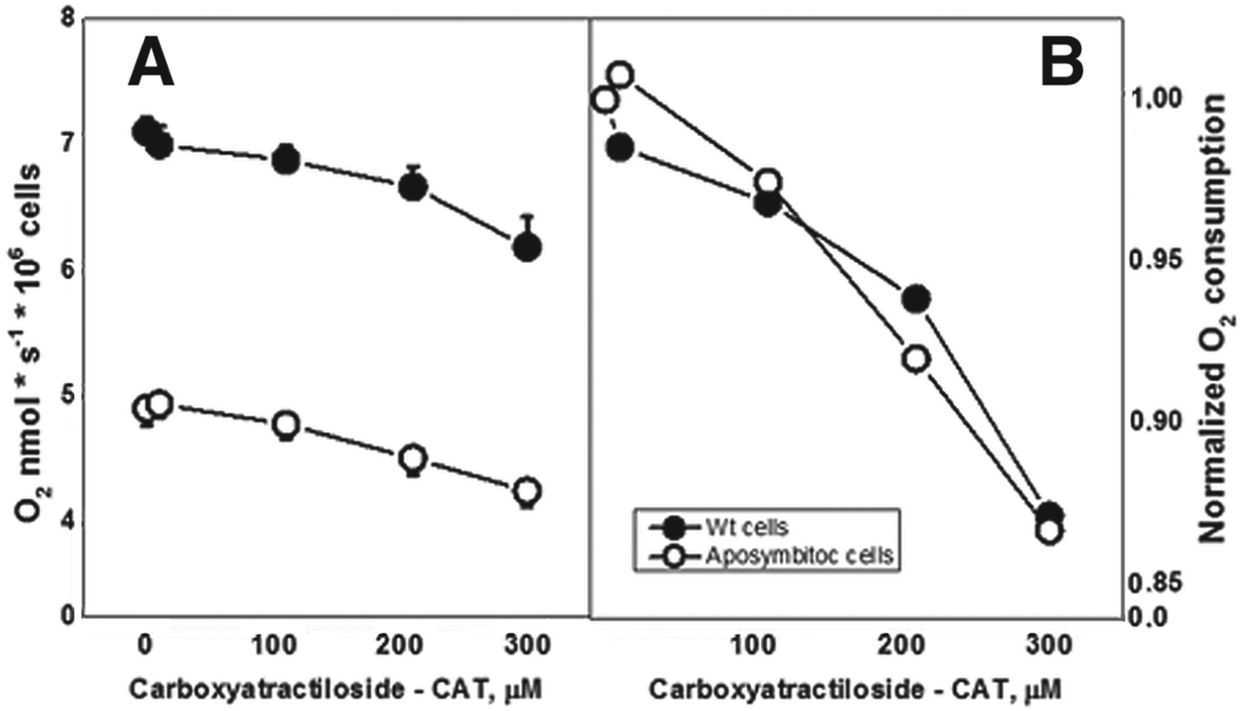
Surprisingly, oligomycin, which inhibits FoF1-ATP synthase, did not reduce oxygen consumption in either strain (P<0·264), even at 5 μg mL−1. In the oligomycin incubation experiments, the Wt cells, which presented 5·4 pmol O2* s−1 (±0·3) before the addition of the compound, still consumed the same rate (5·4 pmol O2* s−1, ±0·2). The aposymbiotic cells presented the same behaviour: 3·4 pmol O2* s−1 before oligomycin (±0·4) and 5·4 pmol O2* s−1 (±0·4) after incubation with oligomycin. However, the use of 300 μ m CAT, an ADP-ATP translocator inhibitor, reduced the oxygen consumption of both cells by 13% (P<0·0001) (Fig. 4).
High-resolution respirometry of Wt and aposymbiotic strains of A. deanei. The titration of CAT is represented in (A) and normalized values of oxygen consumption (0–1) in (B) (n = 3, P<0·0001). CAT affected oxygen consumption in both strains.

Analyses by transmission electron microscopy showed that the different inhibitors used in this study did not affect the ultrastructure of A. deanei (data not shown).
Genomic analysis of sequences that encode the complex subunits of the respiratory chain
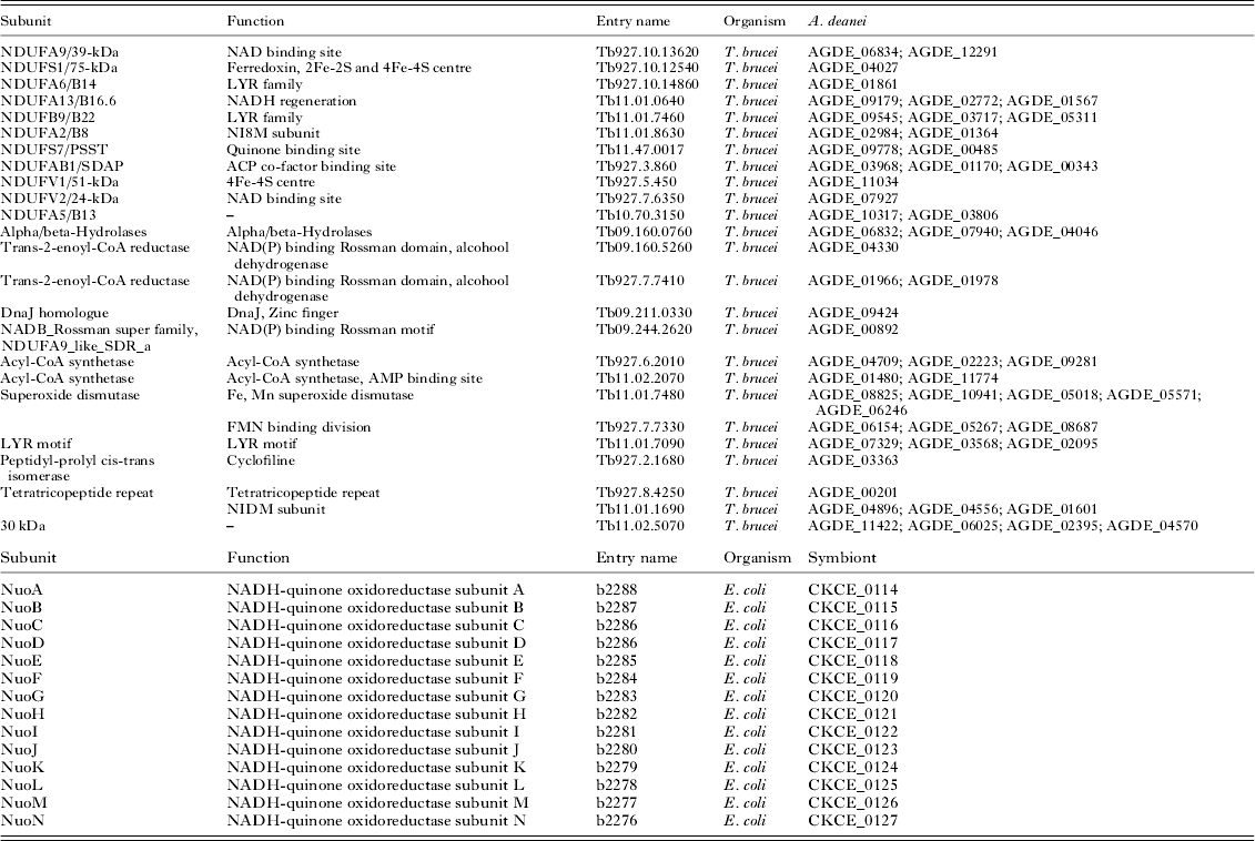
The annotated genome of A. deanei and its symbiont were searched for sequences that encode proteins of the respiratory chain. We considered data that have already been described for trypanosomatids, β-Proteobacteria and other organisms. In relation to complex I, most of the sequences that encode NADH: ubiquinone oxidoreductase were found in A. deanei as functional and structural subunits. Based on T. brucei published data, 33 nucleus-encoded subunits were identified in this symbiont-bearing species, and one sequence has an orthologue in plant cells (Acestor et al. Reference Acestor, Zíková, Dalley, Anupama, Panigrahi and Kenneth2011). Another four subunit sequences were identified in the mitochondrion of A. deanei (data not shown). All NADH: ubiquinone oxidoreductase subunits were found in the endosymbiont (Table 1).
Complex I subunits of A. deanei and its symbiont. Sequences from Tritrypdb.org and NCBI databases are mentioned in the third column. T. brucei and Escherichia coli were used as reference organisms to the host trypanosomatid and symbiont analysis, respectively

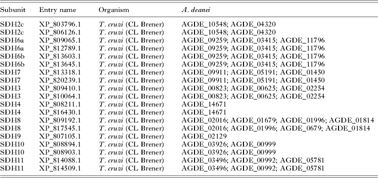
Almost all of the subunits of succinate: fumarate dehydrogenase (SDH) described in T. cruzi were identified in A. deanei; however, the genome lacks SDH1, SDH5 and SDH2n (Morales et al. Reference Morales, Mogi, Mineki, Takashima, Mineki, Hirawake, Sakamoto, Ōmura and Kita2009) (Table 2). With the exception of SDH4 and SDH9, which are present as a single copy, at least two copies of all other subunits are seemingly present. Similarly, only two subunits that compose cytochrome bc oxidase were not present in A. deanei: alfa-MMP and beta-MMP (Table 3). Neither complex was found in the genome of the symbiont when gram-negative bacteria (E. coli and Acidithiobacillus ferrooxidans) were used as reference microorganisms.
Complex II subunits of A. deanei. Sequences from NCBI database are mentioned in the second column. T. cruzi was used as reference organism in these analyses. Sequences related to the complex II were not found in the symbiont genome

Complex III subunits of A. deanei. Sequences from Tritrypdb.org and NCBI databases are mentioned in the second column. T. brucei was used as reference organism in these analyses. Sequences related to the complex III were not found in the symbiont genome

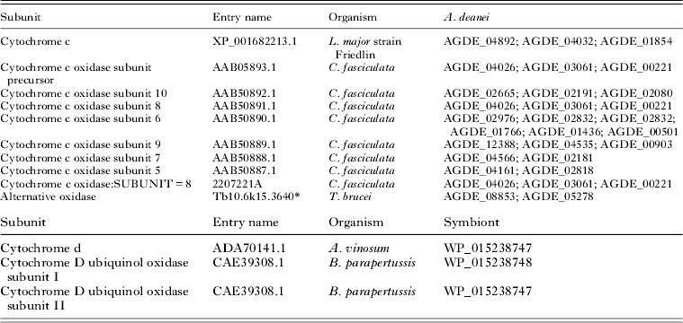
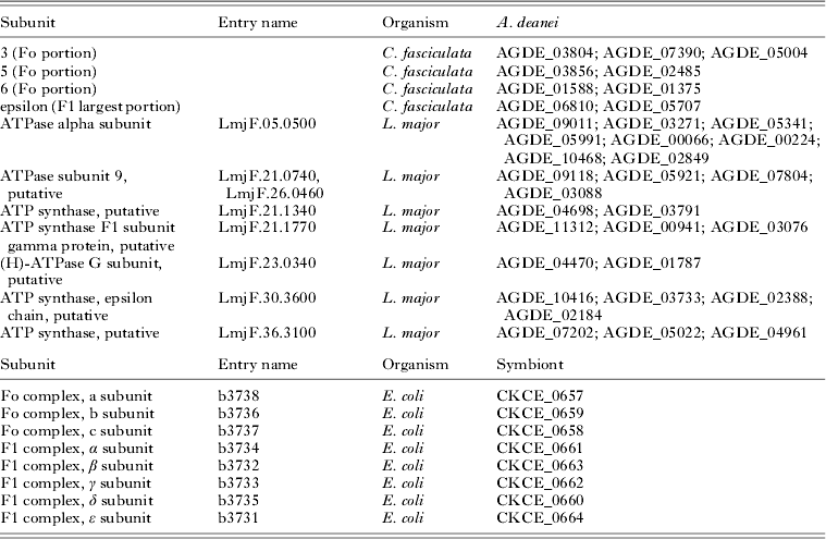
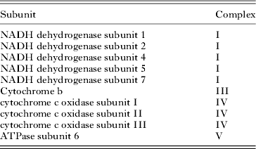
Many subunits of COX that have been reported for C. fasciculata in GenBank (Speijer et al. Reference Speijer, Breek, Muijsers, Groenevelt, Dekker, De Haan and Benne1996) were also found in A. deanei (Table 4). Regarding the A. deanei bacterium, a COX from B. parapertussis was absent, but a cytochrome d oxidase from the same prokaryote was found in the endosymbiont. Interestingly, the trypanosomatid AOX (Tb10.6k15.3640) that has been identified in T. brucei (Tritrypdb.org) was also found in A. deanei. The FoF1-ATP synthase subunits were found in the host genome, as well as in the symbiotic bacterium (Table 5). Moreover, the mitochondrial genome (kDNA) presented 10 sequences similar to complexes I (NADH dehydrogenase subunits 1, 2, 4, 5 and 7), III (cytochrome b), IV (subunits I, II and III), and V (ATPase subunit 6) (Table 6). The whole kDNA genome sequence is identified as KJ778684 at NCBI.
Cytochromes (c and d) and complex IV subunits of A. deanei and its symbiont. Sequences from NCBI database are mentioned in the second column. The cytochrome c of L. major, the COX subunits of C. fasciculata and the AOX of the T. brucei (* = Tritrypdb.org) were used as references in the host genome analyses. The symbiont did not present cytochrome c, neither the cytochrome c oxidase. However, sequences codifying a cytochrome d (A. vinosum) and cytochrome d oxidase subunits (B. parapertussis) were found in the symbiont genome

Complex V subunits of A. deanei and its symbiont. Sequences from TriTrypDB and NCBI databases are mentioned in the second column. Sequences codifying complex V subunits in C. fasciculata and L. major were used as reference in host genome searches. E. coli was used as a reference in the symbiont genome analyses

Mitochondrial sequences in kDNA of A. deanei. The reference sequences are from TriTrypDB database. The second column indicates the complex of the sequence

DISCUSSION
An intensive metabolic exchange occurs between the host trypanosomatid and its symbiotic bacterium. The symbiont provides essential nutrients and metabolic precursors that complete biosynthetic pathways in the host cell, and the protozoan supplies the symbiont with energy and growth factors (Motta, Reference Motta2010). For example, the symbiont contains ornithine carbamoyltransferase, which converts ornithine to citrulline, thus completing the urea cycle of the host (Camargo and Freymüller, Reference Camargo and Freymüller1977; Galinari and Camargo, Reference Galinari and Camargo1978). The urea cycle is involved in amino acid synthesis and also produces intermediate metabolites related to the biosynthesis of pyrimidines, proline and polyamines, which are essential factors in cell growth and differentiation (Herby and Persson, Reference Herby and Persson1990; Alves et al. Reference Alves, Serrano, Maia da Silva, Voegtly, Matveyev, Teixeira, Camargo and Buck2013b ).
The cellular localization of the endosymbiont in A. deanei is also evidence of metabolic exchanges. Surrounded by glycosomes, the endosymbiont presents a surface ATPase, which may indicate that ATP provided by the host is hydrolysed, supporting an active transport mechanism in the bacterial envelope (Motta et al. Reference Motta, Soares, Attias, Morgado, Lemos, Saad-Nehme, Meyer-Fernandes and De Souza1997b ). Such a hypothesis was demonstrated in pathogenic intracellular prokaryotes, such as those belonging to the Rickettsia and Chlamydia genera (Winkler, Reference Winkler1976; Hatch et al. Reference Hatch, Al-Hossainy and Silverman1982). In addition, the symbiont and the host endoplasmic reticulum, where most phospholipid biosynthesis occurs, are closely associated. Inhibitors of phosphatidylcholine biosynthetic pathways affect the symbiont's phospholipid composition (Bernard et al. Reference Bernard, Faustin, Passerieux, Galinier, Rocher, Bellance, Delage, Casteilla, Letellier and Rossignol2006; Palmié-Peixoto et al. Reference Palmié-Peixoto, Rocha, Urbina, De Souza, Einicker-Lamas and Motta2006), and isolated symbionts demonstrate a marked reduction in phosphatidylcholine production, thereby indicating that the bacterium can obtain a portion of this phospholipid from the host cell. In accordance with this idea, the symbiont enhances the glycerophospholipid metabolism of the host protozoan (Azevedo-Martins et al. Reference Azevedo-Martins, Frossard, de Souza, Einicker-Lamas and Motta2007).
Given the accelerated metabolism of the Wt strain and the capacity of isolated symbionts to metabolize, we can suggest that symbiont-containing cells present an increased demand for energy. This point may justify in part the high rate of oxygen consumption of such protozoa in comparison to aposymbiotic cells, as demonstrated experimentally in this work. The increase in oxygen consumption may also be related to an enhanced production of metabolic intermediates by the symbiont. For example, the augmented amino acid production in the host's urea cycle may directly furnish substrates to the Krebs cycle, thereby increasing electron donation to the mitochondrion and its ETS capacity (maximal uncoupled respiration). It is also worth considering that the isolated bacteria are able to respire, thus presenting their own operative and functional ETS. This indicates that the total amount of oxygen consumption in trypanosomatids is increased due to the presence of the symbiont.
The complex I, NADH: ubiquinone oxidoreductase from A. deanei is rotenone non-sensitive. Although many subunits of this complex were identified in the A. deanei genome, the lack of an effect of this inhibitor has been reported in the literature for other protozoa of the Trypanosomatidae family, such as T. brucei (Opperdoes and Michels, Reference Opperdoes and Michels2008). Regarding complex II, many of the 12 subunits identified in T. brucei (Morales et al. Reference Morales, Mogi, Mineki, Takashima, Mineki, Hirawake, Sakamoto, Ōmura and Kita2009) were also found in the A. deanei genome, and the use of TTFA inhibited oxygen consumption in this species. However, this complex appears to be absent in the genome of the endosymbiont. The data presented in Fig. 2 are puzzling. Indeed, the total respiration of the aposymbiotic strain is decreased by the absence of the symbiont, though an increased amount of the TTFA inhibitor is needed to cause 50% inhibition in this strain. This may indicate that a compensatory mechanism for the expression of complex II may be operating and that the presence of the symbiont may suppress this expression. Regardless, it is worth considering that the regulation of ROS production might be directly influencing this complex and that the use of TTFA is known to increase ROS. Further studies are needed to test these hypotheses.
Cytochrome bc oxidase (complex III) genes are absent in the symbiont genome but are present in the host trypanosomatid genome. The genomic data are in accordance with the high-resolution respirometry findings for the specific inhibitor of complex III antimycin A, which was able to inhibit oxygen consumption by A. deanei. However, the amount of antimycin A needed to cause 50% inhibition in the aposymbiotic strain was lower (up to 0·8 μ m) in comparison to the Wt strain. The lack of inhibition observed at higher antimycin A concentrations in the aposymbiotic strain compared with the Wt strain may indicate a compensatory mechanism for the expression of the subunits of complex III. Alternatively, an imbalance in the steady-state levels of ROS caused by the absence of the symbiont could modulate the signalling of redox pathway factors, thereby affecting the expression of other oxidases not sensitive to antimycin A or involved in oxidative phosphorylation. Further studies are needed to clarify these alterations in the bioenergetics in A. deanei.
In contrast, not all subunits that compose mitochondrial complex IV were identified in the A. deanei genome, but the use of KCN completely abolished the protozoan's oxygen consumption. Interestingly, a trypanosomatid AOX was also found in the A. deanei genome, similar to what was previously described for T. brucei (Aslett et al. Reference Aslett, Aurrecoechea, Berriman, Brestelli, Brunk, Carrington, Depledge, Fisher, Gajria, Gao, Gardner, Gingle, Grant, Harb, Heiges, Hertz-Fowler, Houston, Innamorato, Iodice, Kissinger, Kraemer, Li, Logan, Miller, Mitra, Myler, Nayak, Pennington, Phan, Pinney, Ramasamy, Rogers, Roos, Ross, Sivam, Smith, Srinivasamoorthy, Stoeckert, Subramanian, Thibodeau, Tivey, Tratman, Velarde and Wang2010). However, the strong inhibition observed in this work with KCN (at concentrations above 100 μ m, almost 95% of total respiration was arrested) indicates that this AOX is inactive or not expressed in A. deanei. Furthermore, in the high-resolution respirometry of A. deanei during KCN titration, the symbiont-containing and symbiont-free cells presented different sensitivities to this inhibitor, thereby indicating a complex interaction between cyanide and COX sites.
This fact may be related to the amount of COX involved in different cellular states of respiration, which may vary in accordance to the energy requirements. In other words, not all COX units are involved in respiration, and the excess units are considered to be spare COX units that are used to reach maximum respiration (Bernard et al. Reference Bernard, Faustin, Passerieux, Galinier, Rocher, Bellance, Delage, Casteilla, Letellier and Rossignol2006). Thus, titration with KCN in the Wt strain may affect these reservoirs of COX, which may show different KCN affinities. However, the aposymbiotic strain did not show all the slopes detected in the Wt strain (as marked by lines a and b in Fig. 2H). A possible explanation could be that the amount and interactions among COX units were decreased by the absence of symbiotic COX units. In fact, in parallel experiments using isolated symbionts, we were able to detect KCN inhibition of respiration with a single component and with a low affinity for KCN (Fig. 3).
Several subunits of complex V, composed by FoF1-ATP synthase, were found in the genomes of A. deanei and its symbiont. However, oligomycin, a specific inhibitor of this complex, did not affect oxygen consumption by the protozoan. This can be explained by the absence of some Fo subunits in the host that are the target of oligomycin. Conversely, CAT, an ADP-ATP translocator inhibitor, slightly affected the oxygen consumption in both strains. Thus, the data indicate that the A. deanei mitochondrion presents ETS-dependent oxygen consumption that is uncoupled from ATP production.
CONCLUSIONS/FUTURE DIRECTIONS
In this work, we conclude that the presence of the symbiont enhances the trypanosomatid's oxygen consumption. The symbiont-bearing and the symbiont-free strains of A. deanei showed differences in their energetic metabolism and distinct sensitivities when treated with inhibitors that specifically affect each mitochondrial complex of the electron transport chain. The symbiont-containing protozoa appear to be more resistant to these compounds than the aposymbiotic cells. This may be related to the fact that essential biosynthetic routes are completed by the bacterium in the symbiont-bearing trypanosomatids, improving the metabolism of the host. It is also worth considering that differences in O2 consumption may be related to compensatory mutations that may have arisen in the aposymbiotic strain to optimize its respiration in the absence of the symbiont. According to this idea, a single amino acid change in the nucleus-encoded F1 subunit of ATP synthase is able to compensate for energy-demanding activity in dyskinetoplastic cells of T. brucei that lacks the Fo portion, which is essential for electrochemical potential maintenance (Dean et al. Reference Dean, Gould, Dewar and Schnaufer2013).
Another important aspect that should be considered is that the symbiont demands energy molecules from the host cell to maintain optimal physiology. This is most likely related to an enhanced respiration capacity in symbiont-bearing trypanosomatids when compared with aposymbiotic cells and a ROS regulatory activity of the host mitochondria. In accordance with this idea, our genome database searches showed that the symbiont genome lacks important components of the respiratory chain. Further studies are necessary to explain how the absence of complex II in the bacterium can affect the exchange of intermediates between both partners in this symbiotic relationship.
ACKNOWLEDGEMENTS
Special thanks to Dr Ronir Raggio Luiz from Instituto de Estudos em Saúde Coletiva (IESC) for the statistical analyses.
FINANCIAL SUPPORT
This work was supported by Coordenação de Aperfeiçoamento de Pessoal de Nível Superior (CAPES), Fundação Carlos Chagas Filho de Amparo à Pesquisa do Estado do Rio de Janeiro (FAPERJ), Centro Nacional de Desenvolvimento Científico e Tecnológico (CNPq) and Instituto Nacional de Ciência e Tecnologia em Excitotoxicidade e Neuroproteção (INCTEN). This work was also supported by European Research Council under the European Community's Seventh Framework Programme (FP7/2007-2013)/ERC grant agreement no. (247073)10.