Introduction
Understanding how cells respond to their surroundings and external cues is an important factor in the successful design of novel devices and materials for tissue regeneration and engineering. Biologic, physical, and chemical cues have been studied and known to influence neuronal response.[Reference Dvir, Timko, Kohane and Langer1, Reference Bettinger, Langer and Borenstein2] It is a well-established fact that the natural cellular environment in vivo consists of 3-D micro and nano environment and structures. Most in vitro studies, however, are performed on planar cell culture surfaces which are not reflective of the natural environment and therefore, not helpful in the design and development of future neural prosthetics and conduits.
It is also well-established that the presence of electric fields and electrical stimulation, in general, impact the adhesion, proliferation, and regeneration rate of neurons[Reference Borgens, Blight and McGinnis3, Reference Francis, Gluckman and Schiff4, Reference Yao, Shanley, McCaig and Zhao5, Reference McCaig and Rajnicek6, Reference Patel and Poo7] which is to be expected since neural cells are, in fact, electro-active. The electrical conductivity of the material plays a vital role in improving the efficacy and long-term function of existing neural conduits and prostheses. Adding and incorporating electrically conducting components into/onto biologically compatible substrates and scaffolds have been under investigation for decades. Substrates and scaffolds that are not inherently conducting are usually treated and coated with electrically conducting polymers such as polypyrrole and polyaniline to allow for electrical stimulation and interfacing. However, limited thermal and electrical stability, as well as low mechanical strength, has limited the usage of conductive polymers. Furthermore, in most cases, increasing the electrical conductivity to reach the desired level requires increasing the dopant level, which can lower the mechanical strength of these polymers even further.[Reference Kaur, Adhikari, Cass, Bown and Gunatillake8] Carbon-based materials such as single-walled and multi-walled carbon nanotubes have also received a lot of attention as biosensors, scaffolds for tissue engineering, and for the recording of neural signals.[Reference Beg, Rizwan, Sheikh, Hasnain, Anwer and Kohli9–Reference Cellot, Cilia, Cipollone, Rancic, Sucapane, Giordani and Gelain12] Their superior electrical conductivity combined with their mechanical strength has lead to a significant amount of research in these materials. Carbon nanotubes, however, have been known to demonstrate toxicity effects[Reference Khaliullin, Fatkhutdinova, Zalyalov, Kisin, Murray and Shvedova13] and are not ideal for in vivo applications due to concerns regarding toxicity.
A class of porous light-weight materials with a nanometer-scale surface roughness that has been investigated for biomedical applications and more specifically as a neural prosthetic, is the aerogel. A series of in vivo and in vitro studies have demonstrated the biostability, biocompatibility, and suitability of silica-based aerogels as a neuronal scaffold.[Reference Lynch, Skalli and Sabri14–Reference Sabri, Boughter, Gerth, Skalli, Phung, Tamula and Leventis19] More importantly, neurons grown on aerogel substrates showed an increased growth rate when compared with those grown on tissue culture petri dish (control).[Reference Lynch, Skalli and Sabri15] While the exact mechanism of cell–substrate interaction is not yet clearly understood, it does highlight the cells preference for the 3-D topography of the aerogel substrate. Carbon aerogels (CAs) combine the unique 3-D aerogel topography with electrical conductivity, thermal and chemical robustness, and good mechanical properties, not observed in other aerogel materials.[Reference Pekala20, Reference Lu, Nilsson, Fricke and Pekala21] These properties are a direct result of covalently assembling sp2 carbon nanoparticles into an aerogel framework. Consequently, CAs have shown promise in a wide variety of fields including energy storage, catalysis, filtration, chemical sensors, energy generation, sorbents, and electronics.[Reference Li, Zhang, Song, Su, Zhang, Kong, Liu, Jin, Tang and Cheng22] The inherent electrical conductivity of CAs combined with the 3-D surface, the structure makes them an attractive candidate for biomedical applications and its suitability has been investigated for the first time in this study. Unlike other aerogels, CAs can be 3-D printed which would make this type of aerogel highly suitable for bioprinting also. Here, the authors report for the first time an investigation of the behavior of PC-12 cells, a neural analog,[Reference Li, Zhang, Song, Su, Zhang, Kong, Liu, Jin, Tang and Cheng22] on CAs. Acid-catalyzed resorcinol-formaldehyde (ARF) and base-catalyzed resorcinol-formaldehyde (BRF) aerogels were chosen for this study. Results show that while both aerogel types are conducive to neuron adhesion, proliferation, and the development of neurite extensions, PC-12 cells extend significantly longer neurites on BRF aerogels when compared with control, and ARF aerogels.
Materials and methods
Aerogel substrate preparation and synthesis
Preparation of aerogel scaffold
In this study, we were interested in exploring two different pore diameter ranges: >1 µm and <1 µm. To achieve this, two types of aerogels were chosen for this study: (1) ARF and (2) BRF. The conventional ARF aerogel monoliths are synthesized by mixing resorcinol (1.23 g, 0.0112 mol) and 37% formaldehyde solution (1.7 g, 0.0209 mol) in water (1.5 mL), followed by the addition of glacial acetic acid (0.44 g, 0.0007 mol), which was further cured at 80 °C for 72 h. This formulation yields an RF mass ratio of 42% for ARF. For the preparation of BRF aerogel monoliths, 0.625 g of resorcinol (0.005676 mol), and 0.9 g of 37% formaldehyde solution (0.011 mol) in 7.1 g of water and 3 mg (0.000028 mol) of sodium carbonate. This combination of R/F molar ratio of 1:2 and R/C molar ratio of 200, yielded a RF mass ratio of 11wt%. Once gelled, the aqueous solvent (water in this case) was removed by soaking the sample in an acetone bath for 3 days and the solvent was exchanged every 24 h. The samples were then supercritically dried in liquid CO2 at a critical temperature of 55 °C and at a pressure range of 1200–1400 psi.
Carbonization of RF aerogel
CAs were prepared by carbonizing the supercritically dried RF organic aerogels. The aerogels were subjected to a heat treatment process where the samples were heated in a tube furnace under a nitrogen atmosphere at 1050 °C for 3 h with a heating and cooling rate of 2 °C/min. Upon carbonization, the BRF and ARF aerogels turned black and became conductive (at least 100 S/m). Both BRF and ARF aerogels possessed a 3-D architecture resulting in a surface roughness unique to each type of aerogel. BRF and ARF aerogels samples were sterilized as previously described[Reference Borgens, Blight and McGinnis3, Reference Francis, Gluckman and Schiff4] through immersion in 100% ethanol followed by 1 h exposure to 254 nm UV light. Samples were inspected by means of optical and scanning electron microscopy to ensure that the sterilization step did not alter the structure of the ARF and BRF aerogels.
Collagen coating
Aerogels and tissue culture polystyrene (TCPS) substrates previously sterilized were coated with collagen. The thickness of the collagen layer was minimized so as not to mask substrate properties (e.g., stiffness, pore architecture, pore size, etc.).[Reference Lynch, Skalli and Sabri15] These parameters were previously established and the same coating technique and concentration was used in this study.[Reference Lynch, Skalli and Sabri15] Rat tail type I collagen (Invitrogen) was diluted to a final concentration of 0.05 mg/mL in 20 mM acetic acid.[Reference Lynch, Skalli and Sabri14, Reference Lynch, Skalli and Sabri15] The collagen solution was then laid on the substrate to reach a protein density of 4 µg/cm2 and was allowed to sit for 1 h at room temperature in a sterile cabinet. Next, the excess collagen solution was removed with a vacuum line, and the samples were rinsed twice with serum-free RPMI 1640 prior to the cell culture step.
PC-12 cell culture
PC-12 pheochromocytoma cells obtained from the American Type Culture Collection were grown in RPMI 1640 medium supplemented with GlutaMax, HEPES buffer, 10% horse serum, 5% fetal bovine serum, and penicillin/streptomycin. Before each experiment, PC-12 cells plated on a collagen 1 coated TCPS petri-dish cells were “primed” for neural differentiation with 50 ng/mL NGF for 8 days in low serum medium (RPMI 1640 supplemented with GlutaMax, HEPES buffer, 1% horse serum, and penicillin/streptomycin). This medium was replaced every 2–3 days with fresh low serum medium with NGF. After 8 days, the cells were harvested from the dish and frozen in liquid nitrogen until used for plating on aerogel substrates. Details of the method have been described previously.[Reference Tiwari, Vij, Kemp and Kim10] Both 3-day (3-d) and 1-day (1-d) cultures were prepared for BRF and TCPS (control) substrates and only 1-d cultures were prepared for ARF aerogels. The 1-d cell culture was performed first; substrates were fixed and analyzed before the 3-d culture was attempted. Three separate trials (n = 3) were prepared for each experiment and a control was prepared each time and run in parallel. Results were averaged over three separate trials for each set of conditions. An MTT assay was not performed due to concerns with the toxicity of the reagents themselves,[Reference Riss23] instead, all cultures were carefully inspected periodically using a phase contrast microscope for any signs of cell death or stress.
Measurements of neurite
Neurite lengths, cell body area, and branching were measured using NIH Image J software. Neurite selection was performed as described in detail in previous published work by the authors.[Reference Lynch, Skalli and Sabri14, Reference Lynch, Skalli and Sabri15] The overlaid scale bar from scanning electron microscopy (SEM) images was used to calibrate the measurement tools for length measurement. Neurites were treated as vectors and as such were measured with a series of straight line segments using the line segment tool. Neurites were defined as the longest cytoplasmic extensions originating from the cell body and short branches originating from these extensions were not considered for measurement.
Scanning electron microscopy
Samples were fixed in 2.5% glutaraldehyde (Tousimis, Rockville, MD) in 0.1 M sodium cacodylate buffer for 2 h followed by 2 × 10 min washes with 0.1 M sodium cacodylate buffer. Samples were then immersed in an aqueous solution of 1% osmium tetroxide (Electron Microscopy Sciences, Hatfield, Pa) for 1 h followed by 2 × 10 min washes in sodium cacodylate buffer and then dehydrated by a series of 10 min each washes in 10%, 30%, 50%, 70%, 90%, and 100% ethanol. Finally, all substrates were allowed to fully air dry before sputter deposition of 10 nm gold/palladium film (when appropriate) and imaging either with Nova NanoSEM 650 Field Emission Scanning Electron Microscope (FEI Co.) or a Phenom desktop SEM. The FEI Nano NanoSEM 650 ESEM (FEI, Hillsboro, OR, USA) scanning electron microscope was equipped with a through lens detector in secondary electron (SE) operation and was used for elemental analysis. The imaging was carried out in immersion mode at a primary electron energy of 15 keV. The working distance was adjusted to 5.0 mm.
Statistical analysis
Statistical analysis was performed using a two-tailed student's t-test. Significance (*, **) is defined as P < 0.05, and P < 0.005, respectively. In all cases, error bars represent standard error of mean. Neurite length, cell area, number of neurites per cell and number of branches per neurite were calculated for each substrate for three separate trials. As mentioned previously, Image J software was utilized in this study and the entire substrate area was evaluated in each case. Measurements were averaged and the standard deviation was calculated.
Results and discussion
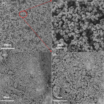
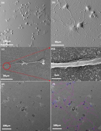
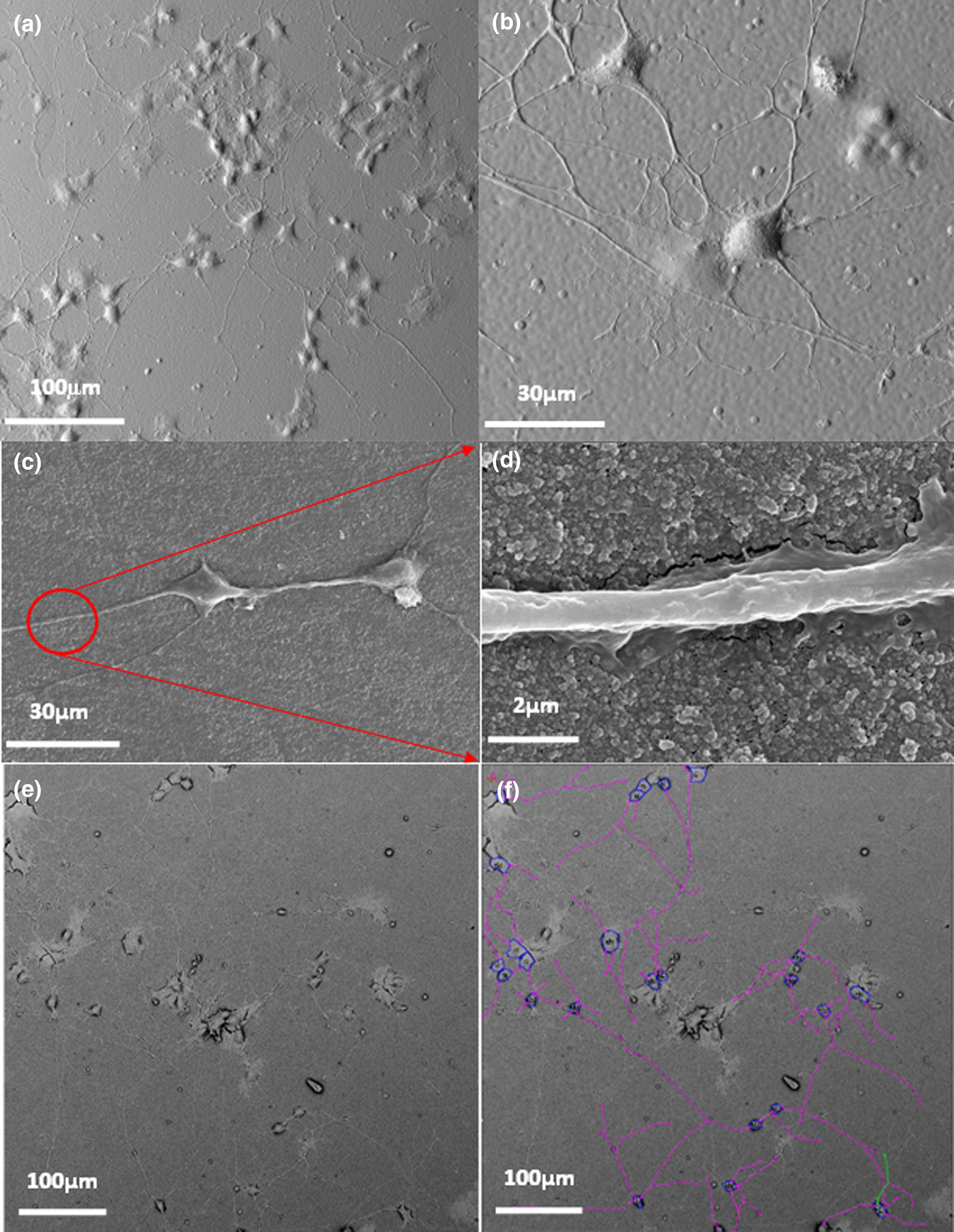
Pristine ARF and BRF aerogels did not support adhesion and proliferation of PC-12 cells. A layer of collagen was necessary for the attachment of cells to both types of substrates. While cell adhesion on pristine aerogels did not take place, cells had a healthy appearance and did not suggest any toxicity effects from the aerogel material. Figure 1 shows SEM images of the ARF [Figs. 1(a) and 1(b)] and BRF [Figs. 1(c) and 1(d)] aerogels at different magnifications prior to collagen coating. The larger pore structure of ARF aerogels can be clearly seen in the images while BRF aerogels show a pore structure below 1 µm size. Representative images of 3-d and 1-d cell culture on aerogels are shown in Fig. 2. After 3-d in culture, PC-12 cells on BRF aerogels [Figs. 2(a) and 2(b)] show a significant amount of proliferation, neurite outgrowth, and connectivity indicating that collagen-coated BRF aerogels are suitable substrates for neuronal scaffold applications. Images from the 1-d culture preparation [Fig. 2(c) and 2(d)] show a reduced number of branching and connectivity, as expected. A close up of the surface [Fig. 2(d)] shows the 3-D nature of the collagen-coated aerogel surface. Another SEM image of PC-12 cells after 3-d in culture on BRF aerogels is shown in Fig. 2e and some of the corresponding Image J tracers used to measure neurite lengths are shown in Fig. 2d.
SEM images of native carbon aerogels: (a), (b) The microscopic structure of ARF aerogel in the absence of any coatings shows a unique porous architecture of the substrate, prior to cell culture stage and fixing. The pore structure shows pores with diameters >1 µm. (c), (d) SEM images of BRF aerogels showing a much smaller pore size range for these aerogels compared with ARF, typically <1 µm.

Adhesion and proliferation of PC-12 cells on carbon aerogels: (a), (b) SEM images showing neurite extensions and connectivity after 3 days (3-d) in culture. (c) The number of neurites is significantly reduced when cells remain in culture only for 1 day (1-d). The 1-d culture was initially performed to evaluate cell–substrate interaction and possible toxicity effects. The 3-D texture of the collagen coating can be seen in (d). (e) SEM image of 3-d culture of cells on BRF aerogel and the corresponding Image J measurement markers are shown in (f).
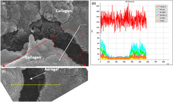
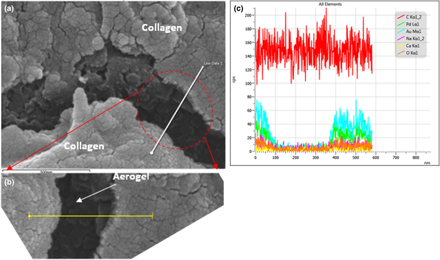
In some instances, upon inspection of the substrates with SEM, fissures, most likely due to the fixing protocol, were observed in the collagen layer and the aerogel substrate underneath was exposed. This was confirmed by using the SEM in SE mode and performing elemental analysis in regions were cracks in the collagen film had exposed the underlying aerogel. Figure 3 shows one such example where analysis was performed across a break in the collagen layer [Fig. 3(a)] identified by the straight white line. The corresponding elemental analysis graph is shown in Fig. 3b identifying the exposed region as the region with little to no coverage of Au or Pd. A higher magnification image of this region shows the 3-D structure of the underlying aerogel substrate [Fig. 3(c)]. Regions such as this one were used to estimate the thickness of the collagen layer in all cases.
Collagen Distribution on Aerogels: (a) A SEM image of a break in the collagen layer used to estimate the collagen thickness. (b) Magnified region of (a), arrow indicating the aerogel structure underneath. (c) SE mode of SEM showing the carbon aerogel beneath the collage film.
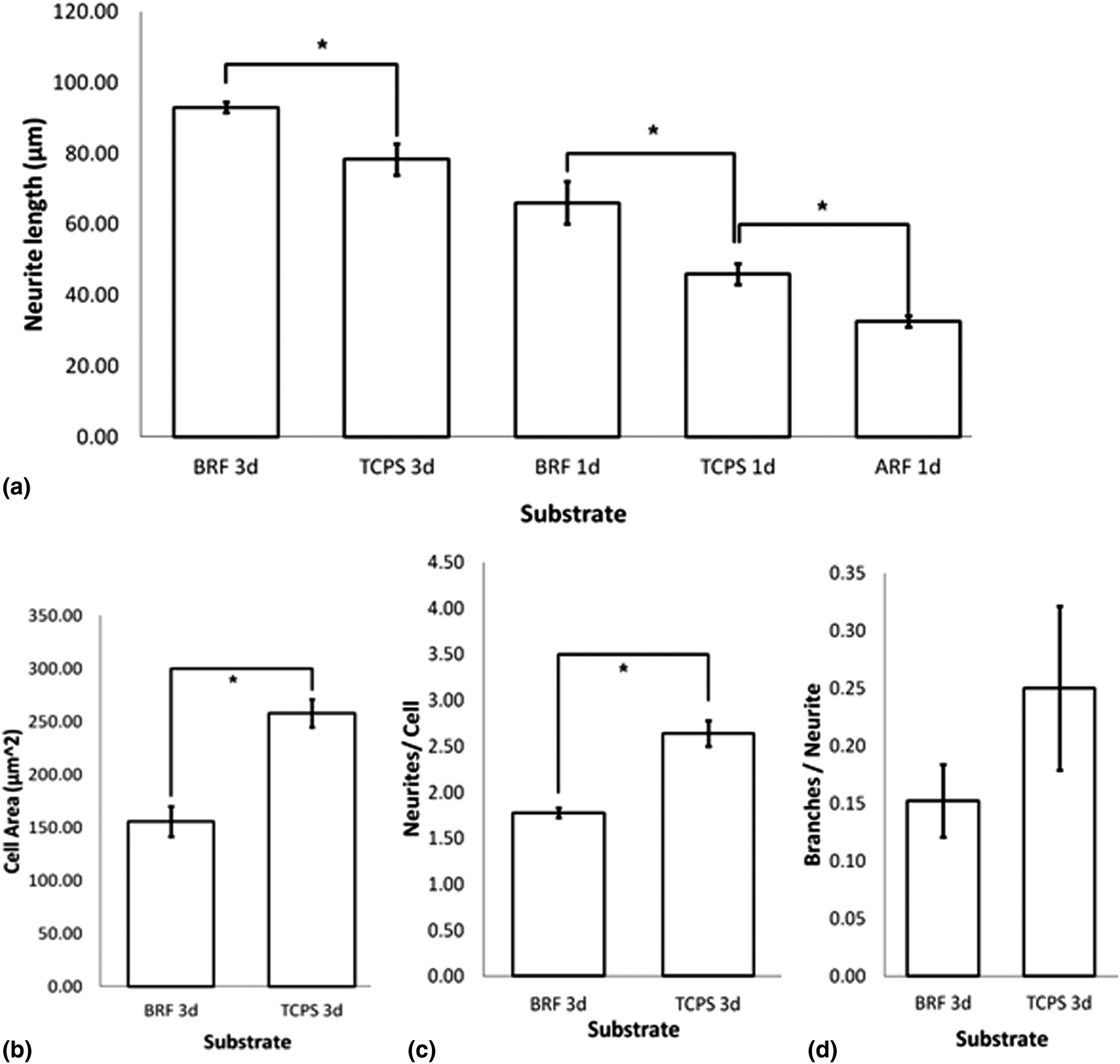
A key parameter used to characterize and understand the influence of substrate property (ies) on neuron behavior is the average neurite length. In the case of the 1-d culture period, PC-12 cells grown on BRF aerogels extended neurites approximately 20% longer than those measured on TCPS and, 45% longer than those extended on ARF substrates [Fig. 4(a)]. After analyzing the 1-d results, it was decided to only prepare 3-d cultures for the BRF aerogels and the TCPS (control). The trend observed for the 1-d culture repeated itself in the case of the 3-d cultures. PC-12 cells grown on BRF aerogels hand longer neurites when compared with the control, TCPS. Further inspection of the behavior of PC-12 cells on BRF aerogels showed that the cell area, on average, was 45% greater on TCPS, than on BRF aerogels [Fig. 4(b)] indicating a significant amount of cell spreading on TCPS than on BRF aerogels. Similarly, the number of neurites per cell [Fig. 4(c)], and, the number of branches extended [Fig. 4(d)] were greater on TCPS than on BRF aerogels. These results are consistent with previously published work on silica aerogels.[Reference Lynch, Skalli and Sabri15]
Cell responses to Carbon Aerogel Substrate: (a) After 1 day (1-d) cell culture all cells were viable on both ARF and BRF aerogels, however, cells on ARF aerogels showed the slowest growth rate while cells on BRF aerogels had the longest neurites among all the substrates. (b) Cell spreading was significantly less on BRF aerogels compared with control. The number on neurites/cell and, (c) (d) the number of branches extended on BRF aerogels was less than the control TCPS.

Discussion
It has been increasingly recognized that topographic features of the nanoscale order have a profound influence on cell functionalities[Reference Allison, Baker, Lynch and Sabri16, Reference Allison, Baker, Lynch and Sabri17] and by carefully manipulating these features, it is possible to elicit the type of response that can accelerate healing and recovery. Typically, two types of external stimuli are used to gain control of the response of neurons on engineered substrates: physical cues and externally applied bias (electric or magnetic). The electrical conductivity of CAs makes these types of aerogels particularly attractive for the application discussed here. Previous work on silica aerogels demonstrated that neurons extend longer neurites on silica aerogels, and in the presence of externally applied DC bias cells show a preferential orientation compared with 0 V applied bias.[Reference Lynch, Skalli and Sabri14, Reference Lynch, Skalli and Sabri15] One drawback of working with silica aerogels is the non-conducting nature of the material. Cells (to various degrees) respond to applied electric fields. In the case of CAs, due to the electrically conducting nature of the material, it is possible to direct a current in the substrate and will be the focus of future studies.
PC-12 cells are anchorage-dependent cells[Reference Ishii, Ishimori, Mori, Uto, Fukuda, Urashima and Nishimura24] and given that they proliferate and differentiate only after successful initial anchoring, it can be concluded that both ARF and BRF aerogels provide the necessary anchoring sites needed for the initial adhesion. Although both types of substrates supported neurite outgrowth (after an initial coating of a thin collagen film), our results show significant differences between the neuronal responses for BRF compared with ARF aerogels. The response of PC-12 cells to ARF aerogels was significantly different than their response to BRF aerogels. Given the identical composition of the CAs derived from ARF and BRF aerogels, properties such as surface contact angle, electrical conductivity, and stiffness (all very close between the two aerogels) are not expected to be the cause for the differences in the response of PC-12 cells. The pore size and pore geometry, however, vary significantly between the two aerogel types, leading to differences in the apparent surface roughness that the cells are exposed to. We believe that the aerogel surface appears purely isotropic to the cells and are identified by one characteristic parameter only, the surface roughness. The surface roughness is also believed to be the cause of the differences in cell spreading (quantified by measuring cell area) when compared with TCPS. PC-12 cells extending longer neurites on patterned and textured substrates has been observed in previous studies[Reference Lynch, Skalli and Sabri14, Reference Yang, Co and Ho25–Reference Haq, Anandan, Keith and Zhang27] however, the majority of previous studies[Reference Yang, Co and Ho25–Reference Haq, Anandan, Keith and Zhang27] have studied surfaces with periodic and well-defined grooves, pillars and dot arrays and results cannot be compared directly with the study presented here. Aerogels have an intrinsically unique surface pattern that is 3-D at the nanometer scale and does not resemble the surface of other materials used in tissue scaffolding studies. It is reasonable to assume that not only the surface roughness, but also the substrate stiffness affects the cell spreading, a number of neurites/cell, and branching that takes place. The exact correlation between these parameters is not fully understood and is currently under investigation. Future studies will attempt to develop a better understanding of the role that these parameters play and the design of electrically active substrates.
Acknowledgment
F. Sabri would like to thank FedEx Institute of Technology for partial financial support. F. Sabri would also like to thank the Integrated Microscopy Centre (IMC) at the University of Memphis for assistance with microscopy. This work was performed under the auspices of the US Department of Energy by Lawrence Livermore National Laboratory under Contract DE-AC52-07NA27344. M. Worsley and S. Chandrasekaran would like to acknowledge LDRD award 16-ERD-051 and 13-LW-099 for funding support.