1. Introduction
The earliest known representatives of the main Palaeozoic coral orders Tabulata and Rugosa are from the Lower and Middle Ordovician, respectively (Webby et al. Reference Webby, Elias, Young, Neuman, Kaljo, Webby, Paris, Droser and Percival2004b ). The origin of the Rugosa has been widely discussed but is not well understood. It is now widely accepted that the Rugosa is a monophyletic taxon that had its origins in a non-skeletal anthozoan (Scrutton, Reference Scrutton and House1979a , Reference Scrutton1997; Neuman, Reference Neuman, Oliver, Sando, Coates, Macintyre, Bayer and Sorauf1984; Webby et al. Reference Webby, Cooper, Bergström, Paris, Webby, Paris, Droser and Percival2004b ), with the first representatives resembling Primitophyllum primum Kaljo, Reference Kaljo, Kiparisova, Markovskii and Radchenko1956a . Primary morphological developments in the Rugosa include bilateral symmetry of the epithecate corallum, which is conical at least in the early growth stages; vertical septa typically of two orders, inserted serially in four quadrants; and horizontal tabulae and peripheral dissepiments in most (cf. Hill, Reference Hill1981; Scrutton, Reference Scrutton1997). No new fossils with the potential for shedding light on the early evolution of the Rugosa have been found since the discovery of Hillophyllum priscum by Webby in Reference Webby1971.
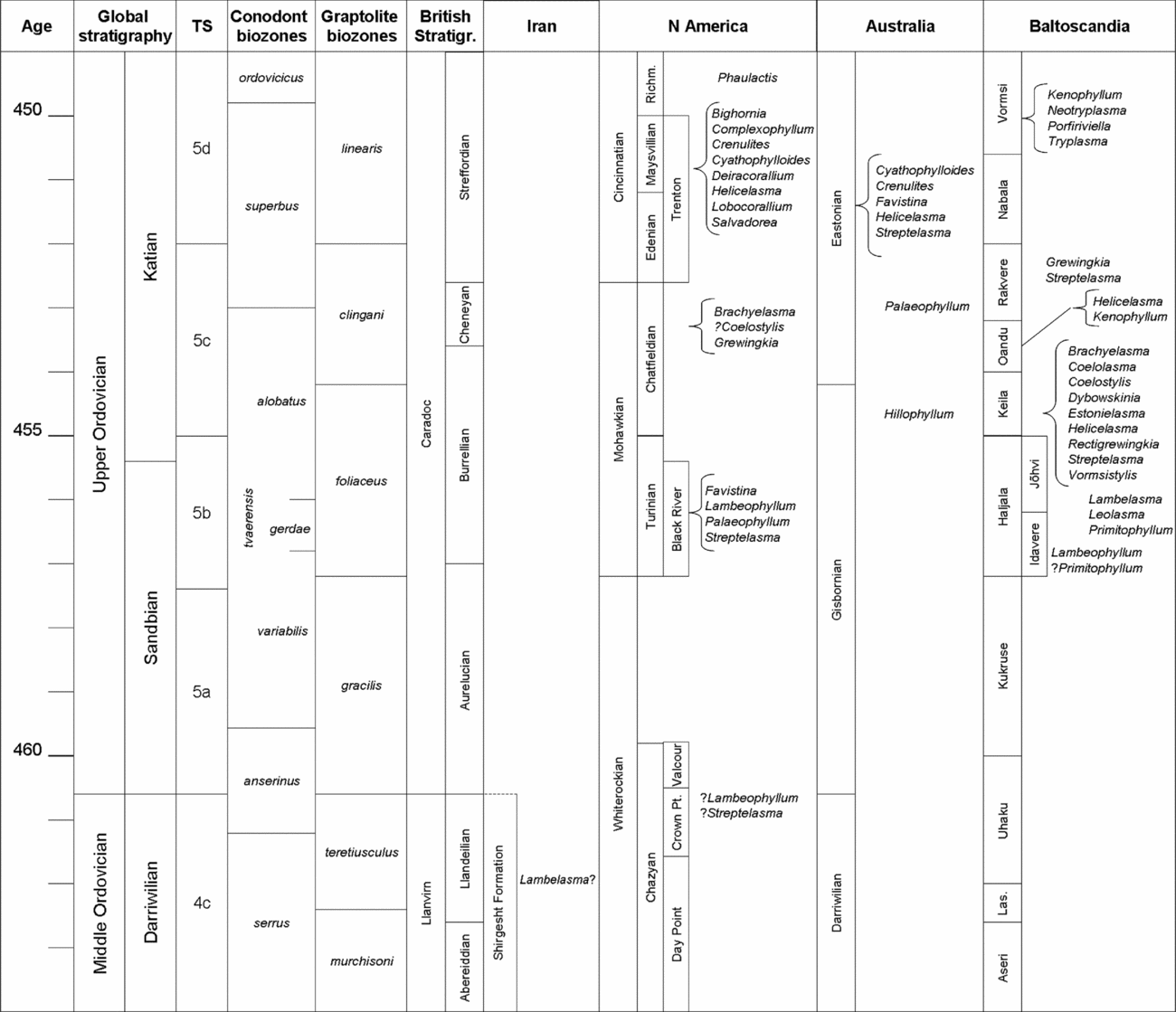
Undoubted Rugosa appeared during late Sandbian or early Katian time in Australia, Baltoscandia and North America (Fig. 1; cf. Webby et al. Reference Webby, Elias, Young, Neuman, Kaljo, Webby, Paris, Droser and Percival2004b ). Two taxa have been recorded from Baltoscandia: Primitophyllum primum (Kaljo, Reference Kaljo, Kiparisova, Markovskii and Radchenko1956a ), which appears morphologically close to a hypothesized ancestral form of rugose corals (Weyer, Reference Weyer1980), and Lambelasma (Weyer, Reference Weyer1980), both from the Haljala Regional Stage (upper Sandbian). Contemporaneous North American faunas comprise Favistina, Lambeophyllum, Palaeophyllum and Streptelasma, which often occur in sediments originating in the shallow lagoons of an extensive epeiric sea (Stumm, Reference Stumm1963; Elias, Reference Elias1983). An earlier record of Lambeophyllum and Streptelasma from the Crown Point Limestone Formation (uppermost Darriwilian to lowermost Sandbian) of Vermont has been reported but remains unsubstantiated (Welby, Reference Welby1961, Reference Welby1962; cf. Webby et al. Reference Webby, Cooper, Bergström, Paris, Webby, Paris, Droser and Percival2004b ). The oldest rugose coral in Australia, Hillophyllum priscum (Webby, Reference Webby1971), is from the lower Eastonian Regional Stage (lower Katian; Webby et al. Reference Webby, Cooper, Bergström, Paris, Webby, Paris, Droser and Percival2004b ).
Comparative upper Middle to lower Upper Ordovician stratigraphy and first occurrences of rugose coral genera in Australia, Baltoscandia and North America. Age is in Ma. TS – time slices, after Webby et al. (Reference Webby, Cooper, Bergström, Paris, Webby, Paris, Droser and Percival2004a ). Richm. – Richmondian, Las. – Lasnamägi. Data from: Cocks, Fortey & Rushton (Reference Cocks, Fortey and Rushton2010), Copper (Reference Copper1986), Elias (Reference Elias, Barnes and Williams1991), Hall (Reference Hall1963), Hill (Reference Hill1981), Ivanovskii (Reference Ivanovskii1965a , Reference Ivanovskii b , Reference Ivanovskii1969), Kaljo (Reference Kaljo, Kiparisova, Markovskii and Radchenko1956a ), Langenheim et al. (Reference Langenheim, Barnes, Delise, Ross and Staunton1956), Nelson (Reference Nelson1981), Neuman (Reference Neuman1977, Reference Neuman, Oliver, Sando, Coates, Macintyre, Bayer and Sorauf1997), Pestana (Reference Pestana1960), Ross et al. (Reference Ross, Adler, Amsden, Bergstrom, Bergstrom, Carter, Churkin, Cressman, Derby, Dutro, Ethington, Finney, Fisher, Fisher, Harris, Hintze, Ketner, Kolata, Landing, Neuman, Sweet, Pojeta, Potter, Rader, Repetski, Shaver, Thompson and Webers1982), Scrutton (Reference Scrutton and House1979a , Reference Scrutton1997), Stumm (Reference Stumm1963), Sweet & Bergström (Reference Sweet, Bergström and Bassett1976), Webby (Reference Webby1971, Reference Webby, Webby and Laurie1992), Welby (Reference Welby1961, Reference Welby1962), Weyer (Reference Weyer1973, Reference Weyer1984).

Two specimens resembling rugose corals have been found recently in Central Iran. Owing to the uniqueness and limited nature of the fossil material it was decided not to undertake conventional serial sectioning but to attempt to describe the specimens using a new, non-destructive technique. One of the specimens was analysed using synchrotron X-ray tomography at Diamond Light Source in Didcot, UK. The analysis was successful and the resulting images showed that the specimen is indeed a rugose coral and allowed it to be identified as potentially belonging to the Ordovician genus Lambelasma. The purpose of this publication is to describe the technique and the fossils. The fossils are of Darriwilian age and therefore pre-date all previously described Rugosa. The implications of this discovery for the early evolution of the Rugosa are discussed.
2. Geological setting
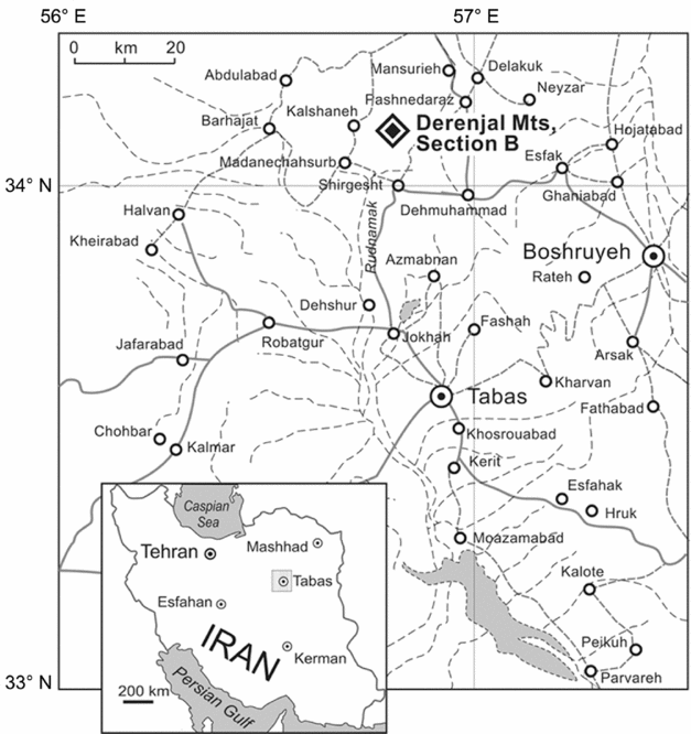
The new rugose corals were recovered from the upper part of the Shirgesht Formation (Section B, Unit B5, sample F-35 of Ghobadi Pour & Popov, Reference Ghobadi Pour and Popov2009) on the western side of Dahaneh-Kalut in the Derenjal Mountains, approximately 65 km north of the city of Tabas (Fig. 2). An outline of the geology of the area can be found in publications by Ruttner, Nabavi & Hajian (Reference Ruttner, Nabavi and Hajian1968) and Bruton, Wright & Hamedi (Reference Bruton, Wright and Hamedi2004). A detailed description and faunal logs for the Ordovician part of the Shirgesht Formation are given by Ghobadi Pour et al. (Reference Ghobadi Pour, Williams, Vannier, Meidla and Popov2006), Ghobadi Pour & Popov (2009) and Ghobadi Pour & Turvey (Reference Ghobadi Pour and Turvey2009).
Map of Iran with sample locality in the Derenjal Mountains near Shirgesht, north of Tabas.

The Darriwilian age of the fossiliferous horizon containing the rugose corals (Unit B5) is defined by the occurrence of stratigraphically diagnostic ostracods, including Aechmina? cf. ventadorni Vannier, Reference Vannier1986a , Cerninella aryana Williams, Vannier & Meidla, Reference Williams, Vannier, Meidla, Ghobadi Pour, Williams, Vannier, Meidla and Popov2006 and Ordovizona amyitisae Williams, Vannier & Meidla, Reference Williams, Vannier, Meidla, Ghobadi Pour, Williams, Vannier, Meidla and Popov2006 (in Ghobadi Pour et al. Reference Ghobadi Pour, Williams, Vannier, Meidla and Popov2006; Ghobadi Pour, Williams & Popov, Reference Ghobadi Pour, Williams and Popov2007). Aechmina? ventadorni occurs in the Hustedograptus teretiusculus graptolite Biozone (Vannier, Reference Vannier1986a , Reference Vannier b ; Vannier, Siveter & Schallreuter, Reference Vannier, Siveter and Schallreuter1989). The ostracods Cerninella aryana and Ordovizona amyitisae are endemic to Iran, but they are also found in Alborz, where they occur in the same horizon as the conodont Lenodus pseudoplanus, which is the index species for the mid Darriwilian conodont Biozone (Ghobadi Pour, Williams & Popov, Reference Ghobadi Pour, Williams and Popov2007). Unit B5 pre-dates the earliest occurrence of the trilobite Ovalocephalus kanlingensis (Zhang, Reference Zhang1981) in the overlying Unit B6. Ovalocephalus kanlingensis has a wide range from the late Darriwilian to early Katian (Zhou, Yuan & Zhou, Reference Zhou, Yuan and Zhou2010). This taxon was originally described and illustrated as Ovalocephalus aff. obsoletus Zhou & Dean, Reference Zhou and Dean1986 by Ghobadi Pour & Popov (2009) and was later considered as conspecific with O. kanlingensis in the comprehensive revision of the genus by Zhou, Yuan & Zhou (2010). Other components of the associated fauna are podocope ostracods and the trilobite Neseuritinus birmanicus Reed, Reference Reed1906 (Ghobadi Pour et al. Reference Ghobadi Pour, Williams, Vannier, Meidla and Popov2006; Ghobadi Pour & Popov, Reference Ghobadi Pour and Popov2009). Thus the Iranian occurrence of the new rugose corals can be dated as late Darriwilian, probably Hustedograptus teretiusculus graptolite Biozone equivalent or even slightly older (Fig. 1). During the Ordovician, this part of Iran was situated in mid-latitudinal Gondwana (Cocks & Torsvik, Reference Cocks and Torsvik2002).
The specimens are housed at the National Museum of Wales, Cardiff, under accession number NMW 2012.15G.
3. Materials and methods
Two coral specimens were discovered on the same rock. Owing to the rarity of material, one of the new fossils was examined using synchrotron X-ray tomography at Beamline I12 (Joint Engineering, Environmental and Processing), Diamond Light Source, Didcot, UK. Synchrotron tomography has been used successfully for the non-invasive examination of fossils, mainly vertebrates and insects (for example, Tafforeau et al. Reference Tafforeau, Boistel, Boller, Bravin, Brunet, Chaimanee, Cloetens, Feist, Hoszowska, Jaeger, Kay, Lazzari, Mariva, Nel, Nemoz, Thibault, Vignaud and Zabler2006; Martins et al. Reference Martins, Beckmann, Castanhinha, Mateus and Pranzas2011; Perreau & Tafforeau, Reference Perreau and Tafforeau2011). Recently there have been spectacular advances in this technology (Bergmann et al. Reference Bergmann, Morton, Manning, Sellers, Farrar, Huntley, Wogelius and Larson2010). Being able to examine the internal structure of fossils non-invasively over, for example, traditional thin-sections or serial sections, has many advantages; for example, a complete specimen is preserved for future studies or display (Scrutton, Reference Scrutton1979b ; Baars, Reference Baars2011). In this study the technique allowed the specimen to be described in detail without the need for destructive sampling. At the same time it was possible to generate virtual sections in different planes as well as spectacular three-dimensional images, which were extremely useful for visualization and description of the specimen.
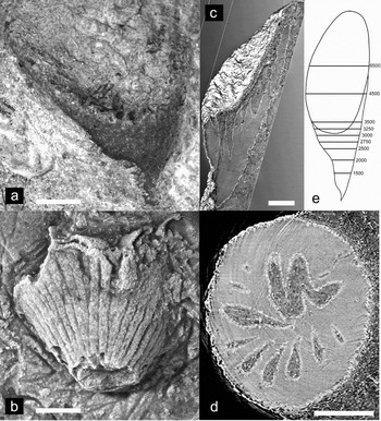
The rock containing the corals is a finely crystalline calcitic-dolomitic carbonate (55% ankerite and/or dolomite, 30% calcite and 15% quartz, as established by X-ray diffraction (XRD) analysis at National Museum of Wales). The calcite most likely has strontium inclusions as, during X-ray tomography, the entire rock was phosphorescing slightly. The analysed coral is partially silicified, being mainly preserved as the original calcite with a thin layer of microcrystalline silica coating the entire surface. The microcrystalline silica is visible in the tomographic reconstructions as a high-contrast outline (Fig. 3c, d). While the contrast between silica and calcite/dolomite was low, it was sufficient to enable visualization of the specimen even though a previous attempt to image the fossil using an experimental CT scanner at University of Wales Hospital Cardiff had been unsuccessful. Unfortunately, the calcitic nature of the septa did not allow determination of the microstructure of the septa.
Lambelasma? sp. (a) Specimen NMW 2012.15G.1 prior to preparation for tomography, embedded in rock matrix. (b) Specimen NMW 2012.15G.2 embedded in rock matrix. (c) Slice (YZ axis) through the three-dimensional reconstruction (specimen NMW 2012.15G.1). (d) Example of reconstruction from tomography, transverse section (XY axis) of specimen NMW 2012.15G.1 at 3000 μm; the concentric rings originating from the top right corner of the image are tomography artefacts. (e) Schematic view of NMW 2012.15G.1 showing orientation and position of transverse sections in μm above tip of the corallum in Figure 4. Scale bar in (a–d) is 1 mm.

The broad-spectrum beam at the Diamond Light Source wiggler source at Beamline I12 was filtered through diamond and silicon carbide before selection of a narrow band of the energy spectrum via a two-crystal monochromator. The fossil was secured to a high-precision rotation stage and images of a cadmium tungstate crystal scintillator were obtained via microscope optics and a digital imaging camera. The energy level of the X-rays was 53 keV, table rotation between projections was 0.03° and scintillator pixel size was 5 μm.
For each angle, six projections were averaged and this was performed for each of the 6000 projections. A set of 20 images without the sample in place was averaged and used as the reference (‘flat field’) image. Correction for the reference image variation, suppression of ring artefacts and reconstruction of the three-dimensional geometry was calculated using a reconstruction computer program (Titarenko, et al. Reference Titarenko, Bradley, Martin, Withers and Titarenko2010) including Titarenko's method for ring suppression (Titarenko, Withers & Yagola, Reference Titarenko, Withers and Yagola2010), and a ‘Graphics Processing Unit’ implementation of filtered back projection was used for tomographic reconstruction. Post processing was undertaken using the three-dimensional graphics software packages ‘ImageJ’ and ‘Avizo’, as well as the ‘Insight Image Registration and Segmentation Tool Kit’ image processing library.
Nonetheless, the low contrast of the sample material, as well as the base resolution of the camera and optics, reduced the clarity of features of less than approximately 20 μm size. In addition, some physical defects to the scintillator crystals resulted in concentric ring artefacts (Fig. 3d). It did not prove possible to remove these artefacts entirely despite attempts to reduce them by interpolation. At the same time, because the corallum walls and septa have material properties very similar to the surrounding rock matrix, stripping the rock data resulted in a loss of information for the fossil structural parts, which meant it was possible to visualize the fossil outline only, in essence only one (the calicular) growth stage. Therefore, the very advantage of the technique – removing the data for the rock matrix to reveal the fossil within – has a drawback if the chemical composition of the fossil is similar to that of the rock matrix.
4. Fossil record of early Rugosa
Coralomorph cnidarians have been reported from as far back as the Early Cambrian; these were reviewed and discussed by Scrutton (Reference Scrutton1997) who concluded that none of the Ordovician groups descended from Cambrian genera. Ordovician groups include the Tabulata, first documented from the Early Ordovician (Webby et al. Reference Webby, Cooper, Bergström, Paris, Webby, Paris, Droser and Percival2004b ), Kilbuchophyllida from the early Katian (Dicranograptus clingani Biozone, Scrutton & Clarkson, Reference Scrutton and Clarkson1991) and Rugosa; the Tetradiida (Darriwilian to Hirnantian; Webby et al. Reference Webby, Cooper, Bergström, Paris, Webby, Paris, Droser and Percival2004b ) were recently re-classified and placed in the Kingdom Plantae (Steele-Petrovich, Reference Steele-Petrovich2011 and references therein). It is generally accepted that the Rugosa appeared during the early Sandbian (Webby et al. Reference Webby, Cooper, Bergström, Paris, Webby, Paris, Droser and Percival2004b ) but there is no evidence that the Rugosa evolved from a lineage of the Tabulata (Scrutton, Reference Scrutton1997). Instead, the Rugosa represent a monophyletic clade, which was evolved most likely by the acquisition of a skeleton by a cnidarian such as a solitary anemone, resulting in a form such as Primitophyllum (Weyer, Reference Weyer1973; Sytova, Reference Sytova1977; Neuman, Reference Neuman, Oliver, Sando, Coates, Macintyre, Bayer and Sorauf1984; Scrutton, Reference Scrutton1997). Data on their origin are scarce and partly contentious, with several reported findings of upper Darriwilian/lower Sandbian specimens unconfirmed (Scrutton, Reference Scrutton1997; Webby et al. Reference Webby, Cooper, Bergström, Paris, Webby, Paris, Droser and Percival2004b ). The Rugosa quickly diversified and by the early Katian the major suborders Calostylina (Lambeophyllum, Lambelasma), Streptelasmatina (Streptelasma), Cystiphyllina (Hillophyllum, Primitophyllum) and Stauriina (Favistina, Palaeophyllum) had been established (Webby et al. Reference Webby, Cooper, Bergström, Paris, Webby, Paris, Droser and Percival2004b ; Fig. 1) and attained a geographical distribution ranging from North America, Baltoscandia, Siberia and China to Australia (Kaljo & Klaaman, Reference Kaljo, Klaaman and Hallam1973; Webby, Reference Webby, Webby and Laurie1992).
It appears reasonable to assume that the Rugosa evolved by addition of morphological features rather than by degeneration. Amongst the known Ordovician rugose genera there was a trend towards steadily increasing morphological complexity, and it is difficult to argue the opposite for hypothetical, unknown forms (cf. Kaljo, Reference Kaljo2004). These early forms are not necessarily derived from each other but potentially interrelated and probably have an earlier common ancestor.
Lambelasma and Primitophyllum from the Haljala Stage of Baltoscandia (Kaljo, Reference Kaljo, Kiparisova, Markovskii and Radchenko1956a ; Weyer, Reference Weyer1983) are approximately contemporaneous with Lambeophyllum, Favistina, Palaeophyllum and Streptelasma from the Blackriveran (upper Sandbian) of North America (Hill, Reference Hill1981). The appearance of the oldest Australian rugose coral, Hillophyllum priscum from the upper Gisbornian (Webby, Reference Webby1971), was placed more recently in the lower Eastonian by Webby et al. (Reference Webby, Cooper, Bergström, Paris, Webby, Paris, Droser and Percival2004b ), which is slightly later than the late Sandbian lambelasmatid-dominated Baltoscandian faunas. Ivanovskii (Reference Ivanovskii1969) and Weyer (Reference Weyer1973) considered Primitophyllum primum to be close to the ancestral form of all Rugosa despite being potentially younger than the Blackriveran rugosans. Primitophyllum primum has very short monacanthine septa and a small conical corallum containing irregularly distributed spines only, which makes it the simplest of the early rugose corals. The species is documented from the Baltoscandian Jõhvi Regional Substage. One of the paratypes of P. primum displays hyposepta (sensu Weyer, Reference Weyer1973; third order septa sensu Scrutton, Reference Scrutton1997), which are not observed in the holotype; Weyer (Reference Weyer1980) therefore doubted conspecificity of the two specimens. Instead, this particular paratype of P. primum may represent a different taxon congeneric to Lambelasma (D. Weyer, pers. comm.) as, typically, only Lambelasma has hyposepta.
There is a possibility that Lambeophyllum cf. L. profundum (Welby, Reference Welby1961) may pre-date even the late Sandbian North American and Baltoscandian faunas, although this has been particularly widely discussed and yet remains hitherto unsubstantiated independently. Welby (Reference Welby1961) mentioned apparently finding Lambeophyllum in the Crown Point Limestone of Vermont (uppermost Darriwilian to lower Sandbian). In addition, he figured one specimen each of Lambeophyllum profundum from the Orwell and Crown Point limestone formations (Chazy Group), together with a specimen of Streptelasma expansum from the Crown Point Limestone and one of Streptelasma corniculum from the Orwell Limestone (pl. II.7–11 in Welby, Reference Welby1962). His images are of poor quality but appear to be of simple rugose corals, but there are no descriptions included. There are no records of any Chazyan rugose coral specimens in the collections of the Harvard University Museum of Comparative Zoology, where Welby had deposited some of the other specimens mentioned in his 1961 note (J. Cundiff, pers. comm.). Nor is there any record in the regional or international geological literature of any other rugose corals from these Chazyan rocks. The lack of original and newly collected specimens from the Chazyan of North America casts doubt on Welby's determinations, especially since these Chazyan exposures and faunas are well known and have been studied intensively for many years by numerous researchers (e.g. Cooper, Reference Cooper1956; Pitcher, Reference Pitcher1964; Kapp, Reference Kapp1974). In addition, the stromatoporoid Stromatocerium recorded by Welby (Reference Welby1961) in the same publication as the supposed Lambeophyllum is common in the Blackriveran but does not occur in the Chazyan (Kapp & Stearn, Reference Kapp and Stearn1975). Owing to these inconsistencies, we agree with Webby et al. (Reference Webby, Cooper, Bergström, Paris, Webby, Paris, Droser and Percival2004b ) that Welby's accounts ought to be discounted as in error.
Aside from Welby's unlikely record of Lambeophyllum in the Crown Point Formation there are taxonomic problems with the genus. Various authors regard Lambelasma Weyer, Reference Weyer1973 and Lambeophyllum Okulitch, Reference Okulitch1938 as synonymous (see review in Weyer, Reference Weyer1983), while at the same time they point out significant differences between Lambeophyllum dybowskii Kaljo, Reference Kaljo1956b and Lambeophyllum profundum (Hall, Reference Weyer1847). North American records of Lambeophyllum were considered so doubtful by Weyer (Reference Weyer1973) that he suggested the name ought not to be used before further detailed studies. Lambeophyllum may be synonymous with Lambelasma but this can only be decided after revision of the genera in question (D. Weyer, pers. comm.). This process was started by Okulitch's definition of the genus in 1938. Elias (Reference Elias1983) examined a collection of approximately 200 specimens identified as Lambeophyllum? from the Trentonian Tyrone Limestone of Kentucky and concluded that these resemble Lambelasma lambei Weyer, Reference Weyer1973 from the Baltic Macrourus Limestone (Keila Regional Stage) but differ sufficiently to retain the genus name Lambeophyllum. Lambelasmatids were diverse in Baltoscandia: the presence of ten genera in the mid Upper Ordovician means that they were initially the dominant rugosan group, to be replaced only later in the Upper Ordovician by streptelasmatids. However, lambelasmatids are not recorded in Ordovician rocks elsewhere except for the records of Lambeophyllum in North America (e.g. Bassler, Reference Bassler1950; Hall, Reference Hall1963; Stumm, Reference Stumm1963; Elias, Reference Elias1983). It follows that Sandbian and, in particular, North American records of Lambeophyllum need to be revisited and revised.
In addition to the records above, there are a number of occurrences of early rugose corals from Asia. Yohophyllum Lin, Reference Lin1965 from the ‘Middle’ Ordovician (Xia Qiao Jia Formation) of Sichuan was thought to be close to the Calostylidae owing to the apparent presence of peripherally porous septa (Lin, Reference Lin1965). Following modern Ordovician stratigraphy this is probably contemporaneous with the Pagoda Formation, which is of Sandbian age, certainly post-Darriwilian (cf. Zhan & Zin, Reference Zhan and Jin2005). However, the lack of depiction of important characteristics, combined with the poor quality of the images and plates, resulted in Weyer (Reference Weyer1973) doubting this affiliation. Instead, Yohophyllum apparently displays characteristics that place it closer to the streptelasmatids, such as a normal septal stereozone. Ningnanophyllum Lin, Reference Lin1965, from the same formation as Yohophyllum, is apparently closely related to Yohophyllum and differs only by the presence of an aulos, the lack of an axial structure and in being colonial. Nevertheless, while published images are of poor quality, they do not appear to support the presence of septal pores (Weyer, Reference Weyer1973). Pending revision of the original material, therefore, the status of these two genera is uncertain, as is the precise age of these genera.
From the above it is apparent that there are taxonomic problems associated with most early rugose corals. This is mainly caused by a paucity of well-preserved fossil material. Detailed descriptions are only achievable after examination of all ontogenetic stages; this has so far been impossible in most of the corals mentioned above. In addition, stratigraphical uncertainties as well as poor published images also hamper many publications about early rugose corals. Notable exceptions are Webby's (1971) description of Hillophyllum priscum and Elias's (1983) review of Lambeophyllum.
5. The earliest rugose coral
The specimens described here considerably pre-date all other known rugose corals, even Welby's (1961) oldest potential and unproven record of Lambeophyllum profundum. The available material is insufficient to describe the early growth stage but the intermediate and late stages are documented here. The septa are broad in subcalicular stages and very short in the calice. The shape of the corallum, the presence of an axial structure and the pinnate-zaphrentoidid arrangement of septa resemble equivalent structures in Baltoscandian lambelasmatid corals. The presence of an axial structure during early growth stages would also place the specimens described here close to the Streptelasmatina, but tabulae are absent and the arrangement of septa in streptelasmatids is regular and not pinnate. The absence of evidence for perforate, monacanthine septa presently would speak against an association with the lambelasmatids but may be a result of preservation in combination with the imaging technique used. We place more emphasis on the zaphrentoidid septal arrangement, the lack of tabulae and synapticulae, and the overall simple construction of the corallum.
Septal insertion of specimen NMW 2012.15G.1 was studied using three-dimensional visualization of X-ray tomography data as well as conventional examination of septal grooves on the exotheca. This indicates a typical rugosan pattern of septal insertion following Kunth's rule (Scrutton, Reference Scrutton1997; Weyer, Reference Weyer2007), in that septa are inserted serially by bifurcation in four loci, on either side of the cardinal septum and adjacent to the alar septa. Unfortunately, the tomography data are inconclusive in the lower (oldest) part of the corallum, and in the specimen NMW 2012.15G.2, which was not examined tomographically, only the calicular part of the corallum is preserved. For this reason, the generic status of the Iranian specimens cannot be securely determined and is therefore considered here as provisional (cf. Bengtson, Reference Bengtson1988), pending discovery of additional material that would resolve uncertainties.
6. Systematic palaeontology
Class ANTHOZOA Ehrenberg, Reference Ehrenberg1834
Subclass RUGOSA Milne-Edwards & Haime, Reference Milne-Edwards and Haime1850
Order STAURIIDA Verrill, Reference Verrill1865
Suborder CALOSTYLINA Prantl, Reference Prantl1957
Superfamily Calostylicae Zittel, 1879
Family Lambelasmatidae Weyer, Reference Weyer1973
Genus Lambelasma Weyer, Reference Weyer1973
Type species. Lambelasma lambei Weyer, Reference Weyer1973. Erratic boulder, probably derived from Baltoscandian Macrourus Limestone; Upper Ordovician, lower Katian.
Lambelasma? sp.
Figures 3, 4
Material. Two coralla from the sample F-35, Shirgesht Formation, west side of Dahaneh-Kalut, Derenjal Mountains, north of Tabas, Central Iran.
Lambelasma? sp. Transverse sections (XY axis) of specimen NMW 2012.15G.1, drawn from tomographic images, from just above the tip (1500 μm) to the calice (6500 μm). Sections are drawn as they appear looking from the calical end towards the apical end of the corallum. Dots indicate position of the cardinal septum. Scale bar is 1 mm.

Description. Small, solitary, erect trochoid coralla, almost straight, up to a maximum of 8.5 mm long and 4.5 mm wide. Description of external features from both NMW 2012.15G.1 and NMW 2012.15G.2; description of internal structures from NMW 2012.15G.1 only. Specimen NMW 2012.15G.2 shows the exterior of the corallum cardinal side with septal furrows and interseptal ridges. Epitheca with obvious longitudinal septal furrows and flat intercalated ridges but with only a few growth lines. Cardinal side slightly convex, apex curved. Calice not everted, very deep, comprising approximately two thirds of the corallum length. Broad peripheral stereozone; no synapticulae. No tabulae; no dissepiments. Septa are of two orders, with minor septa barely projecting beyond peripheral stereozone. In early (sub-calicular) growth stages, major septa thickened, arranged irregularly-pinnately zaphrentoidid, extending to the axis, connected in groups but not forming an axial boss; interseptal chambers present at this stage. The cardinal septum (as indicated in Fig. 4) is prominent in sections 2750–3500 but decreases in length more rapidly during ontogeny than the other major septa. In the same sections, the major septa on the counter side of the corallum appear to be deflected in an anticlockwise direction. The alar fossulae are prominent in sections 2500–3500. Calicular septa regular and rapidly retreating from the axis, leaving a central zone free from septa. Minor septa visible only in the upper part of calice. Septal microstructure is not visible in the specimens examined. The pattern of septal insertion indicates a typical rugosan pattern following Kunth's rule in that metasepta bifurcate normally from the protosepta.
Remarks. Specimen NMW 2012.15G.2 shows a rugosan pattern of septal insertion on both sides of the cardinal septum. The septal arrangement in NMW 2012.15G.1 is characteristically lambelasmatid and pinnate-zaphrentoidid. It is distinguished from members of the Lambelasmatidae by having no axial boss (as Dybowskinia Weyer, Reference Weyer1973), major septa that are not denticulate and lack a prominent counter septum (as Lambeophyllum Okulitch, Reference Okulitch1938), not being fasciculate (as Sogdianophyllum Lavrusevich, Reference Lavrusevich1971) and by having no tabulae/tabellae (as Coelostylinae Weyer, Reference Weyer1973). The closest association is with Lambelasma Weyer, Reference Weyer1973 owing to the pinnate arrangement of septa.
In comparison to Lambelasma atavum Kaljo, Reference Kaljo1958 and L. narvense Weyer, Reference Weyer1984, the Iranian coralla are smaller and interseptal furrows are less rounded. Lambelasma? sp. shows some similarity to Lambelasma lambei Weyer, Reference Weyer1973, in particular the counterseptum and alar septa, which, in both species, are not differentiated while the alar fossulae are obvious. In addition, no septal pores were observed in the Iranian corallum and septa in L. lambei are only weakly porous. The Iranian specimens, however, differ from L. lambei in having the cardinal septum on the convex side of the corallum, as in Coelostylis Lindström, Reference Lindström, Angelin and Lindström1880, but septa are broader than in Coelostylis. Septal arrangement in Lambelasma? sp. is not as regularly pinnate as, and interseptal spaces are longer than, in L. lambei. The cardinal septum is as prominent but not as long as in L. lambei, though it withdraws from the axis more rapidly in the later growth stages in the specimens described here. Septa in Lambelasma? sp. are less numerous and considerably broader than in Lambelasma dybowskii Kaljo, Reference Kaljo1956 and L. narvense, and not as spinose as in L. balticum Weyer, Reference Weyer1997 and L. atavum, whereas minor septa are less prominent than in L. carinatum Weyer, Reference Weyer1993.
Some features typical of Lambelasma, however, were not observed in the two specimens described here. Septal spines (for example, Weyer, Reference Weyer1983, fig. III.12–20; Weyer, Reference Weyer1997, fig. 1) are not present, and there is no evidence for monacanthine septa (discontinuous ends of septa due to their spinose edges). The cardinal side in previously described species of Lambelasma is concave, although it is convex in Coelolasma. The Lambelasmatidae belong to the Calostylicae, which are distinguished from other rugosan groups by having a porous septal structure. It is likely that the absence of evidence for septal pores in NMW 2012.15G.1 is a result of the type of preservation in combination with the data reconstruction technique used, which may have prevented visualization of potential pores.
NMW 2012.15G.1 also undoubtedly exhibits characteristics of streptelasmatids. Deflected septa with fused axial ends are a feature of some species of Streptelasma Hall, Reference Hall1847 (Neuman, 1969, p. 9; Elias, 1982, p. 55). The cardinal side in streptelasmatids is convex as in the specimens described here. The septa of NMW 2012.15G.1 are laminar as those of streptelasmatids. The major septa in early and intermediate growth stages are often dilated in streptelasmatids and form an axial structure and, in later stages, are thin and short. Streptelasma, however, has tabulae that are absent in the specimen examined using X-ray tomography, and the arrangement of septa in Streptelasma is not pinnate.
Despite the absence of evidence of a porous and monacanthine septal structure in NMW 2012.15G.1, we suggest that it is close to the lambelasmatids owing to its overall morphology, which is simpler than in streptelasmatids and lacks tabulae. The Iranian specimens most probably represent a new, not formally named taxon. They are described here under open nomenclature, since they provide insufficient information on early growth stages and septal microstructure. Only one specimen (NMW 2012.15G.1) was examined using X-ray tomography. The second specimen (NMW 2012.15G.2) was situated within 1 cm of the first one in the same rock and the size, appearance of the corallum wall and shape of septal furrows is very much like the first one. We therefore suggest that both specimens belong to the same species, even though only an examination of internal structures would give conclusive evidence.
It appears that the overall shape of the calice of specimen NMW 2012.15G.1 is the result of damage. In successive sections between 3000 μm and 4000 μm it can be seen that some septa still extend into the calice even though the theca is already missing, and septa immediately adjacent to the ‘open’ side of the calice do not give any indication of retreating as they would were the calice to open up naturally; some septa even appear to be ‘floating’ without being connected to the theca (Fig. 4, 3250 μm). The visible part of specimen NMW 2012.15G.2, while missing the apical part, appears to be complete in the upper part of the calice (Fig. 3b) and therefore confirms this conclusion.
7. Conclusion
One of the specimens described here was examined using synchrotron X-ray tomography owing to the rarity of the fossil material. The technique was shown to be a useful tool for the non-invasive examination of partially silicified fossils embedded in a calcareous rock matrix. It was possible to describe the specimen in detail from the three-dimensional reconstructions and therefore preserve the specimen for future studies and display. The type of preservation affected the outcome of this study in that, even though the chemistry of the fossil and that of the rock matrix were very similar, partial silicification resulted in sufficient contrast for a reconstruction of the fossil.
The second important outcome of this study is that the two specimens from the Darriwilian of Iran are unequivocal rugose corals and hence represent the earliest known record of Rugosa. The conical coralla display initially broad septa that are connected axially in the early growth stages but later retreat quickly to reveal a deep calice. This simple construction may represent a morphology that is close to an ancestral form of the Rugosa. In addition, the new coral displays characteristics of both the Lambelasmatidae and Streptelasmatidae and is therefore suggested to be ancestral specifically to these two groups, which it pre-dates by a considerable margin. Lambelasmatidae, together with Primitophyllum, appeared first in Baltoscandia during the upper Sandbian; Streptelasmatidae occurred only slightly later in both North America and Baltoscandia. Lambelasma? provides evidence for the hypothesis that the Rugosa represent a monophyletic clade, with acquisition of a skeleton by a solitary anemone and subsequent addition of further morphological features.
Acknowledgements
The authors are enormously appreciative of the support received from Leonid Popov, National Museum of Wales, for assistance with tomography and considerably improving the manuscript; Diamond Light Source for enabling and funding the X-ray tomography; Dieter Weyer for many helpful late-night discussions on coral morphology and evolution; Stig Bergström, Ohio State University, and Robert Elias, University of Manitoba, for stratigraphical advice; Stephen Daniels, University of Wales Hospital, for an experimental CT scan of the fossil; Tom Cotterell, National Museum of Wales, for XRD analysis of the host rock; Amanda Valentine-Baars, National Museum of Wales, for preparing thin-sections; Jessica Cundiff for checking the records and collections of Harvard University Museum of Comparative Zoology; Robert Owens, National Museum of Wales as well as Robert Elias and an additional, anonymous, reviewer for their constructive comments and improving the manuscript. Christian Baars acknowledges support from National Museum of Wales. Mansoureh Ghobadi Pour thanks National Museum of Wales for logistical support on her visits to Cardiff; her work in Iran is supported by Golestan University.