Introduction
Since the birth of the world’s first mammal produced by in vitro fertilization (IVF) in 1959, many different animals have been successfully born using IVF (Chronopoulou and Harper, Reference Chronopoulou and Harper2015; Niederberger et al., Reference Niederberger, Pellicer, Cohen, Gardner, Palermo, O’Neill, Chow, Rosenwaks, Cobo, Swain, Schoolcraft, Frydman, Bishop, Aharon, Gordon, New, Decherney, Tan, Paulson and LaBarbera2018). Much progress has been made in the field of embryo development and reproductive medicine based on IVF, and the techniques have made great contributions to livestock breeding, the protection of endangered wild animals, and the treatment of human infertility (Chronopoulou and Harper, Reference Chronopoulou and Harper2015; Mandawala et al., Reference Mandawala, Harvey, Roy and Fowler2016; Sirard, Reference Sirard2018). Since the successful birth of the first human baby, Louise Brown, who was conceived by IVF in 1978, IVF has been responsible for millions of births worldwide. Currently, 1–3% of all births every year in the USA and Europe are produced by IVF (Chandra et al., Reference Chandra, Copen and Stephen2014).
Although IVF research has a long history, and significant progress has been made for improving its efficiency, IVF embryos can still show many deficiencies, such as abnormal development and a higher risk of certain diseases (Chronopoulou and Harper, Reference Chronopoulou and Harper2015). Researchers have tested many methods for optimizing the IVF protocol but problems can still occur. Adjusting the culture conditions to be as close to the physiological environment as possible, such as by simulating the internal temperature and composition of oviduct fluid (Xiong et al., Reference Xiong, Wang, Wang, Hua, Zi and Zhang2014), could improve the efficiency of IVF. High ROS levels in the in vitro culture (IVC) medium can be detrimental, but antioxidants could be used to decrease them. The absence of EVs, as found in the genital tract, could be compensated for by the addition of EVs derived from oviduct fluid to improve embryo development (Qu et al., Reference Qu, Zhao, Wang, Zhang, Li, Fan and Liu2019; Zuo et al., Reference Zuo, Niu, Liu, Ma, Qu, Qiao, Su, Zhang and Wang2020). Seminal plasma plays an important role in the process of fertilization and development, and the EVs normally present in seminal plasma are lost during the IVF procedure (Robertson and Sharkey, Reference Robertson and Sharkey2016).
In this study, seminal plasma EVs (SP-EVs) were added to the IVF medium during the procedure and the fertilization rate, cleavage rate, blastocyst rate, apoptosis and ICM/TE ratio were measured to assess the effect of SP-EVs on the development of IVF embryos.
Materials and methods
Reagents, source of mice and animal use statement
Unless otherwise indicated, all chemicals were purchased from Sigma Chemical Co. (St. Louis, MO, USA). ICR mice were used in these experiments. All the mice were housed under a 12-h light/dark cycle. The temperature was kept at 22–25°C, the humidity was 50–60%, and the mice were given food and water ad libitum. All animal procedures were performed in accordance with the regulations on the care and use of experimental animals of Air Force Medical University.
Isolation of EVs from seminal plasma
Male mice were euthanized, and their testes and epididymis were collected and placed in phosphate-buffered saline (PBS). Seminal plasma was squeezed out using forceps under a stereomicroscope. The fluid was centrifuged at 4°C as follows: 300 g for 10 min, discard pellet; 2000 g for 10 min, discard pellet; 10,000 g for 30 min, discard pellet; and 100,000 g for 70 min, discard supernatant. The pellets were resuspended in PBS and centrifuged again at 100,000 g for 70 min. The resulting pellet contained the SP-EVs.
Transmission electron microscopy and nanoparticle analysis of SP-EVs
For transmission electron microscopy (TEM), the EV pellets were resuspended in 20 µl of PBS and pipetted onto 300-mesh grids. The grids were stained with 2% phosphotungstic acid for 5 min and imaged by TEM (JEOL, Tokyo, Japan). Nanoparticle analysis of the EVs was performed using the NanoSight LM-10 (Malvern, Worcestershire, UK) particle analyzer and size and zeta potential were determined according to the manufacturer’s instructions.
IVF and in vitro culture
Semen containing sperm was collected from the cauda epididymis and vas deferens into 1 ml of Krebs–Ringer bicarbonate medium (KRB) buffered with NaHCO3 (prewarmed to 37°C). The sperm suspension was diluted to 2 ml with KRB–HEPES and loaded onto a two-step Percoll gradient (45% and 90% Percoll). The gradient was centrifuged (650 g, 30 min, 25°C). After removing the interface containing immotile sperm and the 45% and 90% Percoll layers, the motile sperm pellet was washed once in KRB–HEPES (430 g, 10 min, 25°C) and then capacitated in KRB–BSA at a concentration of ∼107 sperm/ml for 30 min at 37°C in a 5% CO2 atmosphere. The capacitated sperm were then centrifuged (430 g, 10 min, 25°C), and resuspended in KRB to a concentration of 5–10 × 106 sperm/ml. Females at 8–10 weeks of age were superovulated by serial intraperitoneal injection of 5 IU pregnant mare serum gonadotropin (PMSG) followed 45–54 h later by 5 IU human chorionic gonadotropin (hCG). At 12–15 h after hCG injection, the mice were euthanized, and their oviducts were collected and placed in CZB medium. The oocyte–cumulus complex was carefully removed from the oviduct as a single mass, and, the oocytes were transferred to a drop of fertilization medium [KSOM medium supplemented with 0.3% BSA (KSOM–BSA)] under mineral oil in a 3.5 cm dish. The dish containing the oocytes was placed in an incubator with 5% (v/v) CO2 at 37.5°C for at least 15 min until needed. Oocytes and sperm were co-incubated for 8 h and eggs were considered to be fertilized if they contained two pronuclei and a second polar body. Fertilized embryos were transferred into embryo culture medium, and the medium used for culturing fertilized embryos was KSOM supplemented with amino acids, covered with mineral oil and cultured in 5% (v/v) CO2 atmosphere at 37.5°C.
Treatment of blastocysts with SP-EVs
For the supplemented group, SP-EVs derived from one male was supplemented into one drop of fertilization medium during the in vitro fertilization (IVF) procedure; and also SP-EVs derived from one male were supplemented into one drop of embryo culture medium during the IVC procedure. For the nonsupplemented group, SP-EVs were not supplemented during the IVF and IVC procedures.
Apoptosis detection
Apoptosis was detected using the DeadEnd fluorometric terminal deoxynucleotidyl transferase (TdT) dUTP nick-end labelling (TUNEL) assay (Promega, Madison, WI), as previously described (Jia et al., Reference Jia, Ding, Shen, Luo, Zhang, Zhou, Ding, Qu and Liu2019) and expressed as the percentage of total cells. Blastocysts were fixed in 4% paraformaldehyde at room temperature (RT) for 2 h, permeabilized in 0.5% Triton X-100 (at RT for 5 min) and incubated with fluorescein isothiocyanate (FITC)-conjugated dUTP and TdT at 37°C in the dark for 1 h. The tailing reaction was terminated with 2× saline–sodium citrate (SSC) in the dark at RT for 15 min. Embryos were then incubated with PBS containing 25 µg/ml RNase A in the dark at RT for 30 min. After 4′,6-diamidino-2-phenylindole (DAPI) staining and washing with PBS in the dark, the slides were examined by epifluorescence using a Nikon Eclipse Ti-S microscope and images were captured using a digital camera (Nikon, Tokyo, Japan).
Immunodetection of trophectoderm cells by CDX2
Immunofluorescence was performed as previous described (Qu et al., Reference Qu, Qing, Liu, Qin, Wang, Qiao, Ge, Liu, Zhang, Cui and Wang2017). Trophectoderm cells (TECs) were identified by immunofluorescent detection of CDX2, a specific marker of TECs. Embryos were fixed in paraformaldehyde at RT for 2 h, permeabilized in Triton X-100 at RT for 30 min and blocked in a blocking liquid. The embryos were then incubated overnight with a primary CDX2 antibody (Sigma-Aldrich), followed by incubation with a secondary antibody at RT for 2 h. Then the embryos were washed three times in PBS–polyvinyl alcohol (PBS–PVA) solution and treated with the nuclear stain DAPI for 5 min. After washing and mounting, slides were examined by epifluorescence using a Nikon Eclipse Ti-S microscope and images were captured using a digital camera (Nikon).
Statistical analysis
Fertilization rate, cleavage rate, and blastocyst rate were analyzed by chi-squared (χ2) test. For the in vivo group, blastocysts were collected from the uteri of conceived female mice in the counterpart time of in vitro groups. Apoptosis and ICM/TE ratios were compared using analysis of variance (ANOVA). Statistical analyses were performed using the SPSS software package (SPSS Inc., Chicago, IL, USA). Data were expressed as mean ± SEM. A P-value < 0.05 was considered statistically significant.
Results
Characteristics of EVs derived from seminal plasma(SP-EVs)
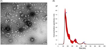
Electron microscopy of the seminal plasma isolate revealed the presence of particles of various sizes (Figure 1A). Nanoparticle analysis gave diameters of the majority of particles ranging from 100 to 200 nm, and a concentration of 6.07 × 10¹⁰ ± 2.91 × 10⁹ particles/ml (Figure 1B).
Presence of extracellular vesicles in seminal plasma. (A) The particles were visualized by transmission electron microscopy (bar, 200 nm). (B) Quantification and size distribution of particles.

Effect of SP-EVs on embryo development in vitro
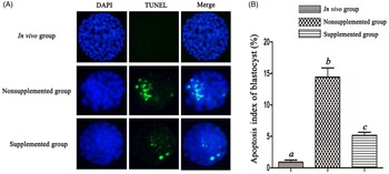
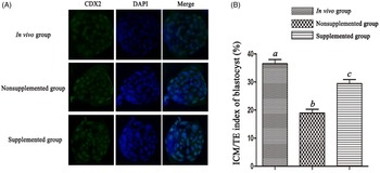
The isolated EVs were tested by incubating them with IVF embryos. There was no significant difference in fertilization rate (69.9% vs 70.8%) and cleavage rate (90.7% vs 90.9%) between the supplemented group and the nonsupplemented group (Table 1). However, the blastocyst rate in the supplemented SP-EVs group was 85.5%, which was significantly higher than in the nonsupplemented group (69.5%, P < 0.05; Table 1). Apoptosis of blastocysts was quantitated by TUNEL assay and was significantly higher in the in vitro groups than the in vivo group, whereas apoptosis in the EV group was significantly lower than in the nonsupplemented group (Figure 2). The ICM/TE index was significantly lower in the in vitro groups than the in vivo group, while the supplemented SP-EVs group showed a significantly higher total number of cells in blastocysts and a higher ICM/TE ratio than the nonsupplemented group (Figure 3).
The effect of EVs derived from seminal plasma on the fertilized rate, cleavage rate, and blastocyst rate of IVF embryo in mice

a,bValues in the same column with different superscripts differ significantly (P < 0.05).
Apoptosis index (%) of blastocysts. (A) Apoptotic blastomeres were detected by TUNEL (green). DNA was stained with DAPI (blue) to visualize all blastomeres. (B) Percent apoptosis of blastocysts in the in vivo group, nonsupplemented group (in vitro) and supplemented group (in vitro). Different letters (lowercase a, b and c) above the bars indicate significant differences at P < 0.05.

Inner cell mass/trophectoderm (ICM/TE) ratios in blastocysts. (A) TE cells were detected using an antibody against CDX2, a marker of TE (green), and DNA was stained with DAPI (blue) to visualize all blastomeres. (B) ICM/TE ratios of blastocysts in the in vivo group, nonsupplemented group (in vitro) and supplemented group (in vitro). The cell numbers in the ICM corresponded to the total cell number minus the cell number in the TE, and the ICM/TE ratio was represented by the cell number in the ICM divided by the cell number in the TE. Different letters (lowercase a, b and c) above the bars indicate significant differences at P < 0.05.

Discussion
Seminal plasma is a complex fluid produced by the accessory glands of the male reproductive tract (Hopkins et al., Reference Hopkins, Sepil and Wigby2017; Druart and de Graaf, Reference Druart and de Graaf2018). In the process of natural fertilization, sperm is transported from the male reproductive tract to the female reproductive tract through seminal plasma where it fertilizes the oocytes (Bromfield, Reference Bromfield2016; Costa et al., Reference Costa, Barbisan, Assmann, Araújo, de Oliveira, Signori, Rogalski, Bonadiman, Fernandes and da Cruz2017). In the past, the seminal plasma has been mainly regarded as a transport medium that provided a nutrient-rich environment to maintain the sperm until the process of genetic material exchange took place (Juyena and Stelletta, Reference Juyena and Stelletta2012; Morrell et al., Reference Morrell, Georgakas, Lundeheim, Nash, Davies Morel and Johannisson2014; Hopkins et al., Reference Hopkins, Sepil and Wigby2017). Recently, more studies have found that seminal plasma plays an important role in signal exchange in male and female reproductive responses (Bromfield, Reference Bromfield2016; Song et al., Reference Song, Li, Li, Fang, Liu, Yang, Meng, Yang and Peng2016; Watkins et al., Reference Watkins, Dias, Tsuro, Allen, Emes, Moreton, Wilson, Ingram and Sinclair2018). Seminal fluids can interact with cells in the oviduct and affect prostaglandin release (Kaczmarek et al., Reference Kaczmarek, Krawczynski, Blitek, Kiewisz, Schams and Ziecik2010). Seminal plasma can also have effects on the uterus and vagina by regulating inflammatory cytokine gene expression in endometrial cells, and cervical and vaginal tissues (Kaczmarek et al., Reference Kaczmarek, Krawczynski, Blitek, Kiewisz, Schams and Ziecik2010; Hartmann et al., Reference Hartmann, Gerner, Walter, Saalmüller and Aurich2018). The antioxidants in seminal plasma can prevent DNA damage in sperm (Dorostghoal et al., Reference Dorostghoal, Kazeminejad, Shahbazian, Pourmehdi and Jabbari2017). The direct communication between seminal plasma proteins and female germ cells can alter the activity of uterine cells causing physiological changes that can increase the success rate of pregnancy and promote healthy offspring (Samanta et al., Reference Samanta, Parida, Dias and Agarwal2018).
Transforming growth factor-β in seminal plasma induced inflammatory cytokinesis in the mother’s endometrium or cervix, resulting in the recruitment of inflammatory immune cells after insemination (Rizo et al., Reference Rizo, Ibrahim, Molinari, Harstine, Piersanti and Bromfield2019). Toll-like receptor (TLR)-4 signalling mediated by seminal plasma plays an important role in the induction of inflammatory cytokine expression and the expansion of the cumulus cell complex (Bromfield and Sheldon, Reference Bromfield and Sheldon2011; Schjenken et al., Reference Schjenken, Glynn, Sharkey and Robertson2015). At present, in many species, including humans, rodents, domestic and wild species, IVF or intracytoplasmic sperm injection (ICSI) does not require seminal plasma to achieve pregnancy; however, the lack of seminal plasma can impair blastocyst development, increase the risk of preterm birth and very low birth weight, and cause delivery complications and severe birth defects (Basatemur and Sutcliffe, Reference Basatemur and Sutcliffe2008; Guo et al., Reference Guo, Liu, Jin, Wang, Ullah, Sheng and Huang2017). Treatment with seminal plasma can improve the pregnancy rate and reduce the incidence of preeclampsia during embryo transfer (Palermo et al., Reference Palermo, Neri, Takeuchi, Squires, Moy and Rosenwaks2008). The phenotype of the offspring could be changed by seminal plasma intervention during pregnancy (Bromfield, Reference Bromfield2014; Bromfield et al., Reference Bromfield, Schjenken, Chin, Care, Jasper and Robertson2014).
Seminal fluid contains an abundant population of extracellular vesicles (EVs) (Eswaran et al., Reference Eswaran, Agaram Sundaram, Rao and Thalaivarisai Balasundaram2018; Skalnikova et al., Reference Skalnikova, Bohuslavova, Turnovcova, Juhasova, Juhas, Rodinova and Vodicka2019), which are membrane-enclosed complexes of proteins, lipids and nucleic acids that facilitate cell-to-cell communication (Mulcahy et al., Reference Mulcahy, Pink and Carter2014). SP-EVs are crucial for sperm maturation, prevention of premature acrosome reaction and capacitation, and to protect sperm from the female immune system (Machtinger et al., Reference Machtinger, Laurent and Baccarelli2016). Depletion of EVs from boar seminal plasma leads to decreased sperm motility, shorter survival time, and declined sperm plasma membrane integrity (Machtinger et al., Reference Machtinger, Laurent and Baccarelli2016). The first EVs identified in human seminal plasma were produced by the prostate and called ‘prostasomes’. They were found to contain at least 140 proteins with antimicrobial, antioxidant and immunomodulatory functions (Siciliano et al., Reference Siciliano, Marcianò and Carpino2008). Prostasomes can fuse with sperm and their contents can promote capacitation and the acrosome reaction (Zelli et al., Reference Zelli, Orlandi, Verstegen, Troisi, Elad Ngonput, Menchetti, Cardinali and Polisca2017). EVs derived from epididymal epithelial cells contain adhesion molecules such as tetraspanins and integrins and milk fat globule-epidermal growth factor 8 protein (MFGE8), which participate in sperm maturation, movement, fertilization ability, and the regulation of reactive oxygen species (Rocha et al., Reference Rocha, Martins, van Tilburg, Oliveira, Moreno, Monteiro-Moreira, Moreira, Araújo and Moura2015; Silva et al., Reference Silva, Frost, Li, Bovin and Miller2017; Barranco et al., Reference Barranco, Padilla, Parrilla, Álvarez-Barrientos, Pérez-Patiño, Peña, Martínez, Rodriguez-Martínez and Roca2019). In addition, seminal plasma EVs contain small RNAs, which regulate gene expression and are involved in cell communication (Barceló et al., Reference Barceló, Mata, Bassas and Larriba2018). In this study, we found abundant EVs in the seminal plasma of mice, and incubating them with mouse embryos in vitro significantly decreased apoptosis.
Previous studies have found that EVs secreted by embryos, could mediate apoptosis, stress responses and cell differentiation (Qu et al., Reference Qu, Qing, Liu, Qin, Wang, Qiao, Ge, Liu, Zhang, Cui and Wang2017). EVs derived from oviduct fluid are necessary for oocyte fertilization and zygote activation (Qu et al., Reference Qu, Zhao, Wang, Zhang, Li, Fan and Liu2019). EVs derived from uterine fluid mediated embryo implantation, material exchange and signal communication between maternal cells and the embryo or fetus (Qiao et al., Reference Qiao, Ge, Ma, Zhang, Zuo, Wang, Zhang and Wang2018). The presence of these EVs can improve embryo development in vitro (Almiñana et al., Reference Almiñana, Corbin, Tsikis, Alcântara-Neto, Labas, Reynaud, Galio, Uzbekov, Garanina, Druart and Mermillod2017; Qu et al., Reference Qu, Qing, Liu, Qin, Wang, Qiao, Ge, Liu, Zhang, Cui and Wang2017, Reference Qu, Zhao, Wang, Zhang, Li, Fan and Liu2019; Qiao et al., Reference Qiao, Ge, Ma, Zhang, Zuo, Wang, Zhang and Wang2018). At present, there have been few reports about the effect of seminal plasma EVs on the development of IVF embryos. In this study, the addition of isolated seminal plasma EVs significantly improved blastocyst development and the ICM/TE ratio, suggesting that SP-EVs derived from seminal plasma could increase the developmental competence of IVF embryos, although this was still inferior to in vivo fertilization. The reason may be that the in vitro embryo culture environment has a relatively high oxygen level and the culture medium cannot fully recapitulate the physiological conditions (Talbot et al., Reference Talbot, Powell, Camp and Ealy2007; Chronopoulou and Harper, Reference Chronopoulou and Harper2015; Urrego et al., Reference Urrego, Bernal-Ulloa, Chavarría, Herrera-Puerta, Lucas-Hahn, Herrmann, Winkler, Pache, Niemann and Rodriguez-Osorio2017). Recent studies have found that oviduct fluid EVs can regulate embryo development, and a lack of these in IVF may also contribute to inferior development (Qu et al., Reference Qu, Zhao, Wang, Zhang, Li, Fan and Liu2019).
In conclusion, we found that EVs derived from seminal plasma reduced blastocyst apoptosis and that supplementation with SP-EVs can significantly increase blastocyst formation and the ICM/TE rate. Our results not only provide new insights into how SP-EVs mediate embryonic development, but also suggest that SP-EVs could be applied to improving the developmental competence of IVF embryos.
Conflict of interest
The authors declare no competing or financial interests.
Financial support
This work was supported by the National Natural Science Foundation of China (No. 31201940).
Ethical standards
All animal use procedures in this study had been applied by The Animal Care and Use Committee of Air Force Medical University.