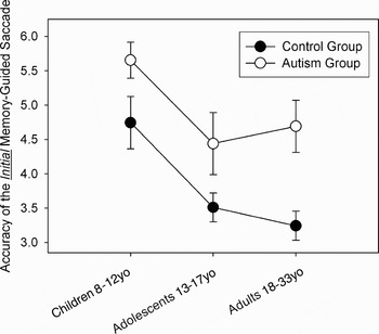
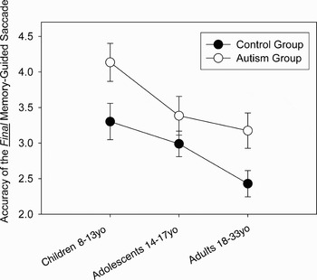
Major psychopathology is now widely recognized to have a neurobiological basis that is distinct to each disorder and underlies its behavioral characteristics, its etiology, and its response to treatment. Neuroimaging has been a revolutionary step in our understanding of neurodevelopmental disorders and psychopathologies, as it provides a window into the possible distinct neural profiles. Two additional areas of research that can provide significant insight into the neural basis of disorders such as autism and other psychopathologies is examining the state of executive function and the developmental profile.
Autism is a neurodevelopmental condition characterized by deficits in language development and social interaction, as well as restricted and stereotyped patterns of behavior, interests, and activity (American Psychiatric Association, 2000). One of the primary broader phenotypes in autism is executive dysfunction (Dawson et al., Reference Dawson, Webb, Schellenberg, Dager, Friedman and Aylward2008), which appears to be present in adulthood as well as throughout development (Bennetto, Pennington, & Rogers, Reference Bennetto, Pennington and Rogers1996; Luna, Doll, Hegedus, Minshew, & Sweeney, Reference Luna, Doll, Hegedus, Minshew and Sweeney2006; Minshew, Meyer, & Dunn, Reference Minshew, Meyer, Dunn, Segakiwutz and Rapin2003; Ozonoff et al., Reference Ozonoff, Cook, Coon, Dawson, Joseph and Klin2004; van der Geest, Kemner, Camfferman, Verbaten, & van Engeland, Reference van der Geest, Kemner, Camfferman, Verbaten and van Engeland2001; Volkmar, Chawarska, & Klin, Reference Volkmar, Chawarska and Klin2005). Executive dysfunction in autism is supposedly supported by brain system abnormalities that compromise complex information processing (Minshew, Sweeney, & Luna, Reference Minshew, Sweeney and Luna2002).
Executive function refers to voluntary planned behavior that is achieved by the representation of a goal in working memory (WM) and inhibition of automatic task-inappropriate responses. Executive function is critical to voluntary decision making. Essential to executive function is the ability to integrate cortical and subcortical systems that are widely distributed throughout the brain. Disrupting function at any stage in this widely distributed circuitry can undermine executive control of behavior. Therefore, abnormalities in brain systems that result in psychopathology usually also affect executive function, and neurodevelopmental disorders such as autism may be particularly vulnerable. These abnormalities in executive function may differ across disorders, thus providing evidence for where in the system the brain abnormalities occur.
The study of development is particularly important for neurodevelopmental disorders like autism. Characterizing developmental profiles of executive function can potentially reveal factors underlying its etiology and provide insight into the nature of neural impairment. Developmental patterns may also have implications for treatment, in that developmental improvement may indicate stages when there is considerable plasticity, and intervention is particularly useful. Identifying when psychopathologies emerge can provide information on which particular brain systems may be impaired and, importantly, which may be spared.
Adolescence is a time of rapid change in physical, social, and cognitive abilities, and thus provides a unique window into autism. In typical children, executive function continues to improve through adolescence (Demetriou, Christou, Spanoudis, & Platsidou, Reference Demetriou, Christou, Spanoudis and Platsidou2002; Luna, Garver, Urban, Lazar, & Sweeney, Reference Luna, Garver, Urban, Lazar and Sweeney2004; Ridderinkhof & van der Molen, Reference Ridderinkhof and van der Molen1997; Swanson, Reference Swanson1999; Zald & Iacono, Reference Zald and Iacono1998) and characterizes mature adult-level decision making. Concurrent with these behavioral improvements are neuroanatomical changes in cortical and subcortical networks (Gogtay et al., Reference Gogtay, Giedd, Lusk, Hayashi, Greenstein and Vaituzis2004; Sowell, Thompson, Holmes, Jernigan, & Toga, Reference Sowell, Thompson, Holmes, Jernigan and Toga1999) including synaptic pruning (Huttenlocher, Reference Huttenlocher1990) and myelination (Pfefferbaum et al., Reference Pfefferbaum, Mathalon, Sullivan, Rawles, Zipursky and Lim1994; Yakovlev & Lecours, Reference Yakovlev, Lecours and Minkowski1967). Understanding the normative course of the brain basis of cognitive maturation, and how it differs in autism, can allow us to better characterize impaired development.
In this review, we will first present the literature on normative brain and cognitive development as a crucial contrast to the literature characterizing impairments in autism. We start with the literature on the development of brain structures and then review findings on two core components of executive function: response inhibition and WM. Each section begins with what is known about the normative development to provide a background for understanding the atypical development found in autism. We then examine behavioral and neuroimaging studies of autism as a prototypical example of how dysmaturation of executive function can provide insight into the integrity of brain systems in a neurodevelopmental disorder
Normative Brain Maturation
Gray matter development
Although brain size and weight approximates adult levels by the first decade of life, maturation continues through adolescence (Caviness, Kennedy, Bates, & Makris, Reference Caviness, Kennedy, Bates, Makris, Thatcher, Reid Lyon, Rumsey and Krasnegor1996; Huttenlocher, Reference Huttenlocher1990; Yakovlev & Lecours, 1967). Magnetic resonance imaging (MRI) volumetric studies have indicated increases in gray matter volume throughout neocortex from childhood to adolescence that then decreases into adulthood (Giedd et al., Reference Giedd, Blumenthal, Jeffries, Castellanos, Liu and Zijdenbos1999; Gogtay et al., Reference Gogtay, Giedd, Lusk, Hayashi, Greenstein and Vaituzis2004; Sowell, Thompson, Tessner, & Toga, Reference Sowell, Thompson, Tessner and Toga2001). Recently, studies have shown that reductions in gray matter volume begin around puberty in the sensorimotor regions followed by a rostral spread over the frontal cortex and caudally over the parietal cortex with continued reductions in temporal regions (Gogtay et al., Reference Gogtay, Giedd, Lusk, Hayashi, Greenstein and Vaituzis2004). Unlike the traditional notion that the human brain develops posterior to anterior with frontal regions developing last, these studies indicate association areas in each lobe are the last to mature. This protracted development includes orbitofrontal cortex and the posterior part of the superior temporal gyrus associated with language development. This pattern indicates that the neural circuits responsible for integrating information across a wide range of areas and functions, thus supporting complex behavior, are the last to mature. Although the neuroanatomical correlate of these decreases in gray matter remains to be confirmed, a likely mechanism that underlies these results is synaptic pruning.
Synaptic pruning
Synaptic pruning refers to the programmed elimination of unused synaptic connections. The connections between neurons multiply rapidly in the first 2 years of life through synaptogenesis. After the first 2 years of life, there is a stage of elimination of synapses (synaptic pruning) that is believed to provide a mechanism for the brain to adapt to an individual's particular environmental needs (Rauschecker & Marler, Reference Rauschecker, Marler, Rauschecker and Marler1987). A more restricted and better specialized synaptic distribution would support more efficient neuronal computations that would, in turn, enhance computational capacity, and increase speed of neuronal transmission of regional circuitry. More efficient widespread processing enables the rapid and complicated computations that support complex behaviors including executive function.
Morphological studies indicate that different parts of the brain undergo pruning at different time intervals. Although area 17 of visual cortex matures in early childhood, synapses in auditory cortex continue to develop into late childhood, and the middle frontal gyrus of the prefrontal cortex (PFC) continues to mature into adolescence (Huttenlocher, Reference Huttenlocher1990; Huttenlocher & Dabholkar, Reference Huttenlocher and Dabholkar1997). These results are frequently cited to indicate that unlike nonhuman primate cortex, which develops concurrently across the brain (Rakic, Bourgeois, Eckenhoff, Zecevic, & Goldman-Rakic, Reference Rakic, Bourgeois, Eckenhoff, Zecevic and Goldman-Rakic1986), the human brain develops in a hierarchical fashion with sensory areas developing before executive regions. However, as mentioned above, the recent MRI studies indicating that gray matter thinning continues in association cortex across lobes indicates that cortical regions that support complex functional integration in each lobe develop last (Giedd et al., Reference Giedd, Blumenthal, Jeffries, Castellanos, Liu and Zijdenbos1999; Gogtay et al., Reference Gogtay, Giedd, Lusk, Hayashi, Greenstein and Vaituzis2004; Paus et al., Reference Paus, Zijdenbos, Worsley, Collins, Blumenthal and Giedd1999; Sowell et al., Reference Sowell, Thompson, Tessner and Toga2001; Toga, Thompson, & Sowell, Reference Toga, Thompson and Sowell2006). The implications are that the late development of executive function and its vulnerability to impairment may not simply reflect abnormalities in prefrontal regions but importantly the ability for the brain to integrate function across cortex.
These increases in efficient regional computational power afforded by synaptic pruning may play a key role in the improvements observed in cognitive maturation, as well as the impairments found in autism. Autism involves the preservation of basic behavioral processes with abnormalities in complex functioning (Minshew et al., Reference Minshew, Sweeney and Luna2002), suggesting that the mechanisms that support complex information processing, such as those subserved by association areas, are particularly vulnerable to abnormalities in this disorder.
White matter maturation
Unlike gray matter development, which is relatively stable by adolescence, white matter development continues through adulthood and in some areas into later phases in the age span (Benes, Reference Benes1989; Yakovlev & Lecours, 1967). Age-related changes in myelination are believed to underlie the protracted maturation of white matter. In the following, we review evidence on the structural development of white matter from histological studies, volumetric MRI, and diffusion tensor imaging (DTI), as well as evidence on the functional connectivity between regions.
Myelination
Myelination is the process of acquisition of myelin, a complex phospholipid outgrowth of the oligodendrocytes in the central nervous system, on neuronal tracts. Histological studies indicate that myelination begins during the second trimester of pregnancy and continues into adult life (Yakovlev & Lecours, 1967) and that myelination in the peripheral nervous system appears to precede development in the central nervous system (CNS; Yakovlev & Lecours, 1967). CNS myelination starts earliest in regions relating to the sensory system rather than the motor system, with regions involved in supporting associative functions and sensory discrimination progressing over decades (Yakovlev & Lecours, 1967). Although histological studies are unique in being able to measure myelin directly, it is important to note, however, that histological studies represent brain areas from data obtained from a discrete sample of a region, and therefore limits the ability to detect changes within regions and to characterize whole brain systems. Recent in vivo MRI methods provide a better view of white matter integrity at the systems level.
MRI volumetric studies of age-related changes in white matter, which provide a whole brain view, indicate a steady increase of white matter volume from early childhood into adulthood with similar rates across different brain lobes, but with slightly steeper trajectories in males compared to females (Giedd et al., Reference Giedd, Blumenthal, Jeffries, Castellanos, Liu and Zijdenbos1999; Lenroot & Giedd, Reference Lenroot and Giedd2006). In these and other studies (Pfefferbaum et al., Reference Pfefferbaum, Mathalon, Sullivan, Rawles, Zipursky and Lim1994; Reiss, Abrams, Singer, Ross, & Denckla, Reference Reiss, Abrams, Singer, Ross and Denckla1996), age-related increases in white matter volume are accompanied by decreases in gray matter volume. Although many of these studies were focused on global white matter changes, the clear visibility of the corpus callosum has facilitated studies in regional white matter development. Pujol, Vendrell, Junqué, Martí-Vilalta, and Capdevila (1993) documented volumetric increases in the corpus callosum up to age 27, and subsequent studies have found regional age-dependent increases, particularly in the posterior region, including the splenium (Giedd et al., Reference Giedd, Rumsey, Castellanos, Rajapakse, Kaysen and Vaituzis1996, Reference Giedd, Blumenthal, Jeffries, Castellanos, Liu and Zijdenbos1999), a finding that again counters the notion of a strictly caudal–rostral maturation pattern of the brain. Some regional differences have been found in the temporal lobe (Paus, Reference Paus1999) and hippocampus (Benes, Turtle, Khan, & Farol, Reference Benes, Turtle, Khan and Farol1994), indicating a protracted development of these areas that continues into the sixth decade of life.
Recently, DTI has been used as an indirect measure of developmental changes in myelination (Klingberg, Vaidya, Gabrieli, Moseley, & Hedehus, Reference Klingberg, Vaidya, Gabrieli, Moseley and Hedehus1999; Paus, Reference Paus1999; Schmithorst, Wilke, Dardzinski, & Holland, Reference Schmithorst, Wilke, Dardzinski and Holland2002; Snook, Plewes, & Beaulieu, Reference Snook, Plewes and Beaulieu2007). DTI is a noninvasive MRI method that measures the coherence of water diffusion in the brain parenchyma. Without anatomic barriers, water typically would diffuse freely in all directions (isotropic diffusion). However, in the boundaries of white matter tracts, diffusion is more restricted and diffusion can predominate in a certain direction (anisotropic diffusion). Therefore, greater coherence of water diffusion reflects better integrity of white matter tracts, to which myelination has been found to be a major contributor (Moseley et al., Reference Moseley, Cohen, Kucharczyk, Mintorovitch, Asgari and Wendland1990; Song et al., Reference Song, Sun, Ju, Lin, Cross and Neufeld2003). A commonly used measure of white matter integrity is fractional anisotropy (FA), which describes diffusion along the axons of the white matter. Whole-brain FA values increase throughout childhood and adolescence, stabilizing in the second or third decade of life; this increase is thought to reflect the known increases in myelination (Ben Bashat et al., Reference Ben Bashat, Ben Sira, Graif, Pianka, Hendler and Cohen2005; Li & Noseworthy, Reference Li and Noseworthy2002; Mukherjee et al., Reference Mukherjee, Miller, Shimony, Conturo, Lee and Almli2001). FA increases with age in the major groups of tracts that provide intracortical and cortical–subcortical connections including the internal capsule, corticospinal tract, arcuate fasciculus, internal capsule, inferior longitudinal fasciculus regions, and corpus callosum (Ashtari et al., Reference Ashtari, Cervellione, Hasan, Wu, McIlree and Kester2007; Barnea-Goraly et al., Reference Barnea-Goraly, Menon, Eckert, Tamm, Bammer and Karchemskiy2005; Cascio, Gerig, & Piven, Reference Cascio, Gerig and Piven2007; Lebel, Walker, Leemans, Phillips, & Beaulieu, Reference Lebel, Walker, Leemans, Phillips and Beaulieu2008; McLaughlin et al., Reference McLaughlin, Paul, Grieve, Williams, Laidlaw and DiCarlo2007; Schmithorst et al., Reference Schmithorst, Wilke, Dardzinski and Holland2002; Snook et al., Reference Snook, Plewes and Beaulieu2007). These tracts support functional integration underlying fine motor control, language processing, and executive control. The role of functional integration in cognition is reflected by the association of FA values across executive regions with diverse measures of cognitive performance, including WM, visuospatial processing, reaction time, reading, and IQ (Klingberg, Reference Klingberg2006; Klingberg, Forssberg, & Westerberg, Reference Klingberg, Forssberg and Westerberg2002; Liston et al., Reference Liston, Watts, Tottenham, Davidson, Niogi and Ulug2006; Mabbott, Noseworthy, Bouffet, Laughlin, & Rockel, Reference Mabbott, Noseworthy, Bouffet, Laughlin and Rockel2006; Nagy, Westerberg, & Klingberg, Reference Nagy, Westerberg and Klingberg2004; Olesen, Nagy, Westerberg, & Klingberg, Reference Olesen, Nagy, Westerberg and Klingberg2003).
Functional connectivity
There are improvements in functional connectivity over development that also support efficient brain function by both enhancing regional computational speed and the ability to integrate information across long distances, which is critical for top-down executive control. The ability to establish widely distributed circuitries through enhanced connectivity is crucial for the maturation of executive function (Goldman-Rakic, Reference Goldman-Rakic1988). Coherence electroencephalography (Thatcher, Walker, & Giudice, Reference Thatcher, Walker and Giudice1987) is a method that has demonstrated the development of corticocortical coupling of discharges in several stages through early adulthood, and may reflect improvements in propagating circuitry. Recently, the integrity of functional connectivity has been assessed using resting-state functional connectivity MRI (rs-fcMRI), which provides correlations of low-frequency blood oxygenation level dependent signal fluctuations between brain regions during rest baseline (while subjects retain fixation between experimental blocks). This method is believed to delineate functional circuits that are present. Using rs-fcMRI, age-related circuit level changes of control networks that underlie executive function have been found to continue to mature through adolescence (Fair et al., Reference Fair, Dosenbach, Church, Cohen, Brahmbhatt and Miezin2007; Thatcher et al., Reference Thatcher, Walker and Giudice1987). Thus, functional connectivity has a long developmental trajectory. Atypical development of functional connectivity could support the generalized impairments found in certain neurodevelopmental disorders such as autism (Williams & Minshew, Reference Williams and Minshew2007).
Taken together, these studies indicate a protracted development of brain structure, including reorganization of association areas and enhancement of structural and functional connectivity, which continues throughout adolescence and involves widespread brain regions. There are several implications of these findings. The emphasis on systems level organization during the developmental stage of adolescence suggests that it is important to focus on brain circuits and the integrity of functional connections. In addition, these findings suggest that this later stage of brain maturation is qualitatively unique, and the developmental course of psychopathologies during this time can potentially inform us regarding underlying brain abnormalities.
Brain Development in Autism
Autism is an example of neurodevelopmental psychopathology that occurs early in development, often diagnosed by 2 or 3 years of age, and is now widely viewed as being of neurobiologic origin (Amaral, Schumann, & Nordahl, Reference Amaral, Schumann and Nordahl2008). There is now a wide literature characterizing a range of neurobiological abnormalities associated with autism. For example, gross morphometric studies using MRI have reported accentuated spaces surrounding the smaller cerebral veins (Virchow–Robin spaces; Taber et al., Reference Taber, Shaw, Loveland, Pearson, Lane and Hayman2004), white matter hyperintensities (Courchesne et al., Reference Courchesne, Karns, Davis, Ziccardi, Carper and Tigue2001), and migrational defects (Piven et al., Reference Piven, Berthier, Starkstein, Nehme, Pearlson and Folstein1990; Schifter et al., Reference Schifter, Hoffman, Hatten, Hanson, Coleman and DeLong1994). Some of these findings point to the association of specific genetic conditions such as fragile X (Levitas et al., Reference Levitas, Hagerman, Braden, Rimland, McBogg and Matus1983) or tuberous sclerosis (Asato & Hardan, Reference Asato and Hardan2004) with autism, which have motivated extensive subsequent studies to investigate the genetic underpinnings of autism. Studies of brain structure in autism have provided evidence of abnormalities in cerebral cortex, limbic structures, and the cerebellum. These abnormalities may indicate disturbances in the elaboration of dendritic and axonal processes, the selective elimination of neuronal processes, and programmed cell death during brain development, which have been associated with a range of social and cognitive impairments (Acosta & Pearl, Reference Acosta and Pearl2003; Amaral et al., 2008). However, other studies indicate that the incidence of these brain structural abnormalities in autism does not exceed the incidence in the general population (Damasio, Maurer, Damasio, & Chui, Reference Damasio, Maurer, Damasio and Chui1980; Filipek, Reference Filipek1999).
Despite the lack of accumulating evidence of structural abnormalities in autism, the common clinical observation of relative macrocephaly in younger children with autism (Aylward, Minshew, Field, Sparks, & Singh, Reference Aylward, Minshew, Field, Sparks and Singh2002; Courchesne et al., Reference Courchesne, Karns, Davis, Ziccardi, Carper and Tigue2001; Sparks et al., Reference Sparks, Friedman, Shaw, Aylward, Echelard and Artru2002) has stimulated volumetric studies in this population. Although adult brain volumes and head circumferences do not differ from healthy controls in the majority of cases (Redcay & Courchesne, Reference Redcay and Courchesne2005), there is evidence for normative brain size in autism at birth (Courchesne, Carper, & Akshoomoff, Reference Courchesne, Carper and Akshoomoff2003; Lainhart et al., Reference Lainhart, Piven, Wzorek, Landa, Santangelo and Coon1997) followed by a rapid growth from 2 to 4 years compared to typical individuals (Aylward et al., Reference Aylward, Minshew, Field, Sparks and Singh2002; Courchesne et al., Reference Courchesne, Karns, Davis, Ziccardi, Carper and Tigue2001; Sparks et al., Reference Sparks, Friedman, Shaw, Aylward, Echelard and Artru2002). These head size differences have been consistently related to increased volume in the lateral ventricles (Piven et al., Reference Piven, Arndt, Bailey, Havercamp, Andreasen and Palmer1995) and uneven expansion across regions in the brain. Early work indicated that the overall size of temporal, parietal, and occipital lobes was larger in autism (Piven, Arndt, Bailey, & Andreasen, Reference Piven, Arndt, Bailey and Andreasen1996). However, more recent work indicates that, although white and gray matter volumes of autistic individuals were enlarged in temporal, parietal, and limbic areas, volumes were increased even more in frontal and especially prefrontal regions, while occipital cortex was found to be normative (Carper & Courchesne, Reference Carper and Courchesne2005; Sparks et al., Reference Sparks, Friedman, Shaw, Aylward, Echelard and Artru2002). From childhood to adolescence, there is an apparent arrest in brain size compared to normative brain development, especially in frontal regions that show early increase in size, resulting in normal brain size by adulthood (Redcay & Courchesne, Reference Redcay and Courchesne2005).The volume enlargement occurs during a period when many autistic behaviors are recognized, suggesting a pathologic process of both early overgrowth of neural and dendritic connections and later deficiency of normal neuronal pruning (Courchesne et al., Reference Courchesne, Karns, Davis, Ziccardi, Carper and Tigue2001; Courchesne, Redcay, Morgan, & Kennedy, Reference Courchesne, Redcay, Morgan and Kennedy2005).
In addition to large differences found in the autistic brain, there has been evidence for regional structural abnormalities in autism including in the corpus callosum, limbic system, and hippocampus (Egaas, Courchesne, & Saitoh, Reference Egaas, Courchesne and Saitoh1995; Hardan, Minshew, & Keshavan, Reference Hardan, Minshew and Keshavan2000). The corpus callosum appears reduced in volume in the posterior region (Egaas et al., Reference Egaas, Courchesne and Saitoh1995; Piven, Bailey, Ranson, & Arndt, Reference Piven, Bailey, Ranson and Arndt1997). There is also evidence of abnormalities in amygdala volume in autism (Abell et al., Reference Abell, Krams, Ashburner, Passingham, Friston and Frackowiak1999; Schumann & Amaral, Reference Schumann and Amaral2006; Sparks et al., Reference Sparks, Friedman, Shaw, Aylward, Echelard and Artru2002); this has resulted in theories emphasizing abnormalities in amygdala as central to autism spectrum disorder, especially to social symptoms (Nacewicz et al., Reference Nacewicz, Dalton, Johnstone, Long, McAuliff and Oakes2006). However, studies of the hippocampus and limbic systems have been limited by small sample sizes, varied age ranges of the subjects, and varied severity of autistic symptoms (Aylward et al., Reference Aylward, Minshew, Goldstein, Honeycutt, Augustine and Yates1999; Piven, Bailey, Ranson, & Arndt, Reference Piven, Bailey, Ranson and Arndt1998; Saitoh, Courchesne, Egaas, Lincoln, & Schreibman, Reference Saitoh, Courchesne, Egaas, Lincoln and Schreibman1995).
Some of the earliest structural differences in autism were documented in the cerebellum. The cerebellum, although crucial for basic integration of motor functions, forms a circuit with the PFC (Middleton & Strick, Reference Middleton and Strick2001), and has been increasingly found to play a role in cognitive function (Akshoomoff & Courchesne, Reference Akshoomoff and Courchesne1992; Mostofsky, Goldberg, & Denckla, Reference Mostofsky, Goldberg and Denckla2000; Ravizza et al., Reference Ravizza, McCormick, Schlerf, Justus, Ivry and Fiez2006). Postmortem histological studies documented a paucity of Purkinje neurons in the vermis of the cerebellum (Bailey et al., Reference Bailey, Luthert, Dean, Harding, Janota and Montgomery1998; Courchesne, Reference Courchesne1997). An early MRI study reported decreased regional cerebellar vermis size (Courchesne, Yeung-Courchesne, Press, Hesselink, & Jernigan, Reference Courchesne, Yeung-Courchesne, Press, Hesselink and Jernigan1988) and decreased ratio of gray to white matter compared to control subjects (Courchesne et al., Reference Courchesne, Karns, Davis, Ziccardi, Carper and Tigue2001). Specific cerebellar regions that were smaller in individuals with autism compared to controls included lobules VI–VII, which are largely composed of gray matter (Courchesne et al., Reference Courchesne, Karns, Davis, Ziccardi, Carper and Tigue2001; Hashimoto et al., Reference Hashimoto, Tayama, Murakawa, Yoshimoto, Miyazaki and Harada1995). However, the groups being compared in these studies were not matched on IQ, which has been reported to have strong effect on vermal volume (Schaefer et al., Reference Schaefer, Thompson, Bodensteiner, McConnell, Kimberling and Gay1996). Studies controlling for IQ did not find differences in vermal lobules in autism (Filipek et al., Reference Filipek, Richelme, Kennedy, Rademacher, Pitcher and Zidel1992; Garber & Ritvo, Reference Garber and Ritvo1992). Although further studies documenting hypoplasia of lobules VI–VII have not been replicated, other studies have shown increased cerebellar volume, which appeared proportional to increases in brain volume (Hardan, Minshew, Mallikarjuhn, & Keshavan, Reference Hardan, Minshew, Mallikarjuhn and Keshavan2001; Herbert et al., Reference Herbert, Ziegler, Deutsch, O'Brien, Lange and Bakardjiev2003). Behavioral studies indicating a consistent impairment in the accuracy of eye movements support a functional impairment in the cerebellar vermis (Luna, Doll, Hegedus, Minshew, & Sweeney, Reference Luna, Doll, Hegedus, Minshew and Sweeney2007; Takarae, Minshew, Luna, & Sweeney, Reference Takarae, Minshew, Luna and Sweeney2004) as do studies indicating decreased recruitment of cerebellum during attention shifting tasks in adolescence (Allen & Courchesne, Reference Allen and Courchesne2003) and adulthood (Townsend et al., Reference Townsend, Courchesne, Covington, Westerfield, Harris and Lyden1999). These results indicate that abnormalities in the cerebellum may also play a role in autism.
White matter in autism
Given the variability of gray matter abnormalities and the range of cognitive and social deficits specific to autism, the possibility of altered connectivity is a new focus of autism research. Volumetric abnormalities in white matter could be attributed to differences in axonal density or organization, abnormalities of myelination, or abnormal glial cell proliferation. Morphometric white matter studies in autism have suggested early overgrowth of white matter in young children with autism, followed by relatively reduced white matter volume in adolescence and adulthood (Courchesne et al., Reference Courchesne, Karns, Davis, Ziccardi, Carper and Tigue2001; Herbert et al., Reference Herbert, Ziegler, Makris, Filipek, Kemper and Normandin2004; Waiter et al., Reference Waiter, Williams, Murray, Gilchrist, Perrett and Whiten2005). In addition, there is evidence for volumetric decreases in the corpus callosum, particularly in the posterior regions (Egaas et al., Reference Egaas, Courchesne and Saitoh1995; Piven et al., Reference Piven, Bailey, Ranson and Arndt1997) undermining interhemispheric connectivity in autism.
White matter volumetric differences could be attributable to changes in axonal density or organization, or from primary abnormalities in myelin. Recent advances in MRI imaging techniques using DTI provide a measure of white matter integrity (of which myelination is a primary contributor) that may provide insight into the possible microstructural mechanisms underlying white matter abnormalities in autism. DTI studies have found evidence for reduced FA in temporal cortex in adult individuals with autism (Lee et al., Reference Lee, Bigler, Alexander, Lazar, DuBray and Chung2007) and throughout cortical and subcortical regions including the ventromedial PFC, anterior cingulate, temporal lobe, amygdala, and the corpus callosum in children and adolescents with autism (Barnea-Goraly et al., Reference Barnea-Goraly, Kwon, Menon, Eliez, Lotspeich and Reiss2004). Because FA increases with age with continued myelination (Mukherjee et al., Reference Mukherjee, Miller, Shimony, Conturo, Lee and Almli2001), it is not clear if autism involves a developmental lag or a persistent reduction in white matter integrity. A large cross sectional study with a broad age range including adults (Keller, Kana, & Just, Reference Keller, Kana and Just2007) found lower FA in regions within and near the corpus callosum and in the right retrolenticular portion of the internal capsule. The posterior limb of the external capsule showed an interaction with age indicating persistent impairment in white matter integrity into adulthood. These impairments in white matter integrity may be more prominent in some subgroups of autism, as findings indicate that abnormal white matter in autism is related to low-performance IQ (Alexander et al., Reference Alexander, Lee, Lazar, Boudos, DuBray and Oakes2007).
Decreased FA in the presence of increased white matter volumes could indicate abnormal connectivity architecture including increases in white matter connections that are not fully myelinated. A surplus of connections whose efficiency is undermined by immature myelination could significantly impact information processing especially for processes such as executive function that rely on the ability to efficiently integrate brain function. Compromised white matter integrity undermines brain functional integration and forms the basis for current theories of abnormal connectivity underlying autism (Hughes, Reference Hughes2007; Just, Cherkassky, Keller, Kana, & Minshew, Reference Just, Cherkassky, Keller, Kana and Minshew2007; Luna et al., Reference Luna, Minshew, Garver, Lazar, Thulborn and Eddy2002; Minshew & Williams, Reference Minshew and Williams2007).
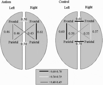
Different lines of evidence support abnormal functional connectivity in autism. For example, individuals with autism are often found to have interictal epileptiform discharges, which do not consistently progress to generalized seizures suggesting differences in overall connectivity (Hughes & Melyn, Reference Hughes and Melyn2005). Similarly, there is magnetoencephalography evidence of decreased gamma oscillations (Wilson, Rojas, Reite, Teale, & Rogers, Reference Wilson, Rojas, Reite, Teale and Rogers2007), and rs-fcMRI evidence of decreased resting state coherence (Cherkassky, Kana, Keller, & Just, Reference Cherkassky, Kana, Keller and Just2006; Murias, Webb, Greenson, & Dawson, Reference Murias, Webb, Greenson and Dawson2007), all of which reflect alterations in interregional connectivity. It is important that studies using functional MRI (fMRI) indicate that underconnectivity is evident during the performance of higher level tasks. Our initial results (see below) of selective decreases in activity in prefrontal and posterior cingulate cortices during an executive task were interpreted as revealing a compromise of functional connectivity of higher order processes (Luna et al., Reference Luna, Minshew, Garver, Lazar, Thulborn and Eddy2002); however, functional connectivity analyses were not performed. Studies where functional connectivity has been assessed by measuring the correlation in activation between two areas consistently show decreased functional connectivity. Several cognitive tasks including sentence comprehension, and WM tasks using letter and face stimuli, indicate compromised functional connectivity of long-range corticocortical connections of the regions used in these tasks. These results have led to a theoretical proposal suggesting that impaired functional connectivity is core to autism (Just, Cherkassky, Keller, & Minshew, Reference Just, Cherkassky, Keller and Minshew2004; Koshino et al., Reference Koshino, Carpenter, Minshew, Cherkassky, Keller and Just2005, Reference Koshino, Kana, Keller, Cherkassky, Minshew and Just2008). Figure 1 illustrates the finding from one of these studies indicating that, during an n-back WM task with letters, high-functioning autistic individuals showed decreased functional connectivity in left prefrontal–parietal circuits compared to controls, suggesting that functional integration is compromised in autism (Koshino et al., Reference Koshino, Carpenter, Minshew, Cherkassky, Keller and Just2005). In addition to WM tasks (Koshino et al., Reference Koshino, Carpenter, Minshew, Cherkassky, Keller and Just2005, Reference Koshino, Kana, Keller, Cherkassky, Minshew and Just2008), decreases in functional connectivity of frontal and posterior regions have been found in response inhibition tasks (Kana, Keller, Minshew, & Just, Reference Kana, Keller, Minshew and Just2007; see below) and sentence comprehension tasks (Just et al., Reference Just, Cherkassky, Keller and Minshew2004; Kana, Keller, Cherkassky, Minshew, & Just, Reference Kana, Keller, Cherkassky, Minshew and Just2006). Reduced connectivity in visual, temporal, and parietal regions has also been found during mental attribution tasks in autism (Castelli, Frith, Happe, & Frith, Reference Castelli, Frith, Happe and Frith2002). Taken together, these results provide evidence for important differences in functional connectivity in autism that may be critical to the limitations in higher order information processing found in this disorder.
The autism group showed significantly less functional connectivity between the left frontal and parietal regions of interest (ROIs) than a control group during a working memory n-back task. Numbers indicate the correlation between areas. From “Functional Connectivity in an fMRI Working Memory Task in High-Functioning Autism,” by H. Koshio, P. A. Carpenter, N. J. Minshew, V. L. Cherkassky, T. A. Keller, and M. A. Just, 2005, NeuroImage, 24, 810–821. Copyright 2005 by Elsevier. Reprinted with permission.

It is important to note that although reduced functional connectivity has been found throughout these studies, much of the circuitry in autism shows functional connectivity that is comparable to controls. This indicates that autism is not associated with a generalized impairment in functional connectivity. Instead, these studies suggest abnormalities in being able to effectively integrate brain circuits when they are used to support complex information processing in a cognitive task. These studies support the proposal that underlying autism are abnormalities that selectively affect higher order behaviors such as executive function and social processing while sparing basic processes that are not as dependent on widely distributed circuitry (Minshew et al., Reference Minshew, Sweeney and Luna2002). This view is also consistent with results indicating excess neurons in certain regions of the brain (such as the frontal regions) at critical periods of development, and the promotion of short connections at the expense of longer distance connections, such as those connecting the frontal and temporal regions in language tasks (Courchesne et al., Reference Courchesne, Pierce, Schumann, Redcay, Buckwalter and Kennedy2007).
Given these white matter differences, as well as morphometric changes in the gray matter in the brain in autism, it appears that there are multiple levels of neuroanatomic involvement suggesting a broad vulnerable network in this neurodevelopmental disorder. These levels include the possibility of decreased synaptic pruning early in life, reflected in increased head size; regional brain abnormalities (e.g., the cerebellum) in adulthood; and decreased connectivity between regions. The timing and developmental course of these neuroanatomical abnormalities provides crucial insight into the mechanisms that are damaged in autism. Because executive function undergoes a long developmental trajectory, studying the alterations in executive function in autism over development provides an opportunity to understand the nature of the impact of compromised neural networks on behavior in autism.
Executive Dysfunction in Autism
Executive functions are not globally impaired in autism. Thus, it is the pattern of deficits across tasks, isolating the specific components of executive function that are damaged, as well as its developmental course, that can elucidate how the system differs in people with autism (for reviews, see Hill, Reference Hill2004; Russo et al., Reference Russo, Flanagan, Iarocci, Berringer, Zelazo and Burack2007). Such behavioral differences may reflect distinct brain circuitry being recruited over development in autism. Although executive functions are traditionally thought to be mediated by the PFC (Duncan, Reference Duncan1986), not all neurodevelopmental disorders that affect executive function clearly involve the frontal lobe. This suggests that intact executive functioning may depend on the flexible use of integrated brain networks in addition to intact PFC function. As such, executive dysfunction in neurodevelopmental disorders such as autism may reflect more generalized brain abnormalities that undermine collaborative neocortical function and connectivity. In the subsequent sections of this paper, we review core skills supporting normative executive functioning, and discuss how the alterations seen in autism may relate to structural and functional differences in the brain, and how this approach may provide a model for study of other neurodevelopmental disorders.
Executive abilities are evident early in development but continue to improve throughout childhood and into adolescence (Demetriou et al., Reference Demetriou, Christou, Spanoudis and Platsidou2002; Luciana, Conklin, Hooper, & Yarger, Reference Luciana, Conklin, Hooper and Yarger2005; Luna et al., Reference Luna, Garver, Urban, Lazar and Sweeney2004). In addition to frontal areas, this protracted maturation of executive function reflects developmental changes in a wide network of areas, including subcortical and cerebellar areas, and their connectivity throughout the brain (Goldman-Rakic, Chafee, & Friedman, Reference Goldman-Rakic, Chafee, Friedman, Ono, Squire, Raichle, Perrett and Fukuda1993). Given the complex neural mechanisms and widely distributed circuitry supporting the higher level processes of executive function, it may be particularly vulnerable to disruption resulting from impaired neurobiological processes in psychopathology. Indeed, executive functions are pervasively affected in many neurodevelopmental disorders, including attention-deficit disorder (Pennington & Ozonoff, Reference Pennington and Ozonoff1996), and Tourette syndrome (Peterson, Pine, Cohen, & Brook, Reference Peterson, Pine, Cohen and Brook2001), across a broad range of ages and functional levels (Ozonoff et al., Reference Ozonoff, Cook, Coon, Dawson, Joseph and Klin2004; Pennington & Ozonoff, Reference Pennington and Ozonoff1996; Rumsey & Hamburger, Reference Rumsey and Hamburger1988; Russell, Reference Russell1997). Thus, probing the integrity of this system behaviorally can provide insight into the integrity of the brain systems supporting it: characterizing its developmental trajectory provides additional information. Although there is evidence that some aspects of executive function are impaired at different ages in autism, its developmental trajectory is not well defined, and it is not clear whether the development of executive function is delayed, persistently low, or truncated. This developmental information can provide insight into aspects of brain maturation that may be particularly intact or impaired in autism. For example, a normative stagelike improvement in late childhood, like that described below, may reflect intact synaptic pruning and myelination. Although still preliminary, behavioral and imaging work using the developmental perspective to provide insight into the deficits in autism is promising (Happe, Booth, Charlton, & Hughs, Reference Happe, Booth, Charlton and Hughs2006; Luna et al., Reference Luna, Minshew, Garver, Lazar, Thulborn and Eddy2002, Reference Luna, Doll, Hegedus, Minshew and Sweeney2007).
Tasks designed to tap executive function, such as in the Wisconsin card sort task and in the tower of London, typically include a range of executive components (set switching, cognitive flexibility, planning). Although these tasks provide us with an overall assessment of executive function, it is important to focus on individual component processes in order to understand the nature of abnormalities in neurodevelopmental disorders. In this review, we focus on response inhibition and WM, two basic components of executive function. Response inhibition allows automatic responses that may not be goal oriented to be suppressed in favor of a goal-driven, voluntary, planned response. WM allows representations to be maintained on-line to guide goal-directed executive behavior. Although response inhibition and WM work in unison to support executive function, their relative contributions can be distinguished (Asato, Sweeney, & Luna, Reference Asato, Sweeney and Luna2006). The study of executive function deficits in autism, and the neural underpinnings of these functions, promises to provide insight into the etiology of autism, especially the integrity of the widely distributed brain circuits.
Response inhibition
Response inhibition is a core executive process that supports goal-directed behavior by allowing the ability to suppress inappropriate automatic responses that are not goal appropriate, filter out distractors, and retain a response set (Bjorklund & Harnishfeger, Reference Bjorklund, Harnishfeger, Dempster and Brainerd1995; Dempster, Reference Dempster1992). Studies of typical adults indicate that successful response inhibition requires the recruitment of a wide range of brain areas that often include frontal and prefrontal areas, parietal cortex, cingulate cortex, basal ganglia, thalamus, and the cerebellum (Luna et al., Reference Luna, Thulborn, Munoz, Merriam, Garver and Minshew2001; Rubia et al., Reference Rubia, Russel, Overmeyer, Brammer, Bullmore and Sharma2001). Impairments in response inhibition affect many individuals with developmental disorders, undermining executive function, most notably in attention-deficit/hyperactive disorder (ADHD), obsessive compulsive disorder (OCD), and autism (Sweeney, Takarae, Macmillan, Luna, & Minshew, Reference Sweeney, Takarae, Macmillan, Luna and Minshew2004). Neuroimaging studies indicate that these deficits reflect abnormalities in frontal circuits including frontostriatal and frontoparietal circuitry (Booth et al., Reference Booth, Burman, Meyer, Lei, Trommer and Davenport2005; Kana et al., Reference Kana, Keller, Minshew and Just2007; Menzies, Chamberlain, et al., Reference Menzies, Chamberlain, Laird, Thelen, Sahakian and Bullmore2007b; Roth et al., Reference Roth, Saykin, Flashman, Pixley, West and Mamourian2007; Rubia, Smith, Brammer, Toone, & Taylor, Reference Rubia, Smith, Brammer, Toone and Taylor2005; Schmitz et al., Reference Schmitz, Rubia, Daly, Smith, Williams and Murphy2006; Suskauer et al., Reference Suskauer, Simmonds, Fotedar, Blankner, Pekar and Denckla2008; Woolley et al., Reference Woolley, Heyman, Brammer, Frampton, McGuire and Rubia2008). In this section, we will first review what is known about the development of response inhibition, and then outline how this development might differ in autism.
Experimental paradigms
Although response inhibition is necessary for most everyday behavior, it is difficult to discriminate it empirically from other executive functions such as WM. However, studies need to isolate different core components of executive function, such as response inhibition, to identify the specific neural substrates that may be compromised in particular psychopathologies. The antisaccade task, used in single-cell studies to delineate the neural circuitry underlying response inhibition (Funahashi, Chafee, & Goldman-Rakic, Reference Funahashi, Chafee and Goldman-Rakic1993), may come the closest to isolating response inhibition from other task demands. In this task, subjects must suppress an automatic eye movement toward a suddenly appearing peripheral target and instead generate a planned eye movement to the opposite location (Hallett, Reference Hallett1978). Inhibitory errors of looking at the visual stimulus are usually corrected with a subsequent eye movement to the correct location indicating that the instruction was understood but that there was a failure to engage processes supporting executive systems. The nonverbal nature of this task makes it less amenable to strategizing, which can undermine or obscure developmental differences. Although in general the antisaccade task has low task demands, it also requires high amounts of inhibition because the response to be inhibited is reflexive.
Other well-established tasks for studying response inhibition include the go/no-go task, the stop signal task and the Stroop Color Word Test. The go/no-go task requires participants to inhibit a button press (no-go response), after subjects have routinely pressed the button (go response) making the button press more automatic and harder to suppress. Similarly, in the stop-signal task, subjects must stop a response that has already begun when an unpredictable “stop-signal” appears. In the traditional Stroop Color Word Test, subjects must inhibit reading a color name (i.e., the word “green” printed in red) and instead name the color of the ink in which the word is written (i.e., red). Although the antisaccade, stop-signal, and go/no-go tasks require inhibiting a prepotent behavioral response, performance on the Stroop task is primarily determined by interference suppression. In other words, the tendency to automatically read the word must be suppressed, and adults can use a number of strategies to do so, such as averting their attention from the word, and using their peripheral vision to identify the color.
Normative development of response inhibition
Response inhibition develops throughout childhood and adolescence (Fischer, Biscaldi, & Gezeck, Reference Fischer, Biscaldi and Gezeck1997; Levin, Culhane, Hartmann, Evankovich, & Mattson, Reference Levin, Culhane, Hartmann, Evankovich and Mattson1991; Luciana & Nelson, Reference Luciana and Nelson1998; Munoz, Broughton, Goldring, & Armstrong, Reference Munoz, Broughton, Goldring and Armstrong1998; Paus, Babenko, & Radil, Reference Paus, Babenko and Radil1990; Ridderinkhof, Band, & Logan, Reference Ridderinkhof, Band and Logan1999; Ridderinkhof, van der Molen, Band, & Bashore, Reference Ridderinkhof, van der Molen, Band and Bashore1997). Although even infants are able to sometimes voluntarily suppress a reflexive response, the ability to do this in a consistent and flexible manner improves through adolescence, as reflected in increases in the proportion of correct inhibitory responses. Preadolescent children in particular have more difficulty inhibiting a response that is reflexive or has been done repeatedly and, even if successful, they may take longer to respond. There are improvements through childhood on several aspects of response inhibition, including the ability to retain visual fixation (Paus et al., Reference Paus, Babenko and Radil1990), perform the go/no-go task (Levin et al., Reference Levin, Culhane, Hartmann, Evankovich and Mattson1991; Luciana & Nelson, Reference Luciana and Nelson1998), attend to the variable of interest in the Stroop task (Tipper, Bourque, Anderson, & Brehaut, Reference Tipper, Bourque, Anderson and Brehaut1989; Wise, Sutton, & Gibbons, Reference Wise, Sutton and Gibbons1975), filter out incompatible cues in the flanker task (Ridderinkhof et al., Reference Ridderinkhof, van der Molen, Band and Bashore1997), and interrupt a planned behavior in stop signal tasks (Greenberg & Waldman, Reference Greenberg and Waldman1993; Ridderinkhof et al., Reference Ridderinkhof, Band and Logan1999; Williams, Ponesse, Schachar, Logan, & Tannock, Reference Williams, Ponesse, Schachar, Logan and Tannock1999).
In particular, performance in the antisaccade continues to show improvement throughout adolescence (Fischer et al., Reference Fischer, Biscaldi and Gezeck1997; Fukushima, Hatta, & Fukushima, Reference Fukushima, Hatta and Fukushima2000; Klein & Foerster, Reference Klein and Foerster2001; Luna et al., Reference Luna, Garver, Urban, Lazar and Sweeney2004; Munoz et al., Reference Munoz, Broughton, Goldring and Armstrong1998). Studies have found improvements from 8 years old, when approximately 50% of trials are errors, to adulthood, with only 10–20% errors (Fischer et al., Reference Fischer, Biscaldi and Gezeck1997; Luna et al., Reference Luna, Garver, Urban, Lazar and Sweeney2004). Our results (Luna et al., Reference Luna, Garver, Urban, Lazar and Sweeney2004) on 245 8- to 30-year-old healthy individuals showed significant decreases in the proportion of trials where subjects erroneously made saccades to the visual target until approximately age 14–15 when performance plateaued. This improvement in the ability to suppress a reflexive saccade to make a voluntary response reflects the maturation of a widely distributed circuitry including frontostriatal regions. It is important to note that although inhibitory abilities are limited early in development, they are present. Therefore, what is developing is not the appearance of an ability that was absent, but the reliable and flexible manner in which we can use this ability. This well-delineated typical development in the antisaccade task, continuing into adolescence, provides a crucial comparison for understanding the time line of autism and other neurodevelopmental disorders associated with response inhibition deficits.
Numerous fMRI studies have provided a characterization of the systems supporting antisaccade performance involving a widely distributed circuitry including the frontal eye field (FEF), supplementary eye fields (SEFs), dorsolateral PFC (DLPFC), posterior parietal cortex, anterior cingulate cortex, basal ganglia, thalamus, and superior colliculus (Burman & Bruce, Reference Burman and Bruce1997; Connolly, Goodale, DeSouza, Menon, & Vilis, Reference Connolly, Goodale, DeSouza, Menon and Vilis2000; Cornelissen et al., Reference Cornelissen, Kimmig, Schira, Rutschmann, Maguire and Broerse2002; Curtis & D'Esposito, Reference Curtis and D'Esposito2003; Doricchi et al., Reference Doricchi, Perani, Incoccia, Grassi, Cappa and Bettinardi1997; Everling, Dorris, Klein, & Munoz, Reference Everling, Dorris, Klein and Munoz1999; Ford, Goltz, Brown, & Everling, Reference Ford, Goltz, Brown and Everling2005; Funahashi et al., Reference Funahashi, Chafee and Goldman-Rakic1993; Gottlieb & Goldberg, Reference Gottlieb and Goldberg1999; Guitton, Buchtel, & Douglas, Reference Guitton, Buchtel and Douglas1985; Kimmig et al., Reference Kimmig, Greenlee, Gondan, Schira, Kassubek and Mergner2001; Matsuda et al., Reference Matsuda, Matsuura, Ohkubo, Ohkubo, Matsushima and Inoue2004; Muri et al., Reference Muri, Heid, Nirkko, Ozdoba, Felblinger and Schroth1998; O'Driscoll et al., Reference O'Driscoll, Alpert, Matthysse, Levy, Rauch and Holzman1995; Raemaekers, Vink, van den Heuvel, Kahn, & Ramsey, Reference Raemaekers, Vink, van den Heuvel, Kahn and Ramsey2005; Schlag-Rey, Amador, Sanchez, & Schlag, Reference Schlag-Rey, Amador, Sanchez and Schlag1997; Sweeney et al., Reference Sweeney, Mintun, Kwee, Wiseman, Brown and Rosenberg1996). Several developmental studies have now provided important information delineating age-related changes in brain function supporting inhibitory control.
Developmental fMRI studies of response inhibition show that prefrontal regions are recruited early in development but, with age, there is evidence for both increases and decreases in the magnitude of recruitment of cortical regions, including PFC (Adleman et al., Reference Adleman, Menon, Blasey, White, Warsofsky and Glover2002; Bunge, Dudukovic, Thomason, Vaidya, & Gabrieli, Reference Bunge, Dudukovic, Thomason, Vaidya and Gabrieli2002; Casey et al., Reference Casey, Trainor, Orendi, Schubert, ystrom and Giedd1997; Rubia et al., Reference Rubia, Overmeyer, Taylor, Brammer, Williams and Simmons2000; Tamm, Menon, & Reiss, Reference Tamm, Menon and Reiss2002). Aspects of the inferior frontal gyrus (Broca area [BA] 45/46) and premotor regions (BA 46) have been found to increase activity in this task with age, presumably reflecting the maturation of brain processes that support improved recruitment of this region (Adleman et al., Reference Adleman, Menon, Blasey, White, Warsofsky and Glover2002; Bunge et al., Reference Bunge, Dudukovic, Thomason, Vaidya and Gabrieli2002; Casey et al., Reference Casey, Trainor, Orendi, Schubert, ystrom and Giedd1997; Luna et al., Reference Luna, Thulborn, Munoz, Merriam, Garver and Minshew2001; Marsh et al., Reference Marsh, Zhu, Schultz, Quackenbush, Royal and Skudlarski2006; Rubia et al., Reference Rubia, Overmeyer, Taylor, Brammer, Williams and Simmons2000, Reference Rubia, Smith, Woolley, Nosarti, Heyman and Taylor2006; Tamm et al., Reference Tamm, Menon and Reiss2002). In contrast, there is also evidence of decreases in the recruitment of distinct frontal regions and frontostriatal circuitry with age, including medial and superior frontal regions (Booth et al., Reference Booth, Burman, Meyer, Lei, Trommer and Davenport2003; Casey et al., Reference Casey, Trainor, Orendi, Schubert, ystrom and Giedd1997; Johnstone, Pleffer, Barry, Clarke, & Smith, Reference Johnstone, Pleffer, Barry, Clarke and Smith2005; Tamm et al., Reference Tamm, Menon and Reiss2002). Decreases in activation with age may reflect a transition from immature diffuse recruitment to more focalized processing (Durston et al., Reference Durston, Davidson, Tottenham, Galvan, Spicer and Fossella2006) or the decrease in difficulty that older subjects experience when inhibiting responses (Luna et al., Reference Luna, Thulborn, Munoz, Merriam, Garver and Minshew2001).
Our own results using the antisaccade task to assess inhibitory control indicate that developmental stage may be crucial in determining the nature of the changes (e.g., regional vs. network activity) in brain function (Luna et al., Reference Luna, Thulborn, Munoz, Merriam, Garver and Minshew2001). We found that from childhood to adolescence, there is increased recruitment of frontal regions as children perform at levels inferior to older subjects, suggesting specialization of frontal circuitry for inhibitory control. However, the pattern of brain development from adolescence to adulthood, when performance is similar, differs from this earlier shift. From adolescence to adulthood, there is actually a decrease in prefrontal recruitment, which may reflect a reduced effort exerted in adults, compared to adolescents, when doing the antisaccade. Instead of a reliance on prefrontal regions, adults tended to recruit a wider circuitry than adolescents, including cerebellar regions. This late change may reflect unique maturational processes that are related to enhanced efficiency in processing information with development, including a transition to more widely distributed brain function.
Thus, our results suggest distinct developmental transitions. From childhood to adolescence, there is an increase in the recruitment of prefrontal circuitry that parallels obtaining adult level performance in adolescence. From adolescence to adulthood, there appears to be a qualitatively distinct transition to a more widely distributed circuitry that may rely less on prefrontal areas and results in similar behavioral performance (Luna et al., Reference Luna, Thulborn, Munoz, Merriam, Garver and Minshew2001). This additional circuitry may support a system that is more flexible to accommodate new demanding tasks. Several recent papers have suggested that, in addition to frontal and parietal areas, networks including thalamus and cerebellum are crucial for mature response inhibition (Rubia, Smith, Taylor, & Brammer, Reference Rubia, Smith, Taylor and Brammer2007; Stevens, Kiehl, Pearlson, & Calhoun, Reference Stevens, Kiehl, Pearlson and Calhoun2007). The integration of cortical systems with subcortical systems and cerebellum emerges in adulthood and seems to support improved response inhibition (Rubia et al., Reference Rubia, Smith, Taylor and Brammer2007; Stevens et al., Reference Stevens, Kiehl, Pearlson and Calhoun2007). These results are similar to those from our own studies, indicating that, from adolescence to adulthood, a more widely distributed circuitry is engaged that may support more reliable and consistent inhibitory performance.
The transition from adolescence to adulthood may be particularly vulnerable to psychopathology that emerges in adolescence such as schizophrenia and affect disorders (Romer & Walker, Reference Pujol, Vendrell, Junqué, Martí-Vilalta and Capdevila2007). However, understanding the dynamics of this late developmental transition can also inform us about the contribution of late brain maturational processes, including synaptic pruning and myelination, to developmental psychopathologies that emerge early, such as autism. There may be inherent plasticity in this period of active reorganization of brain systems. Such periods of plasticity may be especially relevant for treatments in psychopathologies with an early onset but that also demonstrate normative transitions from childhood to adolescence.
Development of response inhibition in autism
Although autism is not characterized as a disorder of inhibitory dysfunction, there is evidence for its disruption. The inability to voluntarily inhibit repetitive behavior in autism has been associated with impaired executive functions (Lopez, Lincoln, Ozonoff, & Lai, Reference Lopez, Lincoln, Ozonoff and Lai2005; South, Ozonoff, & McMahon, Reference South, Ozonoff and McMahon2007). Response inhibition is also affected in other developmental disorders such as ADHD (Barkley, Reference Barkley1997; Castellanos et al., Reference Castellanos, Marvasti, Ducharme, Walter, Israel and Krain2000; Klein, Raschke, & Brandenbusch, Reference Klein, Raschke and Brandenbusch2003; Ross, Harris, Olincy, & Radant, Reference Ross, Harris, Olincy and Radant2000) and OCD (Menzies, Achard, et al., Reference Menzies, Achard, Chamberlain, Fineberg, Chen and del Campo2007a; Penadés et al., Reference Penadés, Catalan, Rubia, Andres, Salamero and Gasto2007; Ruchsow et al., Reference Ruchsow, Reuter, Hermle, Ebert, Kiefer and Falkenstein2007). Thus, a better understanding of the development of the neural circuitry underlying response inhibition, and how it is abnormal in autism, is crucial to understanding and treating not only autism but also other developmental disorders. The comparison of typical and atypical development has long been argued to be crucial for integrating neurobiological function and behavioral measures for a better understanding of both types of development (Cicchetti & Cannon, Reference Cicchetti and Cannon1999).
There are mixed results regarding the consistency of inhibitory abnormalities in autism (for reviews, see Hill, Reference Hill2004; Russo et al., Reference Russo, Flanagan, Iarocci, Berringer, Zelazo and Burack2007), with only a few imaging studies done on this topic to date (Kana et al., Reference Kana, Keller, Minshew and Just2007; Schmitz et al., Reference Schmitz, Rubia, Daly, Smith, Williams and Murphy2006). The developmental perspective provides an important view to make sense of this ambiguous literature. In addition to the paradigms themselves, both chronological age and level of functioning (and the control groups chosen on this basis) may influence whether a deficit in response inhibition is evident in people with autism. Although many studies examine a wide range of ages in autism, they tend to collapse across age in order to increase power, losing the insight that developmental data can provide. Studies of people with high functioning autism, who have a normal IQ, may provide the simplest window into developmental questions, as mental retardation adds another level of complexity, and most of the studies reviewed use this population. In the following sections, we review the behavioral results on response inhibition in autism, followed by the neuroimaging results and speculate on what these findings might reflect in terms of brain function.
The antisaccade task has most reliably shown a basic deficit in response inhibition in autism (Goldberg et al., Reference Goldberg, Lasker, Zee, Garth, Landa and Tien2002; Luna et al., Reference Luna, Doll, Hegedus, Minshew and Sweeney2007; Minshew, Luna, & Sweeney, Reference Minshew, Luna and Sweeney1999). This may reflect a specific impairment in the suppression of a prepotent reflexive/automatic response. This is in contrast to tasks such as Stroop and the Flanker task, where interference suppression is predominant, and which involve attention switching mechanisms that may be intact in autism. Interference suppression may provide the opportunity for high-functioning people with autism to compensate, possibly by using attention switching to suppress interference from distractors, because attentional processes seem relatively intact (Johnson et al., Reference Johnson, Robertson, Kelly, Silk, Barry and Daibhis2007; Pascualvaca, Fantie, Papageorgiou, & Mirsky, Reference Pascualvaca, Fantie, Papageorgiou and Mirsky1998), or using other strategies (e.g., blurring vision or suppression of language processes on the Stroop).
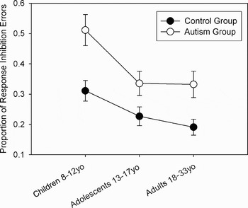
In Luna et al. (Reference Luna, Doll, Hegedus, Minshew and Sweeney2007), we tried to elucidate the developmental trajectory of response inhibition in autism through adolescence into adulthood. The goal was to distinguish whether development is typical, delayed, or arrested at a particular developmental level. As in previous studies, we found that the ability to inhibit a saccade toward a light was impaired in autism across development. However, developmental improvement in inhibitory control, and the age at which performance stabilized, was similar in individuals with autism and typically developing controls matched for gender, age, and IQ (see Figure 2). These results suggest that, although the overall degree of inhibitory control is impaired throughout development, the maturational processes that support inhibitory improvement from childhood to adolescence are intact. That development during this first transition occurs in people with autism indicates that the brain maturational processes that predominate during this period, chiefly synaptic pruning and myelination in association areas of frontal and temporal regions, are relatively intact in autism. Importantly, these results suggest that the plasticity associated with maturation from childhood to adolescence is also available in autism, implying a protracted window for affecting executive function. Despite this development from childhood to adolescence, autistic individuals are still impaired as adults (Goldberg et al., Reference Goldberg, Mostofsky, Cutting, Mahone, Astor and Denckla2005; Luna et al., Reference Luna, Doll, Hegedus, Minshew and Sweeney2007; Minshew et al., Reference Minshew, Luna and Sweeney1999). Thus, the changes from adolescence to adulthood in brain circuitry underlying the antisaccade (Luna et al., Reference Luna, Thulborn, Munoz, Merriam, Garver and Minshew2001), from more effortful processing requiring frontal circuitry to more automated processing using a wider network including basal ganglia, thalamus, and cerebellum, may never become mature in autism.
Mean (±1 SEM) proportion of trials with antisaccade response inhibition failures. Although the autistic group demonstrates impaired response inhibition throughout development, they do show developmental improvements similar to controls. Open circles depict the responses from the autistic group while solid circles depict the responses from the control group. From “Maturation of Executive Function in Autism,” by B. Luna, S. K. Doll, S. J. Hegedus, N. J. Minshew, and J. A. Sweeney, 2007, Biological Psychiatry, 61, 474–481. Copyright 2007 by Elsevier. Reprinted with permission.

Evidence from go/no-go tasks have been somewhat mixed, probably reflecting task differences and the age of the participants. An early study showed a deficit in response inhibition in children with autism (Ozonoff, Strayer, McMahon, & Filloux, Reference Ozonoff, Strayer, McMahon and Filloux1994); a similar pattern was evident in Happe et al. (Reference Happe, Booth, Charlton and Hughs2006), although performance improved into adolescence. Johnson and colleagues (2007) found that children with autism made more response inhibition errors (“commission errors”), but had normal sustained attention, on a go/no-go task. In contrast, Raymaekers, Antrop, van der Meere, Wiersema, and Roeyers (Reference Raymaekers, Antrop, van der Meere, Wiersema and Roeyers2007) found performance was not impaired in children with high-functioning autism; however, this group did find differences in adults on a similar but faster task (Raymaekers et al., Reference Raymaekers, Antrop, van der Meere, Wiersema and Roeyers2007; Raymaekers, van der Meere, & Roeyers, Reference Raymaekers, van der Meere and Roeyers2004). The studies from Raymaekers and colleagues could reflect that (a) the children's task was too easy and thus not sensitive to the inhibitory limitations in autism or (b) the deficit is more evident in adults than children (i.e., “snowballs” over development). Studies of younger lower functioning children with autism on motor tasks that require inhibition to succeed, but also have a number of planning components (i.e., pull a knob), indicate that these groups do not perform more poorly than control groups at a similar verbal mental age (Griffith, Pennington, Wehner, & Rogers, Reference Griffith, Pennington, Wehner and Rogers1999; Hughes & Russell, Reference Hughes and Russell1993; Russell, Jarrold, & Hood, Reference Russell, Jarrold and Hood1999).
In contrast to the deficit on the antisaccade and mixed findings on go/no-go, performance on the Stroop task, both the traditional Word-Color Test and a nonverbal day/night Stroop for younger children, does not appear to be impaired in autism (Eskes, Bryson, & McCormick, Reference Eskes, Bryson and McCormick1990; Goldberg et al., Reference Goldberg, Mostofsky, Cutting, Mahone, Astor and Denckla2005; Russell et al., Reference Russell, Jarrold and Hood1999). Similarly, both stop-signal tasks and tasks examining negative priming appear to be at a normal level in people with high-functioning autism across a wide range of ages (Brian, Tipper, Weaver, & Bryson, Reference Brian, Tipper, Weaver and Bryson2003; Ozonoff & Strayer, Reference Ozonoff and Strayer1997). However, in children, variants of the stop-signal task (i.e., change task, circle drawing task) appear to be impaired in autism, with slower inhibition of a prepotent response and more difficulty inhibiting an ongoing response (Geurts, Verte, Oosterlaan, Roeyers, & Sergeant, Reference Geurts, Verte, Oosterlaan, Roeyers and Sergeant2004; Verte, Geurts, Roeyers, Oosterlaan, & Sergeant, Reference Verte, Geurts, Roeyers, Oosterlaan and Sergeant2006).
Thus, a review of the literature on response inhibition in autism suggests that the paradigm itself substantially affects whether a deficit is evident. Impairment was reliable in the antisaccade task, variable in the go/no-go and stop signal tasks, and not evident in the Stroop or negative priming tasks. It is likely that the most important issues that affected whether or not a deficit was evident was whether the response was prepotent and/or whether alternate attentional strategies could be used (as in the Stroop). In addition, the age, level of functioning, and the inclusion of individuals with Asperger syndrome may influence whether impairment is evident.
Although there have been a substantial number of behavioral response inhibition studies in autism, with varying results, only two neuroimaging studies have examined the neural underpinnings of response inhibition in this disorder (Kana et al., Reference Kana, Keller, Minshew and Just2007; Schmitz et al., Reference Schmitz, Rubia, Daly, Smith, Williams and Murphy2006). Schmitz et al. (Reference Schmitz, Rubia, Daly, Smith, Williams and Murphy2006) utilized both a go/no-go task (considered a motor inhibition task in this paper) and a “spatial Stroop” (considered a cognitive interference inhibition task) in a neuroimaging study of 10 high-functioning adults with autism predominantly including individuals with Asperger syndrome and 12 control adults. There were no behavioral differences on these tasks between groups. In typical adults, these tasks activate distinct frontostriatial pathways (Rubia et al., Reference Rubia, Smith, Brammer, Toone and Taylor2005). The autism group demonstrated greater activation than controls in several relevant areas, including left middle inferior and orbitofrontal gyrus on correct no-go trials, and left insula on correct responses to the spatial Stroop. They also found increased gray matter in similar frontal areas using neuroanatomical measurements, which these authors hypothesize represent neurodevelopmental abnormalities that affect functional activation. The increased activation in the frontostriatal circuitry underlying response inhibition may reflect increased effort and inefficient brain function, or the use of distinct strategies in people with autism.
In contrast to this increased activation, Kana and colleagues (2007) found that activation is decreased in frontostriatal circuits in autism. Because these investigators posit that the discrepancies in the findings on response inhibition may reflect task demands, they utilized both a “simple” go/no-go task and one with a memory demand (“one-back”) in 12 high-functioning participants with autism and matched controls. Again, there were no behavioral differences between groups on these tasks. In this study, decreased activation was evident on both tasks in the anterior cingulate cortex. In addition, an analysis of functional connectivity suggested that there was decreased connectivity between the cingulate gyrus/insula, right middle and inferior frontal regions, and inferior parietal lobule, particularly in the one-back condition (see discussion above of white matter differences). Kana et al. (Reference Kana, Keller, Minshew and Just2007) propose that these results indicate decreased inhibitory control in people with autism, reflecting a lack of connectivity between relevant regions. As described in the previous section there are now several studies providing evidence for compromised functional connectivity during cognitive tasks, which may limit inhibitory control. Although the behavioral components of these imaging studies show similar performance in individuals with autism, it is still important to investigate if the brain systems supporting equivalent performance are also similar. In normative development, by adolescence, inhibitory control can appear similar to that of adults; however, the brain function supporting mature performance in adolescence indicates important differences. These differences reflect immaturities in brain systems, resulting in increased prefrontal activity and possibly indicating difficulty with inhibitory control. Similarly, differences in brain activation in autism, even in the face of similar behavioral performance, could reflect either increased effort or alternate strategies in autism, resulting from brain function that is not optimal.
Two neuroimaging studies of response inhibition in autism both suggest abnormalities in frontostriatal pathways but in opposite directions. Kana and colleagues suggest that this difference in findings reflects the fact that the Schmitz et al. (Reference Schmitz, Rubia, Daly, Smith, Williams and Murphy2006) paper included a majority of participants with Asperger syndrome, who may utilize different strategies including more verbal ones, whereas Kana et al. (Reference Kana, Keller, Cherkassky, Minshew and Just2006) included only high-functioning participants with autism. More neuroimaging studies have been done on response inhibition in ADHD, probably reflecting that deficits in this population in response inhibition are generally more reliable than in autism (Geurts et al., Reference Geurts, Verte, Oosterlaan, Roeyers and Sergeant2004; Happe et al., Reference Happe, Booth, Charlton and Hughs2006; Johnson et al., Reference Johnson, Robertson, Kelly, Silk, Barry and Daibhis2007). These studies often find decreased activation in frontostriatal circuitry in individuals with ADHD (Booth et al., Reference Booth, Burman, Meyer, Lei, Trommer and Davenport2005; Rubia et al., Reference Rubia, Smith, Brammer, Toone and Taylor2005; Suskauer et al., Reference Suskauer, Simmonds, Fotedar, Blankner, Pekar and Denckla2008). The comorbidity of autism and ADHD (Matson & Nebel-Schwalm, Reference Matson and Nebel-Schwalm2007) may play an important role in the variability of results. Subject diagnosis, including whether the participant has Asperger syndrome and/or comorbid ADHD, may contribute to the uneven results across studies.
Our own preliminary fMRI data using the antisaccade task indicate that high-functioning individuals with autism, not including individuals with Asperger syndrome, showed decreased recruitment of frontoparietal regions during adolescence and adulthood. The autism group performed more poorly than the control group. However, recruitment of frontoparietal regions did increase from adolescence to adulthood in the autism group, much like the pattern in typical development. Development continues apace in autism until adulthood, although it is delayed throughout the trajectory. The decreased recruitment of frontoparietal regions is supported by the finding of decreased connectivity between frontal and parietal areas described above (Koshino et al., Reference Koshino, Carpenter, Minshew, Cherkassky, Keller and Just2005, Reference Koshino, Kana, Keller, Cherkassky, Minshew and Just2008).
Summary
These results suggest that response inhibition is impaired in autism, in particular, the ability to inhibit prepotent motor responses, such as the antisaccade task. This ability to inhibit motor responses requires rapid and efficient integration of executive regions, and is not particularly amenable to strategy formation; thus, the impairments in inhibitory control in autism may be due to limitations in effectively recruiting executive circuitry. These deficits may affect executive processing more generally and not be specific to response inhibition. In addition, there may be compensatory mechanisms in autism that allow for response inhibition to be effective under some circumstances, such as the Stroop task and other paradigms that allow attentional and verbal strategies in contrast to motor inhibition, as in the antisaccade. This possibility is supported by the fact that those studies that included subjects with Asperger syndrome, who have normal or high verbal fluency, do not appear to show inhibitory deficits in brain function.
Of importance, developmental results add a new dimension to understanding the basis of inhibitory limitations in autism. Our findings indicating that there is a developmental improvement from childhood to adolescence, similar to typical development, has important implications (Luna et al., Reference Luna, Doll, Hegedus, Minshew and Sweeney2007). First, these results provide evidence that mechanisms underlying this stage of development, including the transition to prefrontally mediated behavior, may be preserved in autism. Second, the fact that autistic individuals are still impaired as adults (Goldberg et al., Reference Goldberg, Mostofsky, Cutting, Mahone, Astor and Denckla2005; Luna et al., Reference Luna, Doll, Hegedus, Minshew and Sweeney2007; Minshew et al., Reference Minshew, Luna and Sweeney1999) suggests that the late developmental shift in neural circuitry, from more effortful processing that requires frontal circuitry to more automated, posterior processing reflecting a wider network including basal ganglia, thalamus and cerebellum, is limited. Similarly, it is possible that the final frontostriatal pathway discussed in Stevens et al. (Reference Stevens, Kiehl, Pearlson and Calhoun2007) never becomes fully functional in autism. Another roadblock to mature response inhibition and other executive functions may be decreased white matter tracts or functional connectivity in autism (see review above).
Spatial WM
WM, like response inhibition, is a core component of executive function that continues to develop into adolescence, and appears abnormal in autism. WM is defined as the ability to temporarily maintain and manipulate information on-line to guide goal-directed behavior (Baddeley, Reference Baddeley1992). In light of the difficulty individuals with autism have in flexibly responding to their environments and in organizing complex responses, it has been hypothesized that a WM deficit is a characteristic of autism spectrum disorders. There are many types of WM (e.g., phonological, object, spatial location) that are believed to be supported by distinct storage buffers (Baddeley, Reference Baddeley1986). The present review will focus on WM for spatial location, given that the other WM types involve additional impairment in autism including verbal and object processing abnormalities. We will present developmental studies that provide behavioral and brain functional characterization of spatial WM and its neural correlates. As before, we will first review the literature on normative development to provide a background for our description of the cognitive impairments and corresponding neural pathology that characterizes autism spectrum disorders as symptoms persist across childhood, adolescence, and adulthood. Although a range of paradigms will be discussed, we will highlight several common paradigms: the oculomotor delayed response (ODR) task (memory-guided saccade task), the self-ordered search task, and spatial span.
Experimental paradigms
The ODR task (memory-guided saccade task) is a basic task that has been used in single-cell studies to characterize the neural basis of cognitive control (Hikosaka & Wurtz, Reference Hikosaka and Wurtz1983b), and therefore, is particularly amenable for understanding the brain basis of WM. In the ODR task, subjects retain fixation on a central stimulus while a peripheral target briefly appears in an unpredicted location in the periphery and subjects are asked to remember the location of the target. After a varied delay period where subjects must retain the target location in WM, the fixation is extinguished. This cues the participant to make an eye movement, in the absence of any visual stimulus, to the remembered location. This response is thus voluntary and guided exclusively by the representation in WM, providing a very direct probe of this process. WM status is assessed by the accuracy of the initial saccade and final eye position, whereas general processes guiding voluntary behavior are assessed by the latency to initiate the WM response.
Self-ordered search tasks require participants to remember spatial locations searched within a trial and on previous trials to guide subsequent responses. One widely used self-ordered search task is the spatial WM test from the computerized Cambridge Neuropsychological Testing Automated Battery (CANTAB; Luciana & Nelson, Reference Luciana and Nelson1998). Participants search for a token hidden behind an array of squares by touching the stimuli on the screen. They are informed that the location of the token will change with each trial, which requires retaining and updating in WM the previous target locations. An organized search strategy can help to minimize the load associated with searching and is indexed by a “strategy score” that estimates the number of search sequences that began with the same square (Owen, Downes, Sahakian, Polkey, & Robbins, Reference Owen, Downes, Sahakian, Polkey and Robbins1990; Robbins et al., Reference Robbins, James, Owen, Sahakian, Lawrence and McInnes1998). The spatial span task of the CANTAB battery also assesses WM. This is a self-ordered pointing task that requires participants to replicate a sequence where squares changed color in order by using a touch screen. The span becomes increasingly large from two squares to nine based on the examinee's success on prior trials. These tasks have been successfully used to characterize normative development of spatial WM (Conklin, Luciana, Hooper, & Yarger, Reference Conklin, Luciana, Hooper and Yarger2007; Luciana et al., Reference Luciana, Conklin, Hooper and Yarger2005; Luciana & Nelson, Reference Luciana and Nelson1998, Reference Luciana and Nelson2002).
Normative development of spatial WM
Although there is evidence for WM abilities in infancy (Diamond, Reference Diamond and Diamond1990; Diamond & Goldman-Rakic, Reference Diamond and Goldman-Rakic1989) indicating that core abilities are in place early in life, there is continued improvement through adolescence reflecting a protracted maturation of executive function (Luciana & Nelson, 1998; Swanson, Reference Swanson1999; Vuontela et al., Reference Vuontela, Steenari, Carlson, Koivisto, Fjällberg and Aronen2003). Although young children have basic maintenance capacities in place, the mechanisms underlying precision, production of complex responses, and processing despite interference do not mature until late teen years (Hale, Bronik, & Fry, Reference Hale, Bronik and Fry1997; Luna et al., Reference Luna, Garver, Urban, Lazar and Sweeney2004; Takarae, Minshew, Luna, Krisky, & Sweeney, Reference Takarae, Minshew, Luna, Krisky and Sweeney2004) or early adulthood (Zald & Iacono, Reference Zald and Iacono1998). The development of WM is thought to support the emergence of complex information processing (Case, Reference Case1992; Dempster, Reference Dempster1981; Nelson et al., Reference Nelson, Monk, Lin, Carver, Thomas and Truwitt2000).
Developmental studies using the ODR task are consistent with the broader WM literature indicating the functional ability and speed of WM processing have a protracted developmental trajectory into adolescence. Luna et al. (Reference Luna, Garver, Urban, Lazar and Sweeney2004) showed that the accuracy of the initial WM response appears to mature around 15 years of age, but the accuracy of subsequent corrective eye movement responses that require greater precision continues to mature into the second decade of life at around 25 years of age (Luna et al., Reference Luna, Garver, Urban, Lazar and Sweeney2004).
Various studies using self-ordered search task have documented age-related improvements in WM that continue into early adulthood (DeLuca et al., Reference DeLuca, Wood, Anderson, Bucanan, Proffitt and Mahony2003; Luciana et al., Reference Luciana, Conklin, Hooper and Yarger2005; Luciana & Nelson, Reference Luciana and Nelson1998, Reference Luciana and Nelson2002). Although children can perform equivalently to adults in the two- and three-item searches, there are continued improvements on searches and strategy scores with increases in memory load and interference into early adulthood (DeLuca et al., Reference DeLuca, Wood, Anderson, Bucanan, Proffitt and Mahony2003; Luciana & Nelson, Reference Luciana and Nelson1998). Similar results have been found with spatial span tasks where the ability to withstand increasing load and interference in spatial span tasks continue to improve into adulthood (Luciana et al., Reference Luciana, Conklin, Hooper and Yarger2005). Together, these studies using self-ordered search tasks and the spatial span tasks show that basic aspects of spatial WM are present early in development, but there is protracted development when self-monitoring, interference, and incorporation of ongoing information is required.
In typically developing adults, WM utilizes an extended neural network that consistently includes the DLPFC (Carlson et al., Reference Carlson, Martinkauppi, Raemae, Salli, Korvenoja and Aronen1998; D'Esposito et al., Reference D'Esposito, Detre, Alsop, Shin, Atlas and Grossman1995; McCarthy et al., Reference McCarthy, Blamire, Puce, Nobre, Bloch and Hyder1994; Owen et al., Reference Owen, Herrod, Menon, Clark, Downey and Carpenter1999; Owen, Evans, & Petrides, Reference Owen, Evans and Petrides1996). In addition, widely distributed neural activation in the inferior parietal sulcus, anterior cingulate cortex (ACC), basal ganglia, and cerebellum are associated with higher level components of spatial WM (Carlson et al., Reference Carlson, Martinkauppi, Raemae, Salli, Korvenoja and Aronen1998; Casey et al., Reference Casey, Cohen, O'Craven, Davidson, Irwin and Nelson1998; Courtney, Petit, Maisog, Ungerleider, & Haxby, Reference Courtney, Petit, Maisog, Ungerleider and Haxby1998; Curtis, Rao, & D'Esposito, Reference Curtis, Rao and D'Esposito2004). Neuroimaging studies using the ODR task also show involvement of DLPFC, ventrolateral PFC, inferior parietal cortex, ACC, cortical eye fields, basal ganglia, insula, thalamus, and lateral cerebellum (Hikosaka & Wurtz, Reference Hikosaka and Wurtz1983a; Sweeney et al., Reference Sweeney, Mintun, Kwee, Wiseman, Brown and Rosenberg1996). The ODR task is unique because single-cell recordings of nonhuman primates have supported inferences from imaging studies. Specifically, delay-dependent cells involved in maintenance have been identified in the DLPFC, the inferior parietal cortex, cortical eye fields, basal ganglia, and thalamus (Funahashi, Inoue, & Kubota, Reference Funahashi, Inoue and Kubota1997), suggesting multiple regions are involved in similar functions throughout the cortex and may form integrated functional circuits.
Pediatric neuroimaging studies have found evidence for the ability to recruit core brain systems underlying WM as well as developmental differences in network systems. Initial studies comparing children to adults using the n-back task where subjects must remember the order of letters or the spatial location of a target indicate that children, like adults, recruit prefrontal and parietal regions involved in WM (Casey et al., Reference Casey, Cohen, Jezzard, Turner, Noll and Trainor1995; Nelson et al., Reference Nelson, Monk, Lin, Carver, Thomas and Truwitt2000; Thomas et al., Reference Thomas, King, Franzen, Welsh, Berkowitz and Noll1999). However, the magnitude of activation in different areas of the circuitry has been found to change with age. Studies suggest that children rely on a circuitry including premotor, cerebellum, and striatal regions instead of the ventral prefrontal and inferior temporal regions evident in adulthood (Ciesielski, Lesnik, Savoy, Grant, & Ahlfors, Reference Ciesielski, Lesnik, Savoy, Grant and Ahlfors2006). From adolescence to adulthood, however, there are continued developmental refinements in the circuitry supporting WM including decreased recruitment of prefrontal regions with age (Crone, Wendelken, Donohue, van Leijenhorst, & Bunge, Reference Crone, Wendelken, Donohue, van Leijenhorst and Bunge2006; Klingberg et al., Reference Klingberg, Forssberg and Westerberg2002; Luna, Garver, & Sweeney, Reference Luna, Garver and Sweeney2000; Scherf, Sweeney, & Luna, Reference Scherf, Sweeney and Luna2006). Our studies using the ODR task, where subjects have to make an eye movement to a location maintained in WM, found that DLPFC participation increased from childhood to adolescence reflecting improved performance, but then decreased from adolescence to adulthood when performance is similar but the effort is larger in adolescents (Scherf et al., Reference Scherf, Sweeney and Luna2006). Children's inferior performance was accompanied by increased reliance on basal ganglia and insula, whereas adults showed a more widely distributed circuitry including temporal regions. The decreased reliance on prefrontal systems with maturity is believed to reflect decreased effort in performing WM tasks in adulthood compared to adolescence, similar to findings in response inhibition (Klingberg et al., Reference Klingberg, Forssberg and Westerberg2002; Olesen, Macoveanu, Tegner, & Klingberg, Reference Olesen, Macoveanu, Tegner and Klingberg2007; Scherf et al., Reference Scherf, Sweeney and Luna2006).
In addition to changes in prefrontal activation, developmental improvements in WM have been associated with increases in the integrity of white matter in frontoparietal regions. This suggests that maturational improvements in connectivity, including myelination, may play an important role in age-related improvements in WM (Edin, Macoveanu, Olesen, Tegner, & Klingberg, Reference Edin, Macoveanu, Olesen, Tegner and Klingberg2007; Olesen, Macoveanu, Tegner, & Klingberg, Reference Olesen, Macoveanu, Tegner and Klingberg2007). The reliance on integrated cortical systems may be impaired in autism given the evidence of compromised connectivity (see above). These results converge with behavioral results, which provide evidence for WM abilities in infancy but continued improvement into adolescence, indicating that core brain regions are recruited in children but that refinements in the circuitry supporting WM continue from adolescence to adulthood. This development is supported by ongoing neural maturation that facilitates use of widely distributed neural networks with more task-specific neural regions. This highlights a developmental transition toward use of a more functionally specialized, widely dispersed network circuit associated with the executive components of performance, which is consistent with the refinement in behavioral performance across development.
Spatial WM in autism
Similar to response inhibition, there is evidence for abnormalities as well as preservation of WM in autism. This may reflect impairments in core processes underlying WM, and the presence of compensatory mechanisms that can aid in performance. The nature of the WM impairment, its development, and possible neural basis are still not well understood. Thus, we will review studies that are relevant to developmental as well as neurobiological aspects of WM status in autism.
ODR studies of rigorously diagnosed, high-functioning autistic participants and healthy control samples matched in age and gender consistently indicate reduced accuracy of memory-guided saccades reflecting impaired WM (Goldberg et al., Reference Goldberg, Lasker, Zee, Garth, Landa and Tien2002; Luna et al., Reference Luna, Minshew, Garver, Lazar, Thulborn and Eddy2002, Reference Luna, Doll, Hegedus, Minshew and Sweeney2007; Minshew et al., Reference Minshew, Luna and Sweeney1999; Takarae et al., Reference Takarae, Minshew, Luna and Sweeney2004). These deficits in WM-guided eye movement responses in autism have been found in adults (Minshew et al., Reference Minshew, Luna and Sweeney1999) as well as adolescents (Goldberg et al., Reference Goldberg, Lasker, Zee, Garth, Landa and Tien2002; Luna et al., Reference Luna, Doll, Hegedus, Minshew and Sweeney2007). Consistent with the results from response inhibition studies, WM is impaired in autism throughout childhood, adolescence, and adulthood, but there was evidence for developmental improvements in WM from childhood to adolescence similar to the normative sample (Luna et al., Reference Luna, Doll, Hegedus, Minshew and Sweeney2007; see Figure 3). This supports the proposal put forth earlier on the basis of response inhibition studies that there are core abnormalities in executive function that persist throughout development but do not disrupt the programmed plasticity in development found from childhood to adolescence.
Mean (±1 SEM) absolute error of the initial memory-guided saccade (in degrees of visual angle) made to a remembered location in the oculomotor delayed response task. Although the autistic group demonstrates impaired response inhibition throughout development, they do show developmental improvements similar to controls. From “Maturation of Executive Function in Autism,” by B. Luna, S. K. Doll, S. J. Hegedus, N. J. Minshew, and J. A. Sweeney, 2007, Biological Psychiatry, 61, 474–481. Copyright 2007 by Elsevier. Reprinted with permission.

This last stage of development is most pronounced when looking at the final position after corrective eye movements are made to best approximate the remembered spatial location. This corrective final response may be driven by the WM representation or by accurate error detection processes, compared to the initial saccade that involves general executive processes that support voluntary responses of making an eye movement in the absence of a visual target, as well as WM. In normative development, this final response continues to improve past adolescence into adulthood (Luna et al., Reference Luna, Garver, Urban, Lazar and Sweeney2004) as do error processing abilities (Santesso & Segalowitz, Reference Santesso and Segalowitz2008; Velanova, Wheeler, & Luna, 2008). In autism, there is no evidence of this later transition, indicating additional impairments in the ability to establish higher order mechanisms that are supported by widely distributed functional integration (Figure 4).
Mean (±1 SEM) absolute error of the final resting position of gaze (in degrees of visual angle) made to a remembered location in the oculomotor delayed response task. Although the autistic group improves from childhood to adolescence, they do not show the improvement from adolescence to adulthood that is evident in controls. From “Maturation of Executive Function in Autism,” by B. Luna, S. K. Doll, S. J. Hegedus, N. J. Minshew, and J. A. Sweeney, 2007, Biological Psychiatry, 61, 474–481. Copyright 2007 by Elsevier. Reprinted with permission.

Impaired WM in autism is also supported by studies using self-ordered search tasks. Collectively, these studies indicate impaired performance in autism that is magnified with increases in task difficulty and evident throughout development (Goldberg et al., Reference Goldberg, Mostofsky, Cutting, Mahone, Astor and Denckla2005; Landa & Goldberg, Reference Landa and Goldberg2005a; Steele, Minshew, Luna, & Sweeney, Reference Steele, Minshew, Luna and Sweeney2007). These studies show evidence for perseverative errors of returning to incorrect locations (Landa & Goldberg, Reference Landa and Goldberg2005a) as well as for interference susceptibility (Goldberg et al., Reference Goldberg, Mostofsky, Cutting, Mahone, Astor and Denckla2005; Steele et al., Reference Steele, Minshew, Luna and Sweeney2007). Search strategies appear impaired when including adults (Landa & Goldberg, Reference Landa and Goldberg2005a; Steele et al., Reference Steele, Minshew, Luna and Sweeney2007), but can appear intact in childhood; this probably reflects the poor search strategies of children in general (Goldberg et al., Reference Goldberg, Mostofsky, Cutting, Mahone, Astor and Denckla2005). However, there is other evidence for preserved basic aspects of WM, for instance, normative rates of within-search errors in autistic individuals. This intact ability may simply reflect WM performance in the face of basic maintenance demands of the task (Goldberg et al., Reference Goldberg, Mostofsky, Cutting, Mahone, Astor and Denckla2005; Landa & Goldberg, Reference Landa and Goldberg2005a, Reference Landa and Goldberg2005b; Steele et al., Reference Steele, Minshew, Luna and Sweeney2007). This suggests that WM is impaired in autism only when task demands become more complex.
There is additional evidence for intact spatial WM processes in autism. Several studies using n-back tasks, visual search, and self-ordered pointing tasks have found intact WM processes in autism (Geurts et al., Reference Geurts, Verte, Oosterlaan, Roeyers and Sergeant2004; Joseph, McGrath, & Tager-Flusberg, Reference Joseph, McGrath and Tager-Flusberg2005; Ozonoff & Strayer, Reference Ozonoff and Strayer2001). Using a battery of spatial WM tasks where geometrical figures were used as targets, including an n-back task, and visual search task, no impairments were found in autistic individuals (Ozonoff et al., 2001). The normative performance may be explained by relatively easier or simpler demands than those found in the ODR or self-ordered pointing. The n-back had relatively low maintenance demands (one-back task) and the visual search tasks used circular arrangement of target locations that may have facilitated the use of attentional or verbal strategies to assist in performance. However, the self-ordered pointing task, where subjects have to indicate the novel abstract figure in an array of different novel figures on a card that range from 6–12 items, is purportedly less amenable to verbal strategies. It could be argued that verbal strategies of naming the abstract figures could still be made (looks like a star, maze, or snake). The self-ordered pointing task could also be argued to be less difficult than the ODR and self-ordered search tasks because the former has a predetermined number of targets, whereas the ODR task is a measure of accuracy as a continuous variable. Regardless of task differences, the results indicating there is preservation of WM with certain tasks indicates that some aspects of WM are preserved and/or that there are compensatory mechanisms that allow for normative performance. In addition, some of these studies included participants spanning a wide age range, which may have decreased sensitivity to impairments in core WM processes that are developmentally sensitive. Nevertheless, these studies do show that WM can appear normative and that either compensatory mechanisms or components of WM are spared in autism.
Only a handful of neuroimaging studies have been performed characterizing neural processes underlying WM in autism, including verbal and object WM (for a review, see Greene, Braet, Johnson, & Bellgrove, 2008). These studies provide converging evidence that there is impaired activity in crucial regions underlying executive processing, including parietal and frontal regions as well as their functional integration (Koshino et al., Reference Koshino, Carpenter, Minshew, Cherkassky, Keller and Just2005, Reference Koshino, Kana, Keller, Cherkassky, Minshew and Just2008; Luna et al., Reference Luna, Minshew, Garver, Lazar, Thulborn and Eddy2002; Silk et al., Reference Silk, Rinehart, Bradshaw, Tonge, Egan and O'Boyle2006). Adolescents with autism spectrum disorder show significantly less activation in the frontal lobe, including the right inferior frontal gyrus, the right medial frontal gyrus, and ACC, as well as the head of the caudate nucleus, when matching figures in a mental rotation task (Silk et al., Reference Silk, Rinehart, Bradshaw, Tonge, Egan and O'Boyle2006). Similarly, using the ODR task, we found that autistic adults showed decreased activity in the DLPFC and posterior cingulate regions known to be part of an integrated circuitry supporting WM (Luna et al., Reference Luna, Minshew, Garver, Lazar, Thulborn and Eddy2002). Of importance, we also found that much of the circuitry known to support WM was intact in autism including the insula, intraparietal sulcus, basal ganglia, thalamus, supramarginal gyrus, FEF, SEF, presupplementary motor area, ACC, precuneus, and cerebellum, suggesting a specific impairment involving the connectivity of prefrontal with posterior cortex. Koshino et al. (Reference Koshino, Carpenter, Minshew, Cherkassky, Keller and Just2005), using an n-back task where performance was equivalent for autistic and typical groups, found that the autism group showed less recruitment of prefrontal regions including the DLPFC, inferior frontal gyrus, and precentral sulcus. In addition, typicals showed higher functional connectivity in left hemisphere areas supporting verbal strategies, whereas the autistic group were more likely to recruit right hemisphere areas supporting visual processing strategies, suggesting that impaired WM may be associated with differences in using the most appropriate strategies (see Figure 1). These results were supported by an n-back task using face stimuli where again there was underactivity of prefrontal regions as well as temporal regions involved in face processing in a high functioning autism group compared to typically developing individuals (Koshino et al., Reference Koshino, Kana, Keller, Cherkassky, Minshew and Just2008). Again, functional connectivity of frontal regions was limited in autism compared to typically developing individuals, whereas connectivity of posterior regions appeared intact. The specificity of the underactivity in the PFC and its functional integration with other regions may suggest that the processes supported by this region, namely, the ability to use WM representations to form plans that guide executive function, are impaired in autism. Although there are no fMRI studies in autism characterizing activity during the self-ordered search task, it is of note that frontal lesions result in decrements in performance (Owen et al., Reference Owen, Downes, Sahakian, Polkey and Robbins1990; Walker, Husain, Hodgson, Harrison, & Kennard, Reference Walker, Husain, Hodgson, Harrison and Kennard1998), and strategy use (Owen et al., Reference Owen, Downes, Sahakian, Polkey and Robbins1990; Owen, Morris, Sahakian, Polkey, & Robbins, Reference Owen, Morris, Sahakian, Polkey and Robbins1996).
Although these studies converge on impaired function in prefrontal systems, this does not indicate a localized impairment that is exclusive to prefrontal systems given that structural data do not reflect particular abnormalities in this region. Moreover, autistic individuals show clear ability to perform frontally guided executive tasks although not to the level of sophistication of typical adults.
Summary and Conclusions
Structural studies indicate developmental abnormalities in brain maturation in autism including decreased synaptic pruning early in life, reflected in head size; regional brain abnormalities (e.g., the cerebellum) in adulthood; and decreased connectivity between regions later in development indicating a broad range of vulnerabilities that compromise higher order complex information processing while sparing basic behavior. Although brain abnormalities are evident early in autism, the protracted maturation of association areas and their interconnectivity in normative development suggest that abnormalities can continue through the life span in autism affecting the development of executive function.
Executive function is supported by the integration of a widely distributed brain circuitry and continues to mature throughout adolescence. There is evidence for preserved executive abilities in both response inhibition and WM in autism, indicating that some processes that support executive function are, in fact, intact, or that reorganization and compensatory behaviors make them appear that way. Studies that do show deficits in executive function are those that specifically test the integrity of core executive processes: eye movement tasks in particular indicate a basic limitation in autism. Although our behavioral studies indicate improvement from late childhood to adolescence in autism, performance never reaches normative adult levels. Thus, on executive function tasks, adolescents and adults with autism reach the level of typical children in late childhood but do not appear to develop further. This difference becomes more striking when typical development continues into adulthood, as in the accuracy of the final saccade in the ODR. Thus, although behavioral studies indicate that the developmental transition from childhood to adolescence is preserved in autism, the delayed ability throughout development leads to a less sophisticated and flexible use of executive function in adulthood. Functional imaging studies consistently find that executive dysfunction is associated with impaired prefrontal activity and its functional integration with the rest of the brain. Individuals with autism may have limited but not absent frontally guided executive function, as well as a wide range of abnormalities including social and communication processes that are supported by posterior temporal regions.
The neural basis of the executive function deficit in autism is not yet well understood but undermined functional connectivity may be a primary deficit. There may be additional molecular mechanisms that are abnormal, that support specific complex neural computations necessary for core executive processes. The next step in understanding the neural basis of autism is to characterize at better spatial resolution the brain processes required for higher order information processing in a developmental context. Although most studies do not look at developmental change in autism, we have found that an understanding of the developmental course of autism provides unique insight into neurophysiological mechanisms, both those that are intact and impaired. In particular, our evidence of developmental improvement from late childhood to adolescence suggests that neural mechanisms underlying this transitional time (i.e., myelination) might be relatively intact in autism. If so, interventions can target this late, and largely ignored, developmental stage in which there is still substantial improvement in autism on executive function tasks.