Introduction
Partial resection of diseased tissues or organs is commonly employed in clinical treatment, while tissue loss may affect the quality of patients' life or induce complications. Developing strategies for promoting tissue regeneration is hence of great significance. However, the regeneration of some tissues/organs with complicated anatomies such as gastrointestinal (GI) remains greatly challenging by using existing strategies. Since the GI tract contains abundant vascularized structures embedded in multiple tissue layers consisting of different cell types with specific cell organizations (typically one mucosal layer mainly consisting of epithelial cells and two smooth muscle layers with, respectively, longitudinal and parallel smooth muscle cell arrangements), desirable GI tract regeneration requires comprehensive efforts to first promote the reconstituting of vasculatures and subsequently assist the regeneration of different tissue layers, particularly smooth muscle layers with distinct anisotropies in cell arrangement.[Reference Bitar and Zakhem1] In recent decades, tissue engineering scaffolds, as appropriate structural platform for supporting cell ingrowth and tissue remodeling, have attracted extensive attentions for assisting tissue regeneration.[Reference Webber, Khan, Sydlik, Tang and Langer2] Among different techniques for manufacturing tissue engineering scaffolds, electrospinning has shown significant merits in the ease and versatility to form nanofibrous structures resembling the architectures of native extracellular matrix, which have been widely investigated for producing scaffolds with appropriate structural cues to assist the regeneration of multiple human tissues.[Reference Yang, Li, He, Ma, Ni and Zhou3–Reference Zhao, Cui, Wang, Chen, Wang, Zhang, Du and Wang5] However, the regeneration of the GI tract requires not only proper structural cues to direct the organization of different cell types but also suitable biochemical cues activated in on-demand spatiotemporal manners to stimulate specific cell functions. In addition to readily form biomimetic nanofibrous structures, electrospinning is also facile to fabricate scaffolds with enhanced bioactivity through the incorporation and controlled releases of multiple bioactive molecules such as growth factors (GFs).[Reference Tsao, Pandolfi, Wang, Minardi, Lupo, Evangelopoulos, Hendrickson, Shi, Storci, Taraballi and Tasciotti6,Reference Han, Jia, Dai, Yang, Zhao, Zhao, Fan and Yuan7] There is therefore great promise in improving GI tract regeneration by preparing tissue engineering scaffolds with well-defined structures and bioactivities via adapted electrospinning techniques.
In this investigation, scaffolds consisting of two layers of aligned nanofibers with orthogonal fiber orientations (0° and 90°) are formed by sequential electrospinning of two different GF-containing water-in-oil emulsions. The orthogonally aligned nanofibers of the bilayer scaffolds are designed for mimicking the anatomy of the smooth muscle layers of the GI tract. Different GFs, i.e., vascular endothelial growth factor (VEGF) and platelet-derived growth factor (PDGF), were, respectively, incorporated in different scaffold layers by emulsion electrospinning that enables high-efficiency delivery of GFs with well-preserved bioactivity.[Reference Zhao, Lu and Wang8] The releases of these two GFs are controlled in distinct spatiotemporal manners by using nanofibers made of poly(lactic-co-glycolic acid) (PLGA) with different LA:GA molar ratios (50:50 or 75:25) that show distinct degradation rates as carriers, which aims to provide appropriate biochemical cues to direct the early-stage vascularization and the following smooth muscle layer maturation, respectively.[Reference Ferrara, Gerber and LeCouter9,Reference Wells and Spence10] To evaluate the combined effects of the structural and biochemical cues of the bilayer scaffolds, the responses of vascular endothelial cells and smooth muscle cells grown on the bilayer scaffolds were investigated, demonstrating the superior performance of the bilayer scaffolds that have well-defined structures and bioactivities in stimulating cell behaviors with implications for potential GI tissue engineering.
Materials and methods
Materials
Medical grade PLGA (Lakeshore Biomaterials, IN, USA) with different LA:GA molar ratios (50:50 or 75:25), namely PLGA50/50 (inherent viscosity: 0.6–0.8 dL/g) and PLGA75/25 (inherent viscosity: 0.6–0.8 dL/g), were employed for forming different layers of the bilayer scaffolds by emulsion electrospinning and delivering different GFs owing to their superior biocompatibility, suitable biodegradability, and comparable mechanical properties to soft tissues.[Reference Ye, Zhang, Kai, Li and Loh11] GFs including VEGF (recombinant human VEGF165, M w = 38.2 kDa), PDGF (recombinant human PDGF-BB, M w = 24.3 kDa), as well as their corresponding enzyme-linked immunosorbent assay (ELISA) kits (Human VEGF165 Standard ABTS ELISA Development Kit and Human PDGF-BB Standard ABTS ELISA Development Kit), were bought from Peprotech, NJ, USA. All other chemicals were products of Sigma-Aldrich, MO, USA otherwise specifically explained.
Electrospinning
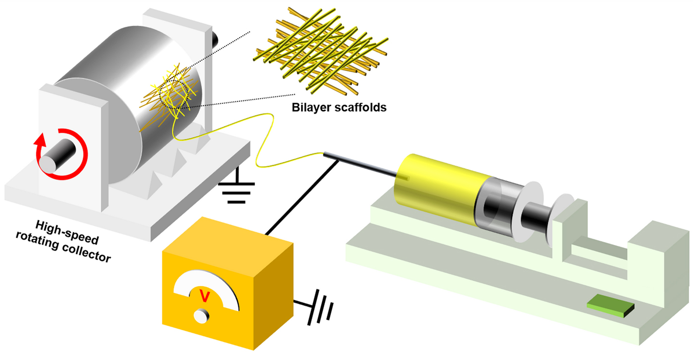
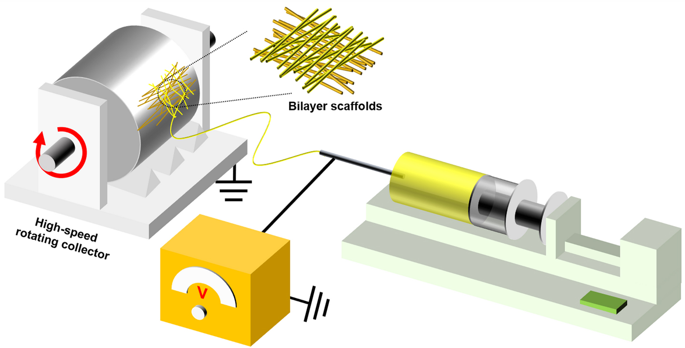
Stable emulsions for electrospinning were first prepared by mixing a polymer solution (either PLGA50/50 or PLGA75/25) and a GF suspension (either VEGF or PDGF) with an addition of 0.05% v/v Span-80 as a surfactant after vigorous mechanical stirring for 10 min. Specifically, 1 mL of VEGF suspension (10 µg/mL) and 1 mL of PDGF suspension (5 µg/mL) were, respectively, mixed with 10 mL of PLGA50/50 solution (15% w/v, in dichloromethane) and 10 mL of PLGA75/25 solution (15% w/v, in dichloromethane) for preparing different emulsions. The emulsions were subjected to electrospinning using an apparatus with a negative-voltage DC high-voltage power supply and a grounded high-speed (3000 rpm) rotating collector (Fig. 1). The applied voltage, emulsion flow rate, and working distance were set at −15 kV, 2.0 mL/h, and 15.00 cm for electrospinning, respectively. During electrospinning, a VEGF-incorporated PLGA50/50 layer was first collected on an alumina film on the collector. After 1 h electrospinning process, the alumina film with the collected electrospun membrane was peeled off, cut into a square with a size of 4 × 4 cm2, and then re-attached onto the collector orthogonally to its original direction. The electrospinning of PDGF-containing PLGA75/25 emulsion was then conducted for another 1 h to form the PDGF-incorporated PLGA75/25 layer. For the resultant bilayer scaffolds, a freeze-drying treatment was performed for completely removing residual solvents.
Schematic illustration of the electrospinning apparatus for fabricating the bilayer scaffolds with orthogonally aligned nanofibers.
Physicochemical characterizations
The morphology and structure of emulsion electrospun scaffolds were examined using a scanning electron microscope (FE-SEM, LEO 1530, Germany). The SEM images at the cross-section and the top and the bottom of the scaffolds were captured for examining the bilayer structure and PLGA75/25 layer and PLGA50/50 layer, respectively. Average fiber diameter and fiber orientation of each layer of the bilayer scaffolds were analyzed using an image analysis software, ImageJ, at three separated experiments (N = 100).
To compare the in vitro degradation properties of the PLGA75/25 layer and the PLGA50/50 layer of the bilayer scaffolds. Emulsion electrospun PLGA75/25 and PLGA50/50 scaffolds were, respectively, prepared at the identical condition to the electrospinning of different layers of the bilayer scaffolds. Immersion treatments were conducted for measuring the scaffold weight loss over different immersion treatment time periods. During immersion treatments, pre-weighed specimens (square-shaped, 1 × 1 cm2) were immersed in immersion medium of phosphate-buffered saline (PBS, pH = 7.4) with supplemented 0.02% w/v sodium azide at individual sealed tubes, which were placed at a 37 °C shaking water bath. The immersion medium was refreshed every 3 days. The immersion treatments were performed for up to 3 weeks. At pre-determined time points, the immersed specimens were taken out, washed, freeze-dried, and then weighed for evaluating their weight loss (N = 6).
In vitro release test
The GF release behaviors were studied through release tests, where PBS (pH = 7.4) with supplemented 0.02% w/v sodium azide, 0.05% v/v Tween-20, and 0.5% w/v bovine serum albumin was used as the immersion medium. VEGF-incorporated PLGA50/50 scaffolds and PDGF-incorporated PLGA75/25 scaffolds were prepared for release tests, which were kept in 37 °C humidity environment for up to 3 weeks. The immersion medium was refreshed every 2 days and collected at pre-determined time points within the 21 days of test periods. The total GF loading and the GF contents in the collected medium were measured using their corresponding ELISA kits at three separated experiments. The encapsulation efficiencies of VEGF and PDGF in different layers of the bilayer scaffolds were calculated (N = 6) according to the actual amounts of encapsulated GFs with respect to the theoretical amounts and the curves demonstrating the cumulative release profiles of different GFs were plotted.
Cell culture
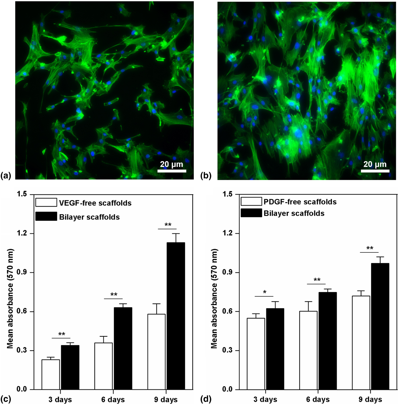
To investigate the influence of structural and biochemical cues of the bilayer scaffolds on vascularization and smooth muscle layer regeneration, human vascular umbilical endothelial cells (HUVECs, ScienCell, USA), and human aortic smooth muscle cells (HASMCs, ScienCell, USA) were, respectively, cultured with different scaffolds., including the bilayer scaffolds, as well as non-woven and GF-free emulsion electrospun PLGA75/25 and PLGA50/50 scaffolds as control. HUVECs and HASMCs used for cell culture were at passage 2 or 3 with a confluence of ~70%, which were trypsinized and then seeded onto the sterilized scaffolds (square-shaped, 2 × 2 cm2) at a cell density of 5 × 10⁴ cells/cm2 on a six-well plate (Corning, USA). After 3 days of culture, HASMCs grown on the bilayer scaffolds and the control (non-woven and PDGF-free emulsion electrospun PLGA75/25 scaffolds) were stained by Alexa Fluor 488 Phalloidin and DAPI to, respectively, label the cellular filamentous actin (F-actin) and nucleus, aiming to assess the effects of topographical cues of scaffolds on F-actin filament alignment. After 3, 6, and 9 days of culture, 3-(4,5-dimethylthiazol-2-yl)-2,5-diphenyltetrazolium bromide (MTT) colorimetric assays were conducted for investigating the proliferation of HUVECs and HASMCs on different scaffolds. The optical density (OD) values at the absorbance of 570 nm of the formazan-dissolving solutions collected from different groups in the MTT assay were recorded using a microplate reader (UVM 340, Asys HiTech GmbH, Australia) and then analyzed for comparing the proliferation of HUVECs and HASMCs on different types of scaffolds.
Statistical analysis
The statistical results are represented as mean ± SD. Statistical analyses are performed using the analysis of varianceANOVA with a Student's t-test. Results with P > 0.05 are regarded as having no significant difference (n.s.), while those with (*) P < 0.05 and (**) P < 0.01 are regarded as significantly different.
Results and discussion
Morphology, structure, and in vitro degradation properties
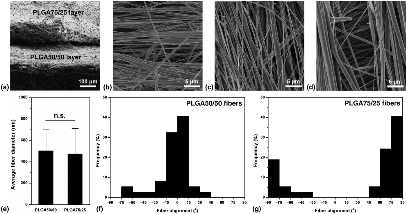
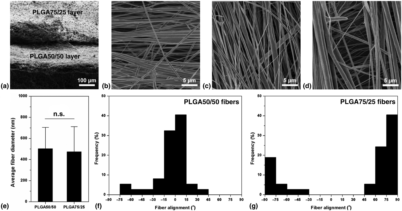
Through a sequential emulsion electrospinning process using a high-speed rotating fiber collector, parallel aligned VEGF-incorporated PLGA 50/50 and PDGF-incorporated PLGA 75/25 nanofibers were, respectively, collected, forming scaffolds with a clear bilayer structure with a thickness of approximately 100 µm for each layer (Fig. 2(a)). Due to the tangential force resulted from the high-speed rotation of the collector, nanofibers in the bottom PLGA50/50 layer (Fig. 2(b)) and the PLGA75/25 upper layer (Fig. 2(c)) both exhibit well-arranged parallel fiber alignment. And both the PLGA50/50 and PLGA75/25 nanofibers show smooth and beadles fiber morphology. The nanofibers in the upper layer are orthogonal to those in the bottom layer (Fig. 2(d)), resembling the anatomy of the smooth muscle layers of the GI tract. Owing to the same processing parameters employed in the emulsion electrospinning to form different layers and little difference in the physicochemical properties between PLGA50/50 and PLGA75/25, PLGA50/50 and PLGA75/25 fibers display similar average fiber diameters of 503 ± 202 and 474 ± 238 nm, respectively, showing no significant difference between them (Fig. 2(e)). In addition, the fiber orientations of the PLGA50/50 and PLGA75/25 fibers at different layers of the scaffolds were analyzed. While the orientation of more than 70% of emulsion electrospun PLGA50/50 nanofibers lays in the range of 0° ± 15° (Fig. 2(f)), most emulsion electrospun PLGA75/25 nanofibers exhibit fiber orientation within the range of 90° ± 15° (Fig. 2(g)). The statistical results further confirm the good alignment and orthogonality of nanofibers at different layers of the bilayer scaffolds.
Morphology and structure of the bilayer scaffolds. (a) The bilayer structure of the scaffolds formed through a sequential emulsion electrospinning process viewed at their cross-section. (b) Morphology of the PLGA50/50 layer. (c) Morphology of the PLGA75/25 layer. (d) The morphology of the bilayer scaffolds viewed from the top, showing clear orthogonal fiber alignment. (e) Average fiber diameters of nanofibers at different layers of the bilayer scaffolds made of PLGA with different LA:GA ratios. (f) Distribution of fiber alignment for the PLGA50/50 nanofibers. (g) Distribution of fiber alignment for the PLGA75/25 nanofibers.
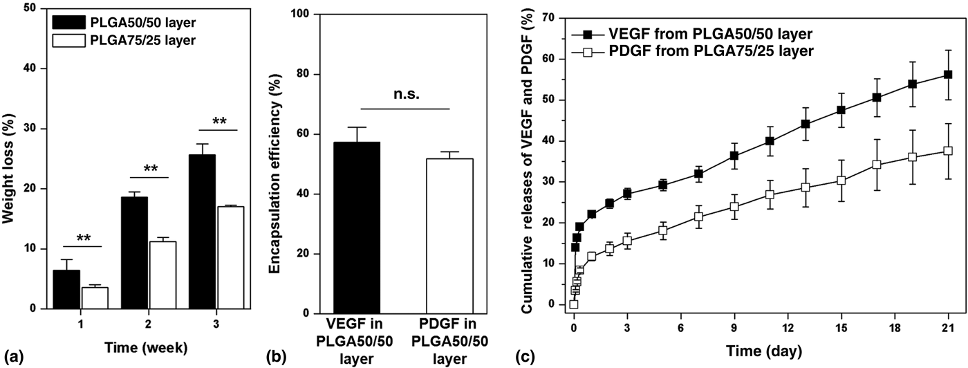
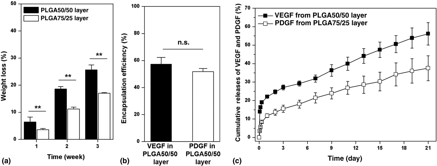
In vitro degradation properties of polymeric delivery vehicles are key factors influencing the release behaviors of GFs incorporated.[Reference Szentivanyi, Chakradeo, Zernetsch and Glasmacher12] For the GF-incorporated bilayer scaffolds, VEGF and PDGF were expected to be released in sequential and sustained manners to offer appropriate biochemical cues for meeting the requirements during GI tract regeneration. To compare the in vitro degradation properties of different layers of the bilayer scaffolds made of PLGA with different LA:GA ratios, monolayer emulsion electrospun PLGA50/50 and PLGA75/25 scaffolds were separately prepared. And immersion treatments for up to 3 weeks were performed for these two types of scaffolds. The results of statistical analyses for the weight loss of different scaffolds upon the immersion treatments reveal that the degradation rate of the PLGA50/50 scaffolds is significantly higher than that of the PLGA75/25 scaffolds (Fig. 3(a)). Since the degradation of PLGA undergoes the hydrolysis of the ester bonds under a bulk erosion mechanism,[Reference Nair and Laurencin13] faster degradation of the scaffolds made of PLGA with a lower LA:GA ratio should arise from the slightly higher wettability and water permeability of the PLGA50/50 than those of PLGA75/25.[Reference Zhou, Zhao, Tsai and Wang14] The distinct degradation rates of emulsion electrospun scaffolds consisting of PLGA with different LA:GA ratios were promising to affect the release profiles of diverse GFs incorporated in different layers of the bilayer scaffolds.
(a) In vitro degradation properties of different layers of the bilayer scaffolds within a 3-week immersion treatment period. (b) Encapsulation efficiencies of VEGF and PDGF, respectively, in different layers of the bilayer scaffolds. (c) In vitro release behaviors of VEGF and PDGF from the bilayer scaffolds for up to 3 weeks.
Encapsulation efficiency and in vitro release behaviors of VEGF and PDGF
For the as-prepared GF-incorporated bilayer scaffolds, the encapsulation efficiencies of VEGF and PDGF, respectively, in the PLGA50/50 layer and the PLGA75/25 layer were investigated. Since emulsion electrospinning can form nanofibers with core–shell structure that provide a desirable reservoir for the encapsulated molecules,[Reference Zhou, Zhao, Tsai and Wang14] both VEGF and PDGF achieve good encapsulation efficiency in each layer of the bilayer scaffolds, attaining 57.27 ± 5.04% and 51.76 ± 2.37%, respectively (Fig. 3(b)). To confirm the modulating effects of different PLGA nanofibers on the release behaviors of GFs incorporated, in vitro release tests were performed for the GF-incorporated bilayer scaffolds for up to 3 weeks. It has been demonstrated that the release behaviors of GFs from biodegradable delivery vehicles were mainly determined by their structure, physicochemical properties, and electrically modulating effects.[Reference Zhao, Lu and Wang8,Reference Szentivanyi, Chakradeo, Zernetsch and Glasmacher12,Reference Freudenberg, Zieris, Chwalek, Tsurkan, Maitz, Atallah, Levental, Eming and Werner15] In our case, there is no significant difference in the structure between the PLGA50/50 and PLGA75/25 fibers, their different physiochemical properties and electrostatically modulating effects should be therefore dominant factors for affecting the release profile of GFs. The isoelectric point (pI) values of VEGF and PDGF are 8.5 and 9.6, respectively. The release of GFs with alkaline pI values has been confirmed to be effectively modulated by scaffolds with negative surface electric charges or functionalized with negative-charged molecules.[Reference Zhao, Lu and Wang8,Reference Freudenberg, Zieris, Chwalek, Tsurkan, Maitz, Atallah, Levental, Eming and Werner15,Reference Zhao and Wang16] The bilayer scaffolds prepared in this study should also possess durable negative surface electric charges resulted from the negative-voltage emulsion electrospinning.[Reference Zhao, Lu and Wang8] The electrostatically modulating effects of the bilayer scaffolds with negative surface electric charges result in sustained release of both VEGF and PDGF from different layers of the bilayer scaffolds, showing two-stage release characteristics with slightly rapid release just within the initial 1 day and sustained release in the following whole release periods (Fig. 3(c)). Importantly, it has been revealed that the release of VEGF from the PLGA50/50 layer of the bilayer scaffolds is faster than that of PDGF from the PLGA75/25 layer (Fig. 3(c)), which should mainly arise from the relatively faster degradation rate of PLGA50/50 than that of PLGA50/50. Owing to the higher molar ratio of hydrophilic GA segments in PLGA50/50, PLGA50/50 should be easier to be swelled than PLGA75/25.[Reference Zhou, Zhao, Tsai and Wang14] Therefore, the difference in the release rate between the VEGF and PDGF is particularly significant in the initial release stage as the swelling of polymeric delivery vehicles plays a dominant role in determining the release of GFs at this stage.[Reference Szentivanyi, Chakradeo, Zernetsch and Glasmacher12] A higher pI value of PDGF than that of VEGF may be an alternative factor that leads to the sequential release behaviors of VEGF and PDGF since PDGF with a higher pI value should exhibit enhanced electrostatic interaction with the scaffolds. In the sustained release stage, the release of VEGF and PDGF from different nanofibers follows a complex diffusion-and-erosion-controlled release model, of which release profiles can be described according to a zero-order equation:[Reference Serra, Doménech and Peppas17]

where M t/M∞ is the fractional growth factor release at time (t) and K is the kinetic constant. The values of K for VEGF and PDGF at their sustained release stages are 1.68 and 1.33 d−1, respectively, further verifying distinct release kinetics of GFs from different nanofibers.
The results of the release tests confirm our hypothesis that the release of VEGF and PDGF can be effectively modulated by using different polymers to form delivery vehicles. It has been demonstrated that the administration of VEGF can effectively promote the self-organization of vascular endothelial cells to form vasculatures.[Reference Ferrara, Gerber and LeCouter9] The initial rapid release of VEGF will satisfy the demands of rapid formation of vasculatures in the early stage of GI tract regeneration.[Reference Wells and Spence10] However, neo-vasculatures cannot be free-standing without the formation of smooth muscle layers at the later stage of GI tract regeneration. The sequentially slow yet sustained release of PDGF will subsequently offer appropriate biochemical cues to direct the maturation of smooth muscle layers.[Reference Tsao, Pandolfi, Wang, Minardi, Lupo, Evangelopoulos, Hendrickson, Shi, Storci, Taraballi and Tasciotti6,Reference Han, Jia, Dai, Yang, Zhao, Zhao, Fan and Yuan7] Overall, the bilayer scaffolds enabling the dual release of VEGF and PDGF in specific spatiotemporal manners will contribute to stimulating the functions of different cells in turn for meeting the requirements of different stages throughout the GI tract regeneration process.
Cell morphology and proliferation
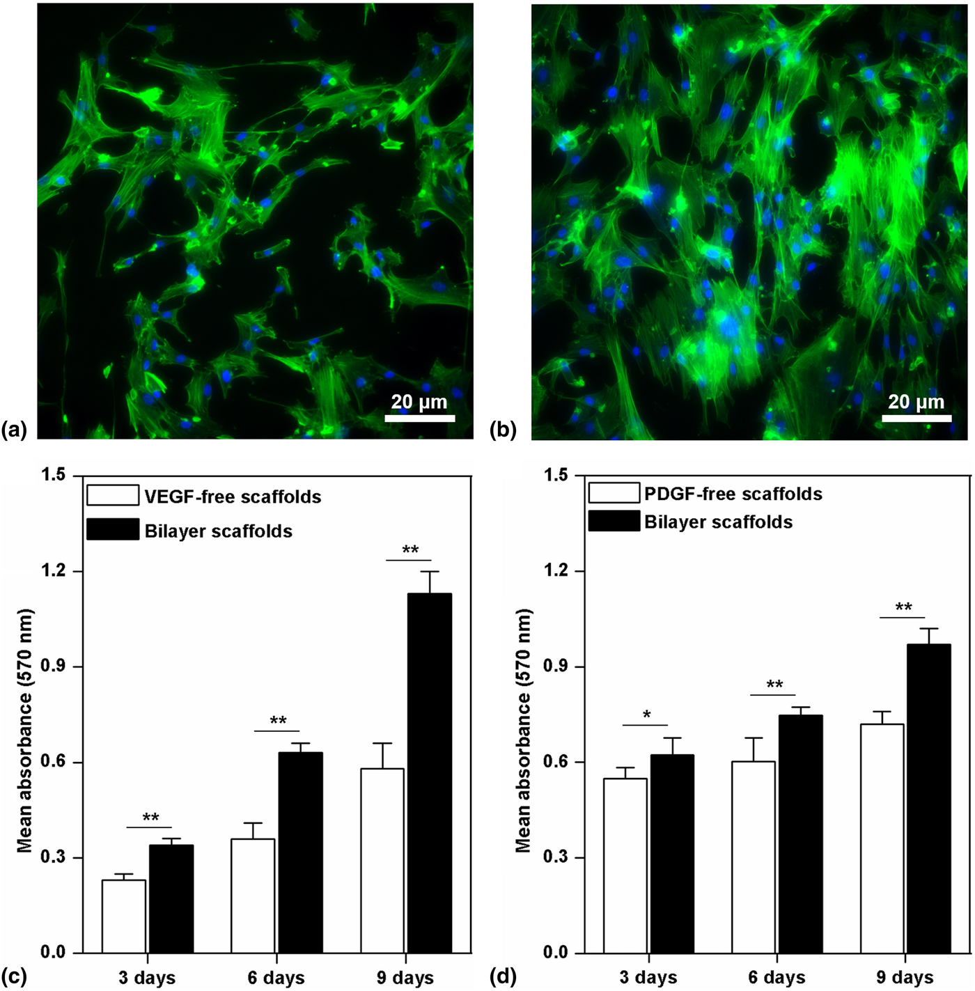
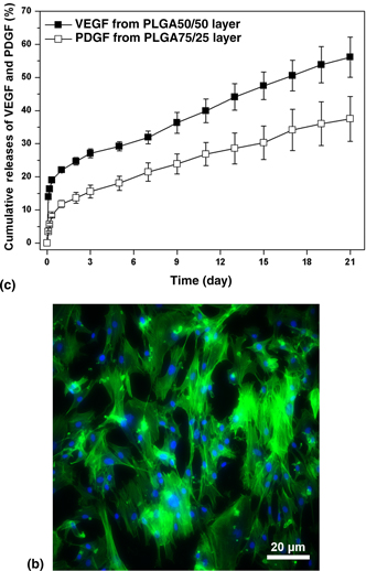
To evaluate the potential of the bilayer scaffolds for GI tissue engineering, the inducements of the structural and biochemical cues of the bilayer scaffolds on the behaviors of vascular endothelial cells and smooth muscle cells (which are important for the vascularization and smooth muscle layer regeneration of the GI tract, respectively) were assessed. The morphology of smooth muscle cells (using HASMC as model cell) was first investigated through the cytoskeleton staining of HASMCs cultured with different scaffolds after 3-day cell culture. While HASMCs on the scaffolds with randomly oriented nanofiber behave well-spread cell morphology with random orientation of F-actin arrangement (Fig. 4(a)), they show obvious directional arrangement for both cell morphology and F-actin alignment along with the nanofiber orientation on the bilayer scaffolds with parallel aligned nanofibers (Fig. 4(b)). The results demonstrate significant inducing effects of the structural cues of the bilayer scaffolds on cell morphology and cytoskeleton development, which shall be promising for promoting the remodeling and maturation of smooth muscle layers with anisotropic anatomies.[Reference Kobayashi, Lei, Wang, Wu and Dunn18] In addition, the effects of biochemical cues (sequentially and sustained releasing VEGF and PDGF) on the proliferation of vascular endothelial cells (using HUVEC as model cell) and smooth muscle cells (using HASMC as model cell) have been also investigated through the MTT assay. The results indicate that HUVECs and HASMCs proliferate well on the GF-free and GF-incorporated scaffolds (bilayer scaffolds), while the proliferation rates of HUVECs and HASMCs on the GF-free scaffolds are both statistically significantly lower than those on the bilayer scaffolds (Figs. 4(c) and 4(d)). The stimulating effects of the bilayer scaffolds with appropriate biochemical cues on the proliferation of vascular endothelial cells and smooth muscle cells have been therefore verified, which shall be promising to assist the early-stage vascularization and the smooth muscle layer remodeling during the regeneration of GI tract or human body tissues with similar anatomy, e.g., blood vessels.[Reference Wells and Spence10,Reference Zhang, Jia, Han, Zhao, Zhao, Fan and Yuan19]
Inducements of the bilayer scaffolds on the morphology and proliferation of smooth muscle cells and vascular endothelial cells. (a) Cytoskeleton staining of HASMCs on the non-woven and PDGF-free emulsion electrospun PLGA75/25 scaffolds 3-day cell culture (green: F-actin, blue: nucleus). (b) Cytoskeleton staining of HASMCs on the upper layer (PDGF-incorporated PLGA75/25 layer) of the bilayer scaffolds after 3-day cell culture (green: F-actin, blue: nucleus). (c) MTT assay results for the HUVECs cultured with the VEGF-free and the dual GF-incorporated bilayer scaffolds. (d) MTT assay results for the HASMCs cultured with the PDGF-free and the dual GF-incorporated bilayer scaffolds (PDGF-incorporated scaffolds). (*) P < 0.05 and (**) P < 0.01.
Conclusions
Bilayer scaffolds with orthogonally aligned nanofibers at different layers were fabricated through a sequential emulsion electrospinning process using PLGA with different degradation properties, in which VEGF and PDGF were, respectively, encapsulated and controlled release in specific sequential and sustained manners. The bilayer scaffolds show appropriate structural and biochemical characteristics to effectively direct and stimulate the behaviors and functions of vascular endothelial cells and smooth muscle cells, which hold promise to assist the early-stage vascularization and the following smooth muscle layer maturation for complex tissue engineering. With these well-defined biomimetic and bioactive properties, it can be envisioned that the bilayer scaffolds can offer promising cell microenvironments for potentially promoting the reconstruction of both structures and functions of many complex human tissues such as GI tract and blood vessels.
Acknowledgments
We would like to acknowledge the financial support from Hong Kong Research Grants Council General Research Funds (HKU717713E, 17201017), National Key R&D Program of China (2017YFA0701303), and the Fundamental Research Program of Shenzhen (JCYJ20170818161757684 and JCYJ20170307164610282).