Implications
Competing processes of milk production and reproduction can be at odds in dairy cows, particularly if the unique metabolic processes that typify early lactation become imbalanced. A potential end result is that cows do not become pregnant during the breeding period. The need to replace infertile cows adds to the cost of production. Understanding basic mechanisms that link the first 60 days of lactation with the subsequent success or failure of establishing pregnancy during the breeding period, therefore, is an important area of research for the dairy industry.
Introduction
Within most dairy production systems, milk or milk solids production per cow is increasing annually. The average United States dairy cow, for example, produces in excess of 9500 kg of milk annually. Peak milk production is in early lactation (within 30 to 60 days after calving) when the uterus is involuting and the ovary is restoring the cyclic processes that lead to oestrus, ovulation, and the formation of a corpus luteum. Competing processes of milk production, uterine involution, and the restoration of ovarian activity can be at odds, particularly if the unique homeorhetic processes that typify early lactation become imbalanced and there is metabolic disease. A potential end result of the competition between high milk production and the reproductive system is that cows do not become pregnant during the breeding period which is typically initiated at ~60 days after calving. Pregnancy failure causes inefficiency in dairy systems because non-pregnant cows do not produce a calf, do not have a subsequent lactation and are replaced with a new (typically 2-year-old) cow. The need to replace infertile cows adds to the cost of production (De Vries, Reference De Vries2006). Understanding basic mechanisms that link the first 60 days of lactation with the subsequent success or failure of establishing pregnancy during the breeding period, therefore, is an important area of research for the dairy industry.
A large number of reviews have been written over the past decade on metabolic processes that link the restoration of ovarian activity with early lactation milk yield (Wathes et al., Reference Wathes, Fenwick, Cheng, Bourne, Llewellyn, Morris, Kenny, Murphy and Fitzpatrick2007; Lucy, Reference Lucy2008; Walsh et al., Reference Walsh, Williams and Evans2011). There is general agreement that restoration of LH pulsatility via release of gonadotropin-releasing hormone (GnRH) from the hypothalamus is a key driver of the process. Hormones such as insulin and IGF1 that hypothetically control GnRH activity at the level of the hypothalamus act synergistically with LH at the level of the ovary. The purpose of this review is to primarily examine factors affecting the establishment of pregnancy during the breeding period via mechanisms that involve the uterus and placenta. This review will specifically focus on glucose because of its dual purpose as a major component of cow’s milk and a molecule that coordinates homeorhetic mechanisms that could possibly impinge upon postpartum uterine health and the subsequent establishment of pregnancy.
Glucose: a central molecule involved in lactation and reproduction
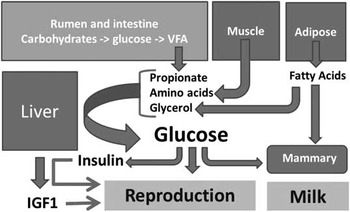
Glucose metabolism presents an interesting challenge for lactating cows (Aschenbach et al., Reference Aschenbach, Kristensen, Donkin, Hammon and Penner2010). The digestive tract of ruminants is specifically adapted to the digestion of forages. Microorganisms in the rumen ferment carbohydrates to volatile fatty acids (VFA) that can be oxidized for energy. In addition to VFA, protein and fat passing into the lower digestive tract are absorbed and used for the synthesis of milk protein and fat. Seventy-two grams of glucose are required for each kg of milk produced (Bell, Reference Bell1995). Most of this glucose is converted directly into lactose (milk sugar). Although glucose is a major product of carbohydrate digestion in the rumen it is rapidly fermented to VFA. Glucose, therefore, must be resynthesized in the liver of postpartum cows via gluconeogenesis (Figure 1). A typical early lactation cow in the United States will produce 50 to 100 kg of milk per day. This equates to a glucose requirement for milk production alone of 3.6 to 7.2 kg/day. Requirements for glucose place a substantial strain on the physiology of dairy cows. Available VFA can only meet about 85% of glucose requirements during early lactation, leaving a 500 g daily deficit for glucose (Bell, Reference Bell1995). Cows undergo a series of homeorhetic mechanisms that are aimed towards elevating blood glucose. In addition to a large increase in hepatic gluconeogenesis shortly after calving, cows assume a state of insulin resistance that redirects glucose to non-insulin-dependent tissues like mammary gland. In spite of these mechanisms, postpartum cows have chronically low blood concentrations of glucose because of failure to meet metabolic requirements for glucose.
Metabolic processes in early postpartum cows that link glucose to the reproductive system. Glucose is synthesized in the liver via gluconeogenesis from substrates arising from rumen fermentation and the catabolism of muscle and adipose tissue. Glucose may ultimately control both circulating insulin (directly) and liver IGF1 production (via insulin-stimulated IGF1 synthesis and secretion). Glucose is required for milk synthesis. Low circulating glucose may impair reproductive processes that are needed to re-establish pregnancy during early lactation.

Glucose may coordinate whole animal metabolism through its capacity to orchestrate changes in circulating concentrations of insulin and IGF1 (Lucy, Reference Lucy2008). Glucose causes insulin release and insulin partitions nutrients towards adipose tissue and muscle (tissue-promoting (anabolic effect) of insulin). Insulin stimulates the liver to release IGF1 into the circulation (Butler et al., Reference Butler, Bork, Pelton, Radcliff, Lucy and Butler2003). As long as glucose remains low, insulin and IGF1 remain low, and cows remain in a catabolic (tissue-losing) state during lactation. Glucose deficiency is only corrected when the mammary gland produces less milk in later lactation, blood insulin and IGF1 increase, and cows partition glucose towards adipose tissue and muscle (an anabolic state). The switch from a catabolic state to an anabolic state is a key regulator of the reproductive axis (Kawashima et al., Reference Kawashima, Matsui, Shimizu, Kida and Miyamoto2012).
Associations between early postpartum glucose and subsequent reproduction
Blood concentrations of glucose decrease in dairy cows after calving. The decrease in blood glucose is hypothetically caused by the initiation of lactation and the associated glucose requirement for milk production. Blood concentrations of glucose were examined in early postpartum cows that either became pregnant or failed to become pregnant later postpartum (Garverick et al., Reference Garverick, Harris, Vogel-Bluel, Sampson, Bader, Lamberson, Spain, Lucy and Youngquist2013). Cows that became pregnant after first artificial insemination (AI) had greater blood concentrations of glucose when compared with cows that did not become pregnant. Significant differences in blood glucose occurred on the day of calving and at 3 days postpartum. Greater blood concentrations of glucose on day 3 postpartum were associated with greater probability of pregnancy at first AI (Garverick et al., Reference Garverick, Harris, Vogel-Bluel, Sampson, Bader, Lamberson, Spain, Lucy and Youngquist2013). A relationship between serum concentrations of non-esterified fatty acid (NEFA) and subsequent pregnancy was detected (cows that became pregnant at first AI had lesser postpartum NEFA during the early postpartum period when compared with cows that did not become pregnant at first AI).
Additional studies published recently have established a link between early postpartum glucose and subsequent reproduction. Effects of lactation on early postpartum nutrient partitioning and the establishment of pregnancy were tested by using a lactating/non-lactating cow model (Green et al., Reference Green, Meyer, Williams, Newsom, Keisler and Lucy2012). Non-lactating cows had greater plasma concentrations of glucose (~+10 mg/dl) compared with lactating cows. Cows that did not get pregnant after first AI at ~60 days postpartum had lesser plasma concentrations of glucose during the first 30 days postpartum when compared with pregnant cows. Later postpartum (30 to 60 days postpartum) there was no relationship between plasma concentrations of glucose and pregnancy.
Green et al. (Reference Green, Meyer, Williams, Newsom, Keisler and Lucy2012) and Garverick et al. (Reference Garverick, Harris, Vogel-Bluel, Sampson, Bader, Lamberson, Spain, Lucy and Youngquist2013) examined association between postpartum blood glucose and pregnancy at first AI. In separate studies performed in Ireland (Cummins et al., Reference Cummins, Lonergan, Evans, Berry, Evans and Butler2012; Moore et al., Reference Moore, Fair, Lonergan and Butler2014), blood concentrations of glucose were examined in dairy cows with similar genetic merit for milk production but with good or poor genetic merit for fertility (Fert+ or Fert−, respectively). Cummins et al. (Reference Cummins, Lonergan, Evans, Berry, Evans and Butler2012) failed to demonstrate differences in circulating glucose postpartum (although early postpartum insulin and IGF1 were greater in Fert+ cows). Subsequent studies of Fert+ and Fert− cows, however, demonstrated greater glucose in Fert+ cows on the day of calving and 1 week later (Moore et al., Reference Moore, Fair, Lonergan and Butler2014). Later postpartum, Fert+ and Fert− cows were similar for blood concentrations of glucose. Finally, in a recent pooled analysis of studies of prepartum nutrition and subsequent postpartum reproduction, Cardoso et al. (Reference Cardoso, LeBlanc, Murphy and Drackley2013) determined that greater blood concentrations of glucose at weeks 3 and 4 postpartum were associated with shorter days to pregnancy.
Studies described in the previous three paragraphs were associative and do not establish cause and effect. Described relationships between blood glucose and pregnancy were for early postpartum blood concentrations of glucose (generally within the 1st month of lactation) and the establishment of pregnancy (several months after the glucose measurements were made). The suggestion is that the early postpartum metabolic profile that includes blood concentrations of glucose is predictive of subsequent postpartum fertility. A key question is how the metabolic profile of early postpartum cows controls reproductive processes leading to pregnancy that occur several months after the early postpartum period.
Confounding of glucose with other metabolites postpartum
Glucose may be a mediator of postpartum reproduction because glucose acts as a substrate for milk production and is an essential molecule for reproductive processes. An obvious limitation to associations involving glucose and reproduction presented in the previous section is that circulating metabolites of early postpartum cows are controlled by endocrine pathways that coordinate several metabolic processes. Uncoupling of the somatotropic axis at calving leads to a decrease in circulating concentrations of IGF1 that exists for at least 60 days postpartum. Demand for glucose during lactation leads to lesser blood concentrations of glucose and insulin. Lesser insulin theoretically prevents (or slows) recoupling of the somatotropic axis through its effects on GH receptor 1A (GHR1A; Butler et al., Reference Butler, Bork, Pelton, Radcliff, Lucy and Butler2003; Lucy, Reference Lucy2008). If a cow is not milked postpartum then blood concentrations of glucose normalize within a few days (Green et al., Reference Green, Meyer, Williams, Newsom, Keisler and Lucy2012; Maillo et al., Reference Maillo, Rizos, Besenfelder, Havlicek, Kelly, Garrett and Lonergan2012; Thompson et al., Reference Thompson, Cerri, Kim, Ealy, Hansen, Staples and Thatcher2012). These same cows with normalized blood glucose, however, have normalized blood concentrations of insulin and IGF1 and a reversal of the metabolic profile that typifies early postpartum cows. It is impossible to say, therefore, whether any change in reproductive function is a consequence of any one hormone/metabolite alone or the collective action of several.
In attempt to address the possibility that glucose is the primary metabolic driver of the entire system, glucose was infused into early postpartum cows in a physiologically relevant manner. Increasing daily doses from 500 to 1500 g/day glucose were administered via jugular infusion by using a constant rate of glucose infusion (Lucy et al., Reference Lucy, Escalante, Keisler, Lamberson and Mathew2013). Glucose infusion increased blood concentrations of insulin and caused a marked decrease in both NEFA and β-hydroxy butyric acid (BHBA). In addition to changes in insulin and circulating metabolites, glucose infusion increased circulating concentrations of IGF1 (Lucy et al., Reference Lucy, Escalante, Keisler, Lamberson and Mathew2013). Insulin may have mediated stimulatory effects of glucose on postpartum IGF1 through its capacity to recouple the somatotropic axis. The magnitude of the IGF1 increase in response to glucose that was observed (10 to 15 ng/ml) approximated the magnitude of IGF1 difference reported elsewhere for cows that have or have not commenced oestrous cycles (Beam and Butler, Reference Beam and Butler1998) or postpartum pregnant v. non-pregnant cows (Taylor et al., Reference Taylor, Cheng, Pushpakumara, Beever and Wathes2004; Green et al., Reference Green, Meyer, Williams, Newsom, Keisler and Lucy2012). The infusion studies demonstrated that a single molecule (glucose) could rapidly reverse the metabolic profile that typifies early lactation. Based on these results, a well-coordinated homeorhetic system that supplies adequate glucose relative to demand in early lactation may be impacting the reproductive systems that are undergoing restoration during the same time period.
Linking the metabolic profile to reproductive processes during the early postpartum period
The state of lowered blood glucose during lactation theoretically compromises the function of tissues that depend on glucose. Associated metabolites such as NEFA and BHBA as well as insulin and IGF1 play a role in controlling tissue function. Based on the data presented above, the first 30 days postpartum may be the most-critical in terms of the impact that metabolites and metabolic hormones have on reproduction. Within this review, two essential processes that occur during the first 30 days postpartum will be examined with respect to metabolites and metabolic hormones – the restoration of ovarian cyclicity and uterine involution.
Restoration of ovarian cyclicity postpartum
With respect to postpartum reproduction, most of the research performed on metabolites and metabolic hormones has focused on the re-initiation of ovarian cyclicity. Cows that have not commenced oestrous cyclicity are by definition infertile because they have not ovulated. Furthermore, fertility generally improves with each successive oestrous cycle before breeding (Peter et al., Reference Peter, Vos and Ambrose2009). There has been, therefore, a traditional focus on understanding mechanisms that control the restoration of ovarian activity before breeding with earlier restoration viewed as favourable. A frequently addressed topic is the positive association between insulin, IGF1, and the day postpartum that cows begin to cycle (Velazquez et al., Reference Velazquez, Spicer and Wathes2008).
The capacity of a variety of metabolites and metabolic signals to act on the hypothalamus and pituitary to increase GnRH and LH pulsatility has been reviewed (Chagas et al., Reference Chagas, Bass, Blache, Burke, Kay, Lindsay, Lucy, Martin, Meier, Rhodes, Roche, Thatcher and Webb2007; LeRoy et al., Reference Leroy, Vanholder, Van Knegsel, Garcia-Ispierto and Bols2008). At the ovary, both insulin and IGF1 promote proliferation, differentiation and survival of follicular cells (Lucy, Reference Lucy2008). Perhaps the most important actions of insulin and IGF1 are observed when either hormone acts synergistically with gonadotropins (either FSH or LH). The synergistic relationship between insulin, IGF1 and gonadotropins is seen for a variety of cellular functions including mitogenesis, cell survival, and steroidogenesis. Glucose controls insulin secretion and ultimately controls hepatic IGF1 secretion via insulin release. Circulating glucose and the insulin/IGF1 systems, therefore, are functionally linked in the whole animal. There are several reviews that have been published recently on the relationships between insulin, IGF1 and postpartum ovarian function (Lucy, Reference Lucy2008; Kawashima et al., Reference Kawashima, Matsui, Shimizu, Kida and Miyamoto2012).
Effects of hormones and metabolites on ovarian cyclicity in postpartum cows are generally thought to be coincident meaning that changes in circulating insulin, IGF1, glucose, etc. exert their effects in real time. The ‘Britt hypothesis’ states that early postpartum hormones and metabolites can exert carry-over effects on the somatic and germ cells (Britt, Reference Britt1991). The Britt hypothesis is based on the fact that ovulatory follicles require ~60 days to fully develop. The oocyte that ovulates around the time of first insemination and somatic cells within the same follicle are initiating their development at calving (or shortly thereafter). Associations between postpartum hormones and metabolites and subsequent reproduction are found early postpartum when the most-extreme homeorhetic adjustments are known to occur. The early postpartum metabolic profile, therefore, may have the capacity to imprint ovarian tissue either through permanent effects on the genome (epigenetic mechanisms) or by changing the chemical composition of the cells themselves.
Perhaps the best-studied example of a metabolic imprint is for postpartum NEFA and its effect on composition of the oocyte and function of follicular cells (Leroy et al., Reference Leroy, Rizos, Sturmey, Bossaert, Gutierrez-Adan, Van Hoeck, Valckx and Bols2011; Van Hoeck et al., Reference Van Hoeck, Sturmey, Bermejo-Alvarez, Rizos, Gutierrez-Adan, Leese, Bols and Leroy2011; Wathes, Reference Wathes2012). The possibility that there are permanent epigenetic modifications to the genome during the early postpartum period that affect long-term developmental competence of follicular cells has not been demonstrated at this time. According to the Britt hypothesis, the only way to correct the metabolic imprint is to allow the imprinted cells (in this case the oocyte, granulosa and theca cells) to exhaust their physiologically relevant lifespan.
Uterine health and immune function
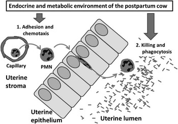
Under normal circumstances, involution (process through which the uterus returns to a normal non-pregnant state) is completed during the 1st month postpartum. During involution, the uterus shrinks in size, re-establishes the luminal epithelium, and immune cells (primarily polymorphonuclear neutrophils (PMN)) infiltrate the uterus to clear residual placental tissue as well as infectious microorganisms (Gilbert et al., Reference Gilbert2011; LeBlanc, Reference LeBlanc2012; Wathes et al., Reference Wathes, Clempson and Pollott2012). Postpartum cows have a depressed immune system particularly during the 1st month after calving. With respect to uterine involution and disease, the current theory is that the metabolic environment in postpartum cows suppresses the innate immune system through effects on PMN function (Graugnard et al., Reference Graugnard, Bionaz, Trevisi, Moyes, Salak-Johnson, Wallace, Drackley, Bertoni and Loor2012; LeBlanc, Reference LeBlanc2012; Ingvartsen and Moyes, Reference Ingvartsen and Moyes2013; Figure 2). In most cases, changes in circulating concentrations of nutrients and metabolites that occur in postpartum cows are exactly opposite to those that would benefit the function of PMN (Table 1). Inhibitory effects of the postpartum metabolic profile can affect traditional aspects of PMN function (migration, oxidative burst, phagocytosis, etc.). There is good agreement between in vitro analyses of PMN function and epidemiological evidence that indicates that an abnormal periparturient metabolic profile predisposes cows to uterine disease during the early postpartum period and infertility later postpartum (Chapinal et al., Reference Chapinal, Leblanc, Carson, Leslie, Godden, Capel, Santos, Overton and Duffield2012).
Model for uterine polymorphonuclear neutrophils (PMN) function in postpartum cows. The PMN migrate into the uterine lumen postpartum where they kill bacteria and clear debris. The endocrine and metabolic environment of postpartum cows may compromise PMN. Cows are predisposed to uterine disease because the PMN fail to efficiently migrate into the uterus or have reduced capacity for phagocytosis and killing.

Hormones and metabolites affected by milk production in postpartum dairy cows, their known effects on polymorphonuclear neutrophil (PMN) function, and relative concentrations (postpartum v. later lactation)

BHBA=β-hydroxy butyric acid; NEFA=non-esterified fatty acid.
Glucose is the primary metabolic fuel that PMN use to generate the oxidative burst that leads to killing activity. Glucose is stored as glycogen within the PMN. Concentrations of glycogen in PMN decrease in a manner that is similar to the decrease in blood glucose postpartum with a lag period of ~7 days (Galvão et al., Reference Galvão, Flaminio, Brittin, Sper, Fraga, Caixeta, Ricci, Guard, Butler and Gilbert2010). The lag in the decrease in PMN glycogen may represent the time required for glycogen loading during maturation of PMN. Galvão et al. (Reference Galvão, Flaminio, Brittin, Sper, Fraga, Caixeta, Ricci, Guard, Butler and Gilbert2010) observed that cows developing uterine disease had lesser concentrations of glycogen in their PMN. Their conclusion was that lesser glycogen reserve led to a reduced capacity for oxidative burst in PMN and uterine disease.
Most of the available data indicate that the metabolic profile of prepartum cows is equally important to that of postpartum cows for subsequent uterine health and (or) the establishment of pregnancy (Castro et al., Reference Castro, Kawashima, van Dorland, Morel, Miyamoto and Bruckmaier2012). In their work in which an index for physiological imbalance was created, Moyes et al. (Reference Moyes, Larsen and Ingvartsen2013) concluded that an index that included NEFA, BHBA and glucose was predictive of postpartum uterine disease especially when the prepartum index was used. In postpartum cows, blood concentrations of NEFA were the best predictor of postpartum disease. Their finding is consistent with the general notion that NEFA can negatively affect immune cell function in postpartum cows. Cardoso et al. (Reference Cardoso, LeBlanc, Murphy and Drackley2013) also concluded that NEFA was the key molecule associated with postpartum disease.
In all likelihood the metabolic profile associated with uterine disease is initiated before calving or shortly thereafter. Perhaps this is not surprising given the relatively acute nature of physiological events at calving and the homeorhetic mechanisms at the initiation of lactation. A cow’s homeorhetic capacity (i.e. capacity for gluconeogenesis, lipid mobilization, etc.) and her inherent resistance to disease are largely manifested after calving but the underlying biology is theoretically in place before calving.
Linking the early postpartum metabolic profile with subsequent pregnancy
Metabolic or uterine disease postpartum extends the interval from calving to pregnancy and increases pregnancy loss (Ribeiro et al., Reference Ribeiro, Lima, Greco, Bisinotto, Monteiro, Favoreto, Ayres, Marsola, Martinez, Thatcher and Santos2013). Arguments presented in the previous paragraph are that the metabolic profile that typifies postpartum cows (elevated NEFA and BHBA and reduced blood glucose) leads to reduced immune cell function that predisposes cows to uterine disease. What is the signal, however, that leads to long-term reproductive failure in cows with relatively short-term postpartum uterine disease? Presumably the disease state of cows is resolved at some stage postpartum but nonetheless there appear to be carry-over effects of the early postpartum period on the establishment of pregnancy. The Britt hypothesis (typically applied to the ovarian follicle) should perhaps be expanded to include the uterus of postpartum cows. The uterus is repaired during uterine involution that occurs primarily within the first 30 days after calving. The process is largely completed before breeding but nonetheless the uterus appears to have a relatively long-term memory (imprinting) for metabolic and disease states of the early postpartum period.
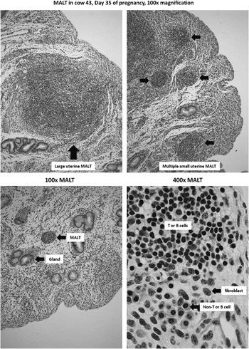
The nature of the metabolic or disease-related imprinting that occurs within the uterus is completely unknown. It is possible that cows with postpartum uterine disease have not fully resolved the infection at the time of first postpartum breeding. Primary methods of assessing uterine health during the breeding period (presence of pus in vaginal mucous or existence of an enlarged uterus) would only detect more extreme cases of prolonged uterine disease. It is possible, therefore, that there are remnants of uterine inflammation in a seemingly normal uterus and that these remnants in some way affect the capacity for cows to become pregnant. Turner et al. (Reference Turner, Healey and Sheldon2012) described aggregates of uterine immune cells in postpartum cows. We have noticed these unique structures as well (Figure 3). We believe that they are analogous to mucosa-associated lymphoid tissue (MALT). Uterine MALT appear as clusters of immune cells within the uterine stroma and uterine caruncular areas. The MALT may be deep within stroma or near the surface epithelium. In unpublished work, MALT were observed within the endometrium of pregnant cows (~100 days postpartum) that had been previously diagnosed with a postpartum uterine infection (Figure 3). Whether the presence of MALT has any implications for pregnancy establishment, maintenance or loss is unknown.
Uterine mucosa-associated lymphoid tissue (MALT) in cow 43 (top panel) and low power and high power magnification of a uterine MALT (bottom panel). Herd records noted a postpartum uterine infection after calving in cow 43. Histological examination of the uterus of the same cow at slaughter (100 days postpartum, top panel) revealed a large number of MALT perhaps implying an immune-cell based carry-over effect of the postpartum uterine infection on uterine morphology of the pregnant uterus. Based on morphology, the MALT appear to be comprised of lymphocytes (either T or B cells, bottom panel).

In addition to potential physical imprinting of the uterus (via resident immune cells), it is possible that proliferation and differentiation of the uterine epithelium is permanently affected by the early postpartum metabolic profile or the disease state of the postpartum uterus. Uterine epithelium, and to a certain extent stroma, is regenerated during uterine involution presumably from stem cells that reside within the uterus. It is possible that the uterine environment that theoretically includes pathogens, PMN, metabolites, and hormones leaves an epigenetic mark on uterine epithelial and stromal cells as they go through the process of differentiating from the stem cell pool. These epigenetic marks would be inherited by daughter cells as they proliferate to occupy uterine epithelium and stroma. This altered epigenome of mature uterine epithelial cells and stroma may in some manner affect the capacity of the uterus to establish pregnancy during the breeding period that occurs several months after the uterine epithelial lining has been established.
Mechanisms linking lactation to pregnancy during the breeding period
Assuming that uterine involution is complete and cows have commenced oestrous cycles then what are the implications of lactation for cows during the breeding period? Several authors have recently addressed potential relationships between normal oestrous cycles and the metabolic profile of cows during the breeding period (Wathes et al., Reference Wathes, Fenwick, Cheng, Bourne, Llewellyn, Morris, Kenny, Murphy and Fitzpatrick2007; López-Gatius, Reference López-Gatius2012). As detailed above, the metabolic profile of later postpartum cows (>30 days postpartum) still involves relatively low concentrations of glucose, insulin and IGF1 although concentrations of NEFA and BHBA have typically normalized.
Oestrous cyclicity during the breeding period
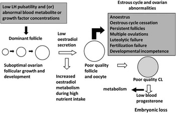
Patterns of oestrous cyclicity for lactating cows are different (appear to be less regular) when compared with oestrous cycle of nulliparous heifers. An existing hypothesis is that the same hormones that control when cows begin to cycle (insulin, IGF1 and LH) have an effect on the periodicity of cyclicity which relates to the functionality of follicles and corpora lutea (Figure 4). As described for early lactation cows, the hormonal environment created by lactation (in this example low blood concentrations of glucose, insulin and IGF1) may potentially affect the capacity of ovarian cells to respond to gonadotropins. In cows that have commenced oestrous cycles, this could potentially affect oestradiol production by follicles as well as progesterone production by corpora lutea. Low blood glucose could potentially compromise a variety of essential metabolic processes in ovarian cells including oocytes that depend on glucose for energy (Clark et al., Reference Clark, Stokes and Thompson2011; Berlinguer et al., Reference Berlinguer, Gonzalez-Bulnes, Contreras-Solis, Spezzigu, Torres-Rovira, Succu, Naitana and Leoni2012). In their recent study of bovine follicles, Walsh et al. (Reference Walsh, Mehta, McGettigan, Browne, Forde, Alibrahim, Mulligan, Loftus, Crowe, Matthews, Diskin, Mihm and Evans2012) concluded that STAR gene expression (the rate limiting enzyme in steroidogenesis) was specifically down-regulated by the metabolic profile found in early lactation (low glucose, insulin and IGF1).
Mechanisms that link metabolism, metabolic hormones and metabolites to oestrous cycle abnormalities, infertility, and embryonic loss in postpartum cows. Follicles and corpora lutea (CL) respond to changing concentrations of gonadotropins, growth factors, and blood metabolites in postpartum cows. Steroids produced by follicles and CL are metabolized by the liver of lactating cows resulting in reduced circulating concentrations. Endocrine failure can lead to oestrous cycle and ovarian abnormalities as well as a poor quality oocyte that may not develop after fertilization. If corpora lutea are compromised and steroid metabolism is greater then blood concentrations of progesterone are low and there is potential for embryonic loss.

Work cited in the previous paragraph indicates that follicles in lactating cows are compromized because lesser glucose, insulin and IGF1 affect the capacity of follicles to respond to LH and synthesize oestradiol. There is the potential for greater steroid metabolism in lactating compared with non-lactating cows that can be explained by greater dry matter intake in cows that are lactating (Wiltbank et al., Reference Wiltbank, Souza, Carvalho, Bender and Nascimento2011). Lesser circulating oestradiol from preovulatory follicles has been implicated in abnormal patterns of follicular growth leading to anovulatory conditions, multiple ovulation and reduced oestrous expression in lactating cows (Figure 4). Likewise, greater steroid metabolism has been implicated as a mechanism leading to low circulating progesterone in lactating cows that could potentially affect early embryonic and foetal development (see subsequent paragraph).
Subnormal luteal function
Most of the work relating early embryonic development to lactation addresses the possibility that low progesterone during the 1st weeks after insemination leads to slower embyronic development that predisposes cows to embyronic loss (Diskin and Morris, Reference Diskin and Morris2008; Lonergan, Reference Lonergan2011). Progesterone stimulates uterine histotroph secretion and lesser uterine histotroph secretion (caused by low progresteorne) leads to slower embyronic development. The theory is that slowly developing embryos fail to reach adequate size to generate an adequate interferon-tau (IFNT) signal to the mother (Robinson et al., Reference Robinson, Hammond, Wathes, Hunter and Mann2008). Pregnancies are lost because the mother fails to recognize the pregnancy and undergoes luteal regression as if not pregnant. Low progesterone during the first week after AI is believed to occur more frequently in lactating cows because of abnormal luteal function or greater steroid metabolism assoicated with lactation. Several authors have recently reviewed mechanisms assoicated with subnormal luteal development and early embryonic loss (Pursley and Martins, Reference Pursley and Martins2011; Wiltbank et al., Reference Wiltbank, Souza, Carvalho, Bender and Nascimento2011; Bridges et al., Reference Bridges, Day, Geary and Cruppe2013).
Glucose as a substrate for the developing embryo and foetus
What other mechanisms in addition to reduced blood concentrations of progesterone potentially explain the rate at which the embryo or foetus grows as well as pregnancy loss is lactating dairy cows? Glucose is typically thought of as a key energy source for ATP production through mitochondrial oxidative phosphorylation. Glucose is not used primarily for metabolic fuel production, however, by either the mammary gland or the pregnancy. In mammary gland, most of the glucose is used to produce lactose (milk sugar). Likewise, in the uterus and placenta most of the glucose is used to supply carbons for the synthesis of cellular components (nucleotides, amino acids, lipids, etc.). This latter phenomenon is known as the ‘Warburg effect’ and typifies proliferating cells (Vander Heiden et al., Reference Vander Heiden, Cantley and Thompson2009).
Green et al. (Reference Green, Meyer, Williams, Newsom, Keisler and Lucy2012) either milked cows normally or dried them off (not milked) immediately after calving. Cows were inseminated at ~60 days postpartum and the foetus and placenta were collected on day 28, 35 or 42. One major conclusion was that for a given day of pregnancy, the foetus and placenta from a lactating cow were smaller (weighed less) than the foetus and placenta from a non-lactating cow. In subsequent work, concentrations of glucose were less in foetal fluids collected from lactating compared with non-lactating cows, perhaps because maternal concentrations of glucose were less during lactation (Lucy et al., Reference Lucy, Green, Meyer, Williams, Newsom and Keisler2012). This latter observation agreed with the general notion that glucose transport to the foetus primarily depends on Glut 1 and Glut 3 transporters with glucose moving down its concentration gradient through facilitative diffusion (Wooding et al., Reference Wooding, Fowden, Bell, Ehrhardt, Limesand and Hay2005). Lesser maternal glucose leads to lesser foetal glucose because the foetus and placenta cannot sequester glucose against a concentration gradient. Furthermore, the foetus and placenta have a limited capacity for gluconeogenesis (Battaglia and Meschia, Reference Battaglia and Meschia1978). The reduction in glucose reaching the uterus and conceptus can potentially affect how the pregnancy develops because the pregnancy depends on glucose as a substrate for tissue synthesis and metabolic energy (Battaglia and Meschia, Reference Battaglia and Meschia1978).
Growth of the foetus and placenta, therefore, depends on the metabolic milieu of cows. Low concentrations of glucose in postpartum cows may predispose cows to pregnancy loss because the foetus/placenta may not have adequate substrate for the creation of new cells. Neither the mammary gland nor the placenta can concentrate glucose via a glucose transporter. Greater blood flow to the mammary gland facilitates greater capacity to extract glucose from the circulation, and sets up the metabolic scenario where the mammay gland has priority over the pregnancy for glucose.
Does slower embryonic or foetal growth lead to pregnancy loss?
What is the evidence that that slower embryonic or foetal growth caused by either low progesterone or low glucose growth actually leads to pregnancy loss? The slower developing embryo or foetus may simply be responding to its environment and adjusting to lesser histotroph secretion (low progesterone environment) or lesser glucose availability (low glucose environment) by growing more slowly. This is, of course, inevitable in as much as a tissue cannot grow faster than its capacity to sequester and use substrates required for growth. Perhaps slower growth is ‘normal’ when substrates for growth are limiting and slower growth may not predispose cows to pregnancy to loss.
Few published studies in cattle have asked the question ‘does slower growth of the foetal/placental unit lead to pregnancy loss?’ Pregnant cows that undergo pregnancy loss have lesser blood concentrations of pregnancy-associated glycoprotein (PAG) leading up to the time that the pregnancy is aborted (Gábor et al., Reference Gábor, Tóth, Ozsvári, Abonyi-Tóth and Sasser2007; Thompson et al., Reference Thompson, Cerri, Kim, Green, Santos and Thatcher2010; Pohler et al., Reference Pohler, Geary, Johnson, Atkins, Jinks, Busch, Green, MacNeil and Smith2013). Lesser blood concentrations of PAG may indicate that cows are pregnant with a small embryo or foetus. These data showing a relationship between smaller embryos or foetuses and pregnancy loss raise the question of cause and effect. Does slow growth eventually lead to death of the embryo/foetus or does an embryo/foetus destine to undergo loss have slower growth.
Conclusions
The endocrine and metabolic environment of lactating cows affects the capacity of cows to become pregnant postpartum. There is ample evidence that the hormones that coordinate homeorhetic adjustments that support lactation can act on the uterus and ovary before and during the breeding period. In addition to the hormonal environment, the metabolic environment created by lactation that includes low blood glucose and elevated NEFA and BHBA impinges upon the ovary as well as the immune system that plays a critical role in restoring uterine health in postpartum cows. Specific mechanisms through which the metabolic environment of early lactation deposits a lasting imprint on uterine and ovarian function is less clear. Also less clear are the mechanisms that link lactation to a predisposition for pregnancy loss in lactating cows. The slow rate of embryonic or foetal growth in lactating cows with low blood progesterone and low blood concentrations of glucose may be an important mechanism explaining pregnancy loss.
Acknowledgements
The authors would like to thank the Fulbright Commission/Education USA, the University of Missouri, and Teagasc, Animal & Grassland Research and Innovation Programme, Moorepark for supporting the research leave granted to MCL during which time this manuscript was prepared and presented.