Introduction
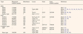
Polydactyly (Gr. Poly = many; dactylos = digit) is one of the most common inherited limb abnormalities with an incidence between 5 and 19 per 10 000 live births (Refs Reference Malik1, Reference Zguricas2). Polydactyly is mainly characterised by supernumerary fingers or toes, ranging from a little of additive soft tissue without bony structure to an integral and fully developed supernumerary digit (s). Most of these malformations are rudimentary skin tags (Refs Reference Malik1, Reference Schwabe and Mundlos3, Reference Christensen4, Reference de la Torre and Simpson5). The involvement of upper limbs has been reported to be more common than the lower limbs, the right hand is more common than the left hand, and the left foot is more common than the right foot (Ref. Reference Malik6). Polydactyly is caused by disturbing the normal procedure of the anterior–posterior axis of the developing limb, with diverse aetiology and variable inter- and intra-familial clinical features (Refs Reference Malik1, Reference Biesecker7, Reference Talamillo8, Reference Materna-Kiryluk9). The disorder can appear as an isolated disorder (non-syndromic polydactyly), in combination with other hand/foot malformations, or as a part of a pleiotropic developmental anomaly syndrome (syndromic polydactyly) (Ref. Reference Biesecker7). Currently, the most acknowledged classification of non-syndromic polydactyly complies with a morphological criterion according to the position of the extra digit (s) in the hand or foot. It can be classified into preaxial polydactyly (PPD), axial (central) polydactyly and postaxial polydactyly (PAP) (Refs Reference Talamillo8, Reference Haber10). PPD is defined as a supernumerary digit affecting the first digits, PAP involves the fifth digits, and the least common type, axial (central) polydactyly, involves duplication of three central digits: the second, third or fourth digits (Refs Reference Talamillo8, Reference Haber10). Additionally, other complex types, including mirror-image polydactyly (MIP), Haas-type polysyndactyly, and palmar/ventral and dorsal type polydactyly, could not fit into the above three types (Refs Reference Malik1, Reference Phadke and Sankar11, Reference Gillessen-Kaesbach and Majewski12). PPD can be further classified into five types, including thumb polydactyly, polydactyly of a triphalangeal thumb, polydactyly of an index finger, polysyndactyly and hallux polydactyly (Ref. Reference Orioli and Castilla13). PAP can be further classified into types A and B, according to the extra digit (s) being either well developed (type A, PAPA) or rudimentary (type B, PAPB) (Ref. Reference Talamillo8). And PAPA can be divided into six genetic types: PAPA1, PAPA2, PAPA3, PAPA4, PAPA5 and PAPA6 (Refs Reference Radhakrishna14, Reference van der Zwaag15, Reference Zhao16, Reference Galjaard17, Reference Mollica18, Reference Mohan19, Reference Kalsoom20). The primary aetiology of polydactyly appears to be genetic, and the environmental factors, diet, infections and other causes are less likely (Refs Reference Christensen4, Reference Chong21). The simple definition belies the complexity and heterogeneity of the disorder, and the different forms show distinguishing genetic entities (Refs Reference Christensen4, Reference Guo22). Non-syndromic polydactyly frequently exhibits an autosomal dominant inheritance with variable penetrance (Refs Reference Talamillo8, Reference Haber10). To date, in human, at least ten loci and four disease-causing genes, including the GLI family zinc finger 3 gene (GLI3, OMIM 165240), the zinc finger protein 141 gene (ZNF141, OMIM 194648), the mirror-image polydactyly 1 gene (MIPOL1, OMIM 606850) and the paired-like homeodomain 1 gene (PITX1, OMIM 602149), have been identified (Table 1).
Loci and disease-causing genes of non-syndromic polydactyly

AD, Autosomal dominant inheritance; AR, Autosomal recessive inheritance; GLI3, the GLI family zinc finger 3 gene; Haas type, Haas-type polysyndactyly; Hallux type, hallux polydactyly; MIP, mirror image polydactyly; MIPOL1, the mirror-image polydactyly 1 gene; PAP, postaxial polydactyly; PAPA1, postaxial polydactyly type A1; PAPA2, postaxial polydactyly type A2; PAPA3, postaxial polydactyly type A3; PAPA4, postaxial polydactyly type A4; PAPA5, postaxial polydactyly type A5; PAPA6, postaxial polydactyly type A6; PITX1, the paired-like homeodomain 1 gene; PPD, preaxial polydactyly; PPD1, thumb polydactyly; PPD2, polydactyly of a triphalangeal thumb; PPD3, polydactyly of an index finger; PPD4, polysyndactyly; ZNF141, the zinc finger protein 141 gene; ZRS, the ZPA regulatory sequence.
In this paper, we review clinical features of non-syndromic polydactyly and summarise the recent progress in the molecular genetics, including loci and genes that are responsible for the disorder, the signalling pathways that these genetic factors are involved in, as well as animal models of the disorder. We hope our review will provide some new insights on the pathogenesis of this complicated disorder and might have implications on genetic counselling such as prenatal diagnosis.
Epidemiology
The incidence of polydactyly is between 5 and 19 per 10 000 live births worldwide, though it varies between genders and among different racial groups (Refs Reference Zguricas2, Reference Bingle and Niswander23, Reference Woolf and Myrianthopoulos24). Twice as many men as women are affected with polydactyly (Ref. Reference Malik1). It was reported that the incidence is about twice more frequent in Chinese male than in white male, and approximately 10–13 times more frequent in Africans than in Caucasians (Refs Reference Bingle and Niswander23, Reference Castilla25). Additionally, about 1.3% of all African Americans are born with polydactyly (Ref. Reference Woolf and Myrianthopoulos24). Non-syndromic polydactyly is detected in 1.01 per 1000 examined live births (Ref. Reference Castilla25). PPD and PAP occur more frequently, while the axial form is extremely rare (Ref. Reference Materna-Kiryluk9). The most common type of polydactyly is partial or complete duplication of thumb/hallux (Ref. Reference Woolf and Woolf26), while complete PAP is uncommon occurring in approximately 0.014% of all live births (Ref. Reference de la Torre and Simpson5).
Genetics of non-syndromic polydactyly
Preaxial polydactyly
PPD is a frequent birth defect featured by extra digit (s) on the tibial/radial side of limb with the incidence as high as 1 in 3000 births. PPD results from aberrant bifurcation of the embryonic preaxial anlage with ectopic sonic hedgehog (SHH, OMIM 600725) expression in the anterior limb bud (Refs Reference Christensen4, Reference Guo22, Reference Semerci27, Reference Light28, Reference Zhang29). PPD seems to have genetic heterogeneity. PPD can be classified into five subtypes: (I) PPD1, characterised by doubling of one or more osteal components of a biphalangeal thumb, (II) PPD2/TPT, characterised by a triphalangeal opposable thumb with or without duplicated thumb, (III) PPD3, characterised by replacement of the thumb by one or two triphalangeal digits accompanied with dermatoglyphic pattern specific for the second finger, (IV) PPD4, characterised by a mild degree of PPD and variable syndactyly involving the third and fourth finger in the hand as well as the second and third toe in the foot, and a special subtype of PPD (hallux polydactyly) characterised by duplication of hallux (Refs Reference Schwabe and Mundlos3, Reference Orioli and Castilla13).
PPD1 (OMIM 174400)
PPD1 is the most common type among the five subtypes of PPD (Refs Reference Materna-Kiryluk9, Reference Chong21). It seems to be more accurate to view PPD1 as a ‘splitting’ of a digit rather than a true digit because the two duplicates are smaller with variations in the size and form than that of a normal thumb in almost all cases (Ref. Reference Chong21). PPD1 follows an autosomal dominant inheritance model with reduced penetrance, and no disease-causing locus or gene has been identified (Ref. Reference Orioli and Castilla13).
PPD2 (7q36, OMIM 174500) and ZRS
In 1994, the disease gene locus for three large kindreds with TPT was mapped to chromosome 7q36 (Refs Reference Heutink30, Reference Tsukurov31). In 1999, PPD2 locus was refined to a region of 1.9-cM after analysing 11 kindreds of different ethnic origin (Ref. Reference Zguricas2). In 2002, a breakpoint for a reciprocal t(5;7) (q11;q36) chromosomal translocation residing in the limb development membrane protein 1 (LMBR1) gene intron 5, disrupting a long-range cis-acting regulator for SHH, was found in a 3-year-old patient with bilateral duplication of the triphalangeal thumb and triplication of the first toe (Ref. Reference Lettice32). In 2003, point mutations (105C>G, 305A>T, 323T>C, 404G>A and 545G>A) in the SHH regulatory factor, ZRS, the zone of polarising activity (ZPA) regulatory sequence, were first reported to segregate with PPD in four unrelated families (Ref. Reference Lettice33). In 2007, a 739A>G transition near the 5-prime end of the ZRS, and a 621C>G mutation in the ZRS of the LMBR1 gene in three families with triphalangeal thumb and PPD were identified (Ref. Reference Gurnett34). Subsequently, point mutations (295T>C, 4909 C>T, 297G>A, 334T>G and 402C>T) in the ZRS were identified to be responsible for PPD2 (Refs Reference Semerci27, Reference Furniss35, Reference Albuisson36, Reference VanderMeer37). Intriguingly, a male with homozygous 4909C>T variant was reported to show no phenotypic difference from heterozygotes. The disease gene locus of a large Chinese pedigree with congenital PPD was mapped to a 1.7-cM interval of 7q36, pathogenic mutation in LMBR1 coding region and ZRS and duplication of ZRS were excluded, which suggested that the disorder has genetic heterogeneity or a mutation was missed (Refs Reference Semerci27, Reference Li38).
Triphalangeal thumb-polysyndactyly syndrome (TPT-PS) belongs to PPD2. It is an isolated limb deformity characterised by pre- and postaxial polysyndactyly of hands and feet (Refs Reference Klopocki39, Reference Balci40). In 1999, the disease gene locus of TPT-PS was mapped to 7q36 by genetic linkage study of a Turkish family (Ref. Reference Balci40). Subsequently, point mutations (739A>G, 621C>G, 4220C>T and 463T>G) and duplications within ZRS were identified in 12 families with TPT-PS (Refs Reference Gurnett34, Reference Klopocki39, Reference Wang41, Reference Farooq42, Reference Sun43). Additionally, point mutations (402C>T, 404G>C and 406A>G) in the ZRS were identified to cause Werner Mesomelic Syndrome (WMS) (Refs Reference VanderMeer37, Reference Wieczorek44, Reference Norbnop45), and duplications of the ZRS can cause syndactyly type IV (Refs Reference Wieczorek44, Reference Wu46).
PPD3 (OMIM 174600)
In 1931, a patient with index finger polydactyly has been reported. Subsequently, in 1962, a family with triphalangeal thumbs (also known as extra index fingers) was described. In 1976, a four-generation Turkish family with index finger polydactyly was reported. PPD3 exhibits an autosomal dominant trait of inheritance, and no disease-causing locus or gene has been identified (Ref. Reference Atasu47).
PPD4 (7p14.1, OMIM 174700) and the GLI3 gene
In 1984, a five-generation family with PPD4 was reported. Twenty-one affected individuals in the family showed variable expression without apparent sex influence, complete penetrance and more severe phenotype in the feet than the hands (Ref. Reference Reynolds48). Heterozygous p.L1216PfsX31 and p.R290X mutations in the GLI3 gene were identified in two families presented with PPD4 (Refs Reference Radhakrishna49, Reference Fujioka50).
Hallux polydactyly(OMIM 601759) and chromosome 2q31.1–31.2
In 2009, a ~3.4-Mb interstitial microdeletion at 2q31.1-31.2 was identified in a three-generation family with duplication of hallux with clinodactyly (Ref. Reference Tsai51). No disease-causing gene has been identified.
Postaxial polydactyly
PAP is featured by duplication of the fifth finger/toe on the ulnar/fibular side of hands/feet (Refs Reference Schwabe and Mundlos3, Reference Umm-e-Kalsoom52). The incidence of PAP is ten times (1/143 versus 1/1339 in live births) more common in the African and African-American population than in their Caucasian counterparts (Ref. Reference Guo22). Non-syndromic PAP usually segregates as an autosomal dominant trait (Ref. Reference Umm-e-Kalsoom52). Based on clinical manifestation, PAP can be divided into PAPA and PAPB. PAPA is featured by a well-developed extra digit, whereas PAPB is characterised by a rudimentary fifth digit frequently appearing as pedunculated postminimus (Refs Reference Schwabe and Mundlos3, Reference Kalsoom20, Reference Umm-e-Kalsoom52). PAPA is a genetically heterogeneous disorder and exhibits as either autosomal dominant or recessive manner of inheritance (Ref. Reference Kalsoom20). To date, six disease gene loci, including PAPA1, PAPA2, PAPA3, PAPA4, PAPA5 and PAPA6, and two genes responsible for PAPA have been identified (Refs Reference Radhakrishna14, Reference van der Zwaag15, Reference Zhao16, Reference Galjaard17, Reference Mollica18, Reference Mohan19, Reference Kalsoom20). Given that both isolated PAPA1 and PAPB are caused by the mutations in the GLI3 gene, they are assigned to the same OMIM number 174200, and PAPB may considered as a variant of PAPA1 in genetics.
PAPA1 (7p14.1, OMIM 174200) and the GLI3 gene
In 1997, the disease gene locus of PAPA1 was mapped to 7pl5-q11.23 by linkage analysis of five-generation Indian family (Ref. Reference Radhakrishna14). Subsequently, a heterozygous mutation, p.A765PfsX14, in the GLI3 gene was identified in a family from Gujarat region of India affected with PAPA1 (Ref. Reference Radhakrishna53). In 2012, a heterozygous mutation, p.R539TfsX12, in the GLI3 gene was identified in a family with broad thumbs with/without big toes, PAP of the hands with/without feet, and variable syndactyly of digits (Ref. Reference Al-Qattan54). Mutation in the C-terminal of the GLI3 gene causes PAPA, mutation in the N-terminal or the zinc finger domain of the GLI3 gene causes Greig cephalopolysyndactyly syndrome, and mutation in the post-zinc finger region, including the protease cleavage site, leads to Pallister–Hall syndrome (PHS) (Ref. Reference Naruse55).
PAPB is characterised by a rudimentary fifth digit frequently appearing as a skin tag or a pedunculated soft tissue devoid of any bone, tendon or nail (Refs Reference Schwabe and Mundlos3, Reference Guo22, Reference Umm-e-Kalsoom52). PAPB exhibits a more complicated trait showing both phenotypes A and B of PAP (Refs Reference Schwabe and Mundlos3, Reference Umm-e-Kalsoom52). Mutations, p.E147X, p.R643X and p. G727R, in the GLI3 gene were identified in three Gujarat pedigrees with dominant PAPA/B (Ref. Reference Radhakrishna49).
PAPA2 (OMIM 602085) and chromosome 13q21-q32
In 1997, the disease gene locus of PAPA2 was mapped to 13q21-q32 with a maximum LOD score of 2.35 at 7-cM by linkage study of a large Indian kindred (Ref. Reference Akarsu56). A 5.59-Mb duplication of 13q31.3-q32.1 was identified in a 27-month boy with a mild phenotype including postaxial polydactyly, proposed as a critical region for PAPA2 (Ref. Reference van der Zwaag15). No disease-causing gene responsible for PAPA2 has been identified.
PAPA3 (OMIM 607324) and chromosome 19p13.2-p13.1
In 2002, the disease gene locus of PAPA3 was mapped to a 4.3-cM critical region between markers D19S1165 and D19S929 on 19p13.2-p13.1 by two-point linkage analysis of a six-generation Chinese kindred with incomplete penetrance of PAPA/B (Ref. Reference Zhao16). However, no disease-causing gene responsible for PAPA3 has been identified.
PAPA4 (OMIM 608562) and chromosome 7q21-q34
In 2003, the disease locus of PAPA4 was mapped to 7q21-q34, with the highest LOD score of 3.34 by genome-wide scan and linkage analysis of a large six-generation Dutch family with an autosomal dominant inheritance and varied phenotype from PAPA or PAPB to PAPA/B with or without co-occurrence of partial cutaneous syndactyly (Ref. Reference Galjaard17). No disease-causing gene responsible for PAPA4 has been identified.
PAPA5 (OMIM 263450) and chromosome 13q13.3-13q21.2
Two Indian families and several affected members from a Sicilian family were presumed to have an autosomal recessive PAPA, which was referred to as PAPA5 (Refs Reference Mollica18, Reference Mohan19). In 2012, PAPA5 was mapped to a 17.87-cM (21.13 Mb) region between markers D13S1288 and D13S632 on 13q13.3-13q21.2 with a maximum multipoint LOD score of 3.84 by linkage and haplotype analysis of a consanguineous Pakistani family with clinical phenotype A (Ref. Reference Umm-e-Kalsoom52). No disease-causing gene responsible for PAPA5 has been identified.
PAPA6 (4p16.3, OMIM 615226) and the ZNF141 gene
In 2013, p.T474I mutation in the ZNF141 gene was identified in a consanguineous Pakistani family showing autosomal recessive PAPA by array comparative genomic hybridisation (aCGH) analysis and exome sequencing (Ref. Reference Kalsoom20). The ZNF141 gene contains four exons and encodes 474 amino acids. The protein, belonging to the C2-H2 family of zinc finger proteins, is expressed in many tissues, including pancreas, lungs, spleen, liver, kidney, skeletal muscles, testis, brain and placentas. Its role in limb embryogenesis is unclear (Ref. Reference Kalsoom20).
Central Polydactyly
Central polydactyly, less prevalent than PPD and PAP, involves duplication of non-border digits, including the second, third or fourth digits (Ref. Reference Haber10). The most common central polydactyly involves the fourth finger, followed by the third finger, and is rarely seen in the second finger. Complete duplication of an entire digit is often hidden within a syndactyly and is referred to as synpolydactyly (Ref. Reference Guo22). No disease-causing locus or gene responsible for central polydactyly has been identified.
Complex types
Complex types include MIP, Haas-type polysyndactyly, and palmar/ventral and dorsal type polydactyly (Refs Reference Malik1, Reference Phadke and Sankar11, Reference Gillessen-Kaesbach and Majewski12).
MIP
This rare variant of polydactyly is characterised by mirror-image duplication of fingers and toes (Refs Reference Kondoh57, Reference Klopocki58). It may occur as an isolated disorder or in association with multiple congenital anomalies. It can exist as a part of Laurin-Sandrow syndrome (LSS, OMIM 135750), which inherits an autosomal dominant trait and is characterised by polysyndactyly of digits, mirror image duplication of toes, nasal defects and loss of identity between fibula and tibia (Refs Reference Skoll59, Reference Lohan60).
In 2002, a breakpoint of t(2;14)(p23.3;q13) at 14q13 in intron 11 of the MIPOL1 gene, resulting in truncated MIPOL1 protein, was identified in one patient with MIP. The MIPOL1 gene contains 16 exons and encodes 442 amino acids. The MIPOL1 protein is weakly expressed in many tissues including adult heart, liver, skeletal muscle, kidney, pancreas and foetal kidney (Ref. Reference Kondoh57). In 2012, two large heterozygous deletions (5.7 Mb and 4.9 Mb) including the PITX1 gene, and an intragenic deletion (p.A256RfsX303) in the PITX1 gene were identified in two fetuses and a child with MIP and tibial hemimelia (Ref. Reference Klopocki58). The PITX1 p.E130K mutation can also result in asymmetric lower-limb malformations (Ref. Reference Gurnett61). The PITX1 gene contains 3 exons and encodes 314 amino acids. Its protein is a bicoid-related homeodomain transcription factor, and the restricted expression of the PITX1 gene in the posterior limb bud indicates that it may act a part in specification of posterior structures, especially of hindlimb identity (Refs Reference Klopocki58, Reference Lanctot62). In 2014, three heterozygous duplications (16 kb, ~47 kb and ~75 kb) in the ZRS region of the LMBR1 gene, were identified in three different families with LSS (Refs Reference Lohan60, Reference Kjaer63).
Haas-type polysyndactyly (OMIM 186200) and ZRS
Haas-type polysyndactyly is characterised by complete cutaneous syndactyly of all fingers and polydactyly, and transmits as autosomal dominant trait with variable expression. It is usually classified as syndactyly type IV (Refs Reference Gillessen-Kaesbach and Majewski12, Reference Rambaud-Cousson64). In 2007, the locus for Haas-type polysyndactyly was mapped to a 17.39-cM region flanked by markers D7S3070 and D7S559 on 7q36 with a maximum two-point LOD score of 1.613 by linkage and haplotype analysis of a Chinese family (Ref. Reference Sato65). Subsequently, genomic duplications involving ZRS were found to be responsible for the Haas-type polysyndactyly in Han Chinese families (Refs Reference Sun43, Reference Wieczorek44).
Palmer/ventral and dorsal polydactyly
This type is very rare. It is characterised by a supernumerary digit arising from the ventral or dorsal side of hand or foot. It may exist as a minor skin annexation or a partially developed digit, or a well-developed digit with or without nail (Ref. Reference Malik1). The inheritance pattern, penetrance estimates, disease-causing locus or gene for this subtype have not been worked out.
Pathogenesis and animal models of polydactyly
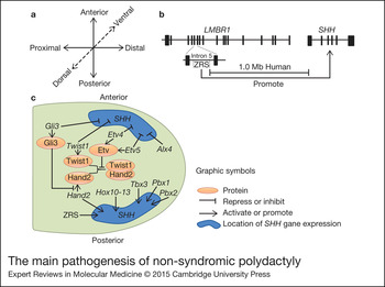
The vertebrate limb bud can develop in three different axes: proximal–distal (PD, shoulder to finger), anterior–posterior (AP, thumb to little finger) and dorsal–ventral (DV, back to palm) (Ref. Reference Zguricas66) (Fig. 1a). Several signalling centres were found to take charge of accurate formation of this three-dimensional structure, for instance, ZPA is responsible for the AP axes. PPD was reported to be the consequence of disruption of normal limb development pattern along the AP axis (Ref. Reference Hill67). Animal models involving mutations in ZRS, the Shh gene, the Gli3 gene and the Pitx1 gene were reported to be related to polydactyly. Animal models carrying human mutations may help to reveal the regulatory interactions of normal and pathologic development, homeostatic maintenance and repair, and thus to prevent and treat intervention (Ref. Reference Grzeschik68).
The main pathogenesis of non-syndromic polydactyly (Refs Reference Zhang29, Reference Lettice33, Reference Zguricas66, Reference Maas and Fallon69, Reference Johnson and Tabin71, Reference Capdevila and Izpisua72). (a) Three axis of limb development. (b) The location relationship of ZRS, the LMBR1 gene and the SHH gene. (c) The signal pathways of several genetic factors and associated proteins of non-syndromic polydactyly in mice. Alx4, the aristaless-like homeobox 4 gene; Etv4, the ets variant 4 gene; Etv5, the ets variant 5 gene; Gli3, the GLI family zinc finger 3 gene; Gli3, the transcriptional activator GLI3 protein; Hand2, The heart and neural crest derivatives expressed transcript 2 gene; Hand2, the heart and neural crest derivatives expressed protein 2; Hox10–13, the hox10 protein gene, the hox11 protein gene, the hox12 protein gene and the hox13 protein gene; LMBR1, the limb development membrane protein 1 gene; Pbx1, the pre B cell leukaemia homeobox 1 gene; Pbx2, the pre B cell leukaemia homeobox 2 gene; SHH, the sonic hedgehog gene, Twist1, the twist family bHLH transcription factor 1 gene; Twist1: the twist-related protein 1; Tbx3, the T-box 3 gene; ZRS, the ZPA regulatory sequence.

ZRS- and Shh-related pathogenesis and animal models
ZRS, located nearly 1-Mb away from SHH within intron 5 of the LMBR1 gene and highly conserved over ~800 bp, is necessary to control the expression of SHH in the limb bud, but not elsewhere in the developing embryo (Refs Reference Lettice33, Reference Maas and Fallon69) (Fig. 1b). SHH is expressed in a small population of mesenchymal cells in the posterior limb bud forming the ZPA and is crucial for limb AP patterning to regulate the identity and number of digits (Ref. Reference Riddle70). SHH asymmetric expression in ZPA in posterior mesoderm is required for appropriate AP limb patterning in the developing limb (Ref. Reference Riddle70), and ectopic anterior SHH signalling has been suspected as a major cause for polydactyly (Refs Reference Johnson and Tabin71, Reference Capdevila and Izpisua72) (Fig. 1c). Mutations in ZRS causing ectopic anterior Shh gene expression have been reported to cause PPD in many species, including mouse, dog, cat, chicken and human (Refs Reference Lettice33, Reference Li38, Reference Lettice73, Reference Sagai74, Reference Masuya75, Reference Park76, Reference Maas and Fallon77).
The chicken oligozugodactyly (ozd) mutation harbours a spontaneous 1654-bp deletion of the ZRS resulting in loss of Shh expression and phenocopying the Shh knockout mouse. The ozd/ozd chickens showed limb defects, including lack of the ulna and all digits in the wing, absence of the fibula and all digits but the first digit in the foot, and morphologically normal bones, while the ozd /+ chicken showed normal phenotype (Ref. Reference Maas78). The Shh −/− mice showed one particular digit in morphology, a well-developed nail and phalanges, and severely truncated and fused intermediate structures of hindlimbs, one distal cartilaginous component, a less-well developed nail and fused middle skeleton of forelimbs (Refs Reference Te79, Reference Kraus80), whereas Shh +/− mice seemed normal (Ref. Reference Chiang81). Both Silkie Breed and Polydactyly (Po) chicken had a single base pair change within the ZRS and showed PPD (Ref. Reference Maas78). Hemimelic extra toe (Hx) mice carry a spontaneous G>A mutation within the Lmbr1 intron 5, and Hx/ + caused PPD and variable foreshortening of the radius/tibia, while Hx/Hx displayed hemimelia and extra toes (Ref. Reference Knudsen and Kochhar82). Among base substitution mutant mice (M100081, M101116, M101117 and M101192) generated by ENU-mutagenesis involving in Lmbr1 intron 5, only M101116 and M100081 mice showed PPD with extra metacarpals, metatarsals or digits, and hemimelia, and the homozygotes were fertile and showed phenotypes resembling that of the heterozygotes, while M101117 and M101192 mice showed no morphologically distinct deformity in limb (Refs Reference Sagai74, Reference Masuya75). Sasquatch (Ssq) implicates a random transgenic insertion, including a Hoxb1/human placental alkaline phosphatase (HPAP) reporter in the intron of the Lmbr1 gene, which lead to a ~20-kb duplication of ZRS and effect the Shh expression (Refs Reference Lettice32, Reference Hill83). The Ssq /+ mice showed normal forelimbs and PPD of the hindlimbs, whereas Ssq/Ssq mice represented more severe polydactyly of the hindlimbs, reduced length of the tibia and an absence of blend between the tibia and fibula, and PPD with mild reduced length of the zeugopod of the forelimbs (Refs Reference Knudsen and Kochhar82, Reference Sharpe84). Hw cats carrying 479A>G mutation in the ZRS showed polydactyly, and Hw/Hw cat did not show a more severe phenotype than Hw /+ (Ref. Reference Lettice73). Mice with overexpression of wild-type Shh in the early limb bud showed polydactyly/proximal deviation of the anterior digits (Ref. Reference Lee85). The doublefoot (Dbf) is a spontaneous mouse mutant with ectopic activation of Indian Hedgehog (Ihh), a close relative of Shh. The Dbf/Dbf mice showed embryonic lethality, and Dbf /+ mice showed polydactyly (6–9 digits per limb) (Refs Reference Talamillo8, Reference Crick86).
Pathogenesis and animal models of transcription factor genes: Gli3 and Pitx1
Factors controlling SHH expression in the limb bud can be characterised as either positive or negative regulators (Ref. Reference Riddle70). Members of the large group of erythroblast transformation specific (ETS) transcription factors are shown to play distinct roles in the spatial pattern of Shh (Ref. Reference Sharrocks87). Occupancy at multiple ETS sites, which bind the factors the GA-binding protein alpha (GABPa) and ETS1, regulates the position of the ZPA boundary (Refs Reference Lettice88, Reference O'Leary89). In mice, the aristaless-like homeobox 4 gene (Alx4), the twist family bHLH transcription factor 1 gene (Twist1), the Gli3 gene, the ets variant 4 gene (Etv4) and the ets variant 5 gene (Etv5) may inhibit ectopic Shh expression in anterior limb bud, and loss-of-function mutants of those genes can cause ectopic Shh expression. The heart and neural crest derivatives expressed transcript 2 gene (Hand2), the T-box 3 gene (Tbx3), the pre B cell leukaemia homeobox 1 gene (Pbx1), the pre B cell leukaemia homeobox 2 gene (Pbx2), the hox10 protein gene (Hox10), Hox11, Hox12 and Hox13 may activate Shh expression in the posterior limb bud, and loss-of-function mutants of those genes can cause reduced or absent Shh expression in this region. Additionally, Gli3 protein directly inhibits the transcription of Hand2. The Shh expression regulators, Twist1, Hand2 and Etv, keep an accurate balance to form AP patterning of the limb, and Twist1 can form homodimers/heterodimers with Hand2, which can be antagonised by overexpressing Etv (Ref. Reference Zhang29) (Fig. 1c). Intriguingly, mice with destroy of one or both Gli3 alleles in embryos lacking Shh can gradually restore limb distal development and digit formation. It indicated that Shh can neutralise Gli3-mediated inhibition of key regulator genes, cell survival and distal progression of limb bud development (Ref. Reference Te79).
The XtH (Extra toes) (Gli3Xt−H ) mice carry a spontaneous large deletion in the A2 region of chromosome 13, containing the Gli3 gene. XtH /+ mice showed PPD, occasionally with PAP, whereas XtH /XtH died before birth or at birth with various malformations, including paddle-shaped feet with eight or nine digits, hemimelia, abnormalities of the brain, spinal cord and sense organs, and oedema (Ref. Reference Naruse55). XtJ (Extra-toes Jackson) (Gli3Xt−J ) mice harbouring an spontaneous more than 30-Kbp intragenic deletion in the Gli3 gene, exhibited polydactyly and craniofacial defects (Refs Reference Naruse55, Reference Rice90). The phenotype of XtJ /+ mice was very similar to that of XtH /+, while XtJ /XtJ mice died in utero or within 2 days after birth with multiple abnormalities, including noticeable synpolydactyly and brain malformations (Ref. Reference Naruse55). The limbs of Gli3 −/− mice embryos showed polydactyly (Ref. Reference Te79). Polydactyly Nagoya (Pdn) implicates the integration of an Early Transposon into the Gli3 gene (Ref. Reference Thien and Ruther91). Pdn /+ mice showed one extra preaxial digit in the hindlimb and the distal phalanx deformity of the first digit in the forelimb, while Pdn/Pdn mice presented with PPD or triplicated metacarpal/metatarsal type in limbs (Ref. Reference Naruse92). Gli3 Δ699 represents the mutation in the end of the Gli3 protein at position 699. Gli3 Δ699/+ mice showed PPD with variable penetrance, while Gli3 Δ699/Δ699 mice displayed central polydactyly and other PHS features (Ref. Reference Bose93). Gli3 Δ68 carries a small deletion from residue 675–743, reducing a half of the Gli3 repressor levels and slightly increasing the activity of full-length mutant protein in the limb. Gli3 Δ68/Gli3 Δ68 mice developed one to two supernumerary partial digits in the preaxial limb, while Gli3 Δ68/+ mice died soon after birth and displayed seven digits (Ref. Reference Wang94). The Gli3P1−4 mice were generated by the replacement of Gli3 genomic sequences encoding the Gli3 protein residues 846–914 by the corresponding human Gli3 gDNA sequence, containing S-to-A mutations at the first four PKA sites. Gli3P1−4/ + mice showed a complete duplication of the first digit of each limb, more severe than that of Gli3Xt /+ or Gli3 +/− mice, while Gli3P1 −4/P1−4 mice mostly died before birth or died soon after birth (Ref. Reference Wang95). The Gli3Mos1 mice carry a p.Y350X mutation. Most Mos1 /+ mice had limb defects resembling polydactyly in Gli3Xt, Mos1/Mos1 mice showed embryonic lethality (Ref. Reference Matera96). Anterior digit pattern deformity (add), a recessive mutation of the Gli3 gene, is an insertion of a transgene about 80 kb of the add integration site, add/add mice showed PPD of the forelimbs and rarely of the hindlimbs, similar to Xt /+ mice (Ref. Reference van der Hoeven97). Intriguingly, distal limbs of Shh −/− Gli3 +/− mice embryos presented with two zeugopodal bones and rudimentary digits form (Ref. Reference Te79). Limbs of Shh −/− Gli3−/− mice were distally complete and polydactylous, which were indistinguishable from the limb of Gli3 −/− embryos in morphology, suggesting that the polydactyly of Gli3 −/− mice is SHH independent (Refs Reference Te79, Reference Litingtung98).
Pitx1 −/− mice embryos showed absence of the ilium and knee cartilage and underdeveloped long bones of the hindlimb, and died at birth, whereas Pitx1 +/− mice were phenotypically normal and fertile (Ref. Reference Lanctot62). Neither Pitx1 −/− nor Pitx1 +/− mice show polydactyly indicating that the situation might be different between humans and mice (Ref. Reference Klopocki58).
Diagnosis and treatment
Accurate diagnosis of polydactyly would benefit from the combination of clinical features, family history, X-ray, three-dimensional sonography detection in pregnancy, and especially molecular genetic analysis, including karyotyping, array comparative genomic hybridisation and sequencing (Ref. Reference Filges99). The primary treatment of polydactyly is surgical removal, whose principle is to remove the least functional component and reconstruct the remaining parts, primarily for psychological, aesthetic and functional reasons (Refs Reference Christensen4, Reference Guo22). The most acknowledged operation time is between 6 and 24 months of age, since the general anaesthesia is safer and the child's structures are better developed (Ref. Reference Chong21). It was also suggested to start shortly after 1 year of age for earlier functional reconstructions and remodelling reasons (Ref. Reference Guo22). Phenotypic diversity requests many different surgical methods, and surgery of PPD requests more attention on reconstructing normal tendon and ligamentum relationships, while the surgery of PAP is generally performed through a racquet-shaped incision commencing at the webbing between the fifth and sixth phalanges and terminating at the base of the fifth metatarsal (Ref. Reference Christensen4).
Conclusions and perspectives
Recent progress in human genetics have successfully revealed the molecular pathogenesis of several familial polydactyly types, and enhanced our understanding of function of the disease-causing genes responsible for embryonic limb development (Refs Reference Materna-Kiryluk9, Reference Jamsheer100). The observation of a wide range of genetic heterogeneity (a single phenotype associated with different genes) and allelic heterogeneity between families (clinically distinct phenotypes related to mutations in the same gene) in this disorder suggest that mechanism besides simple Mendelian inheritance should be considered in some cases (Ref. Reference Grzeschik68). These mechanisms include the roles of background genes, epigenetic factors (somatic mosaicism or X-inactivation) and environmental factors (Ref. Reference Grzeschik68). With the advent of next-generation sequencing technology, including exome sequencing, whole-genome sequencing and RNA-sequence analysis, more novel gene mutations or variants resulting in non-syndromic polydactyly will likely be discovered. Systematic bioinformatics analysis of variants in background genes from the outcome of high-throughput sequencing may help to establish the precise genotype–phenotype correlations of polydactyly. Construction of various genetic-deficient polydactyly animal models could contribute substantially to the understanding of limb developmental pathways, help to clarify the pathogenesis of polydactyly and carry on experimental treatment study. The unveiling of the pathways involved in the development of non-syndromic polydactyly will help to explain diverse clinical phenotypes, strong genetic heterogeneity, and may contribute to the target therapy, including regenerative or cell replacement therapies of this complex condition (Ref. Reference Biesecker7).
Acknowledgements
This work was supported by the National Natural Science Foundation of China (grant numbers 81271921 and 81441033); Construction Fund for Key Subjects of the Third Xiangya Hospital, Central South University; Grant for the Foster Key Subject of the Third Xiangya Hospital Clinical Laboratory Diagnostics (H. Deng); Zhishan Lead Project of the Third Xiangya Hospital (H. Deng); Fundamental Research Funds for the Central Universities of Central South University, China (grant no. 2015zzts318).
Conflict of Interest
None.