Introduction
Sebaceous glands are prominent adnexal components of the skin which are predominantly seen on the face and scalp. Normal glands vary in size from 0.2 to 2 mm in diameter, with the largest appearing in the skin of the nose and the concha of the ear.Reference Batsakis and el-Naggar1 Sebaceous glands secrete sebum.Reference Marx and Stern2 Sebaceous differentiation in the oral mucosa and salivary glands is an expected normal finding; however, sebaceous neoplasms do occur in these regions, albeit rarely and with low recurrence potential.
We present a rare case of sebaceous adenoma of the palate, together with a review of the literature.
Case report
A 38-year-old woman presented with a growth in the hard palate of eight months' duration.
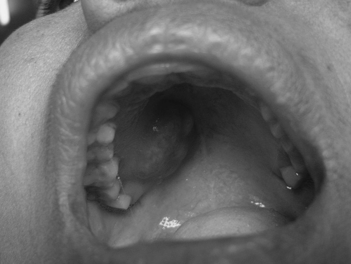
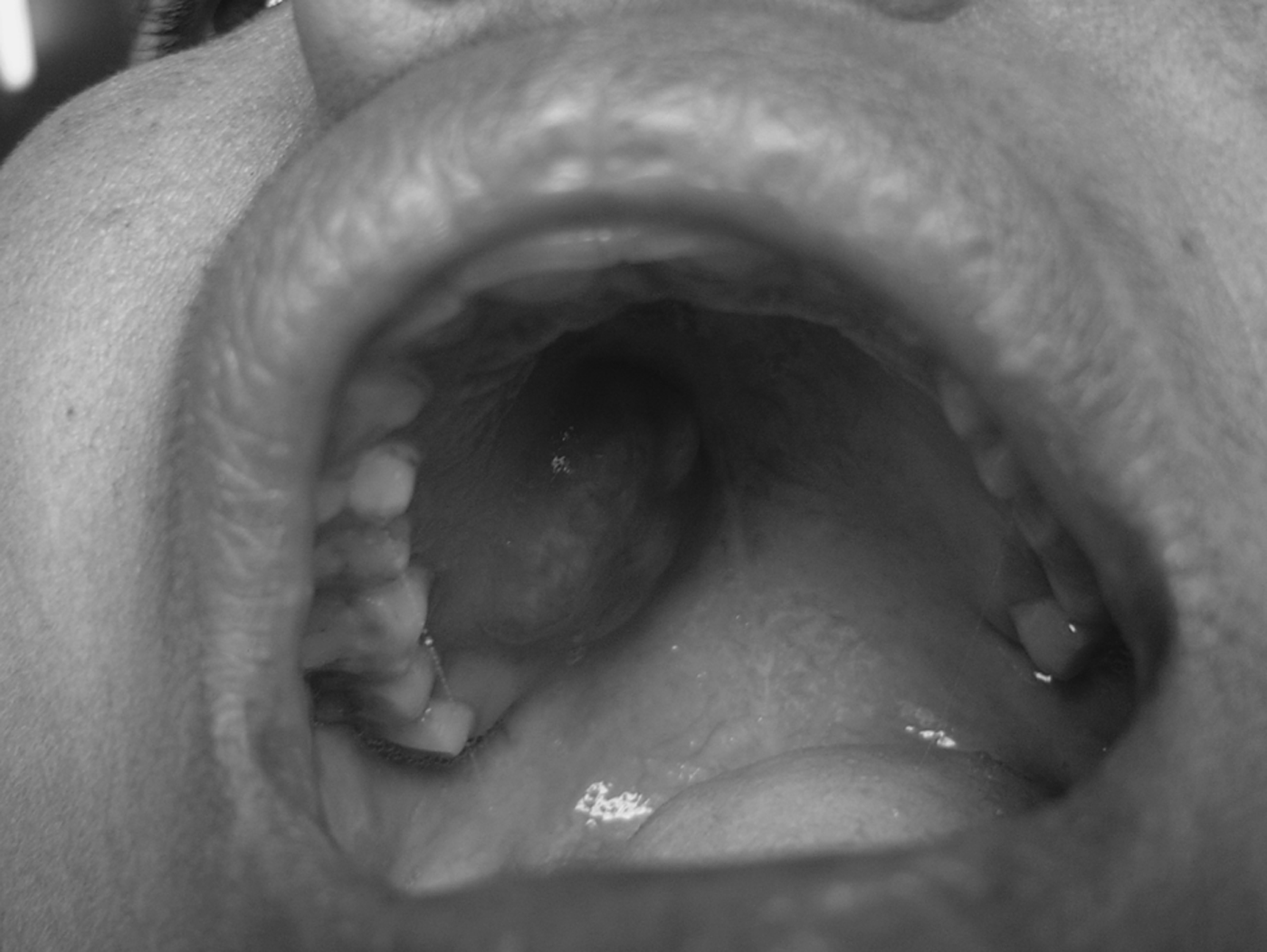
On examination, the lesion was situated in the right posterior part and did not cross the midline. It was a well circumscribed, ovoid, firm, non-tender swelling with a smooth, pink surface, and measured 2 × 1.5 cm (Figure 1).
Photograph of the lesion.
Fine needle aspiration cytology was performed and showed features of a benign lesion; however, the exact nature of the lesion could not be conclusively determined.
Routine haematological and urine examinations were normal.
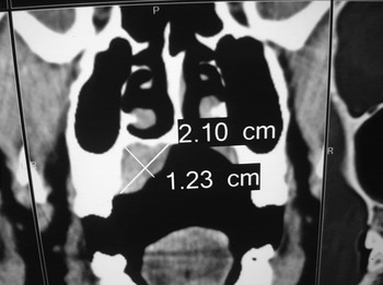
A computed tomography scan showed a well circumscribed, non-enhancing, 2.1 × 1.2 cm lesion confined to the hard palate. Scalloping of the bone was evident without any erosion, suggestive of benign pathology. There was no evidence of calcification or necrosis (Figure 2).
Coronal computed tomography scan showing the tumour, with dimensions as marked.

The palatal mass was excised completely with a 2 mm margin of apparently normal mucosa.
The patient was fed nasogastrically for five days, followed by a soft diet for two weeks.
Macroscopic examination of the excised lesion showed a single, ovoid tissue mass measuring 3 × 2 × 0.3 cm. The cut surface consisted of grey-white tissue.
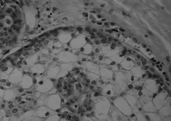
Microscopic examination of haematoxylin and eosin stained sections showed stratified squamous epithelium with stroma showing focal collections of sebaceous cells in lobules. These lobules contained undifferentiated cells in the periphery and mature sebocytes with clear cytoplasm in the centre. Sheets, islands and cystic spaces of squamous epithelial cells and sebaceous cells were seen, without any cellular atypia or mitosis. A focal area of dense lymphoplasmacytic infiltrate was seen. There was no mucoid element. A diagnosis of sebaceous adenoma of the palate was made (Figure 3).
Photomicrograph of the tumour showing a lobule with undifferentiated cells at the periphery and mature sebocytes in the centre. (H&E; ×40)

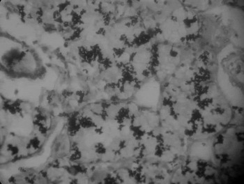
Staining with Sudan Black B stain demonstrated the presence of sebum (Figure 4).
Photomicrograph of tumour preparation stained with Sudan Black B, demonstrating the presence of sebum. (×10)

Immunohistochemical analysis using the Ki 67 marker showed focal positive expression.
The results of special staining and immunohistochemical analysis confirmed the final diagnosis of sebaceous adenoma.
The patient was followed up for six months, over which time her palatal mucosa healed well.
After obtaining informed consent, a diagnostic colonoscopy was performed to exclude Muir–Torre syndrome. The colonoscopy was normal.
Discussion
Sebaceous glands are holocrine glands in which the cell dies and secretions are discharged. These glands are normal skin appendages present throughout the skin, with the exception of the palms and soles.Reference Marx and Stern2 Ectopic sebaceous glands are principally found in the major salivary glands, and are rarely seen in minor salivary glands.Reference Batsakis and el-Naggar1, Reference Gnepp3
In the oral cavity, sebaceous glands normally appear as yellow-white spots, termed Fordyce spots, and are commonly seen on the vermillion border of the lip, the labial and buccal mucosa, and the retro-molar region.Reference Daley4
Sebaceous gland lobules are less common in children because the pilosebaceous system does not reach full development until after puberty.Reference Dent, Hunter and Svirsky5
Sebaceous adenomas are rare, benign tumours arising from sebaceous glands in the oral cavity.Reference Batsakis and el-Naggar1 They account for 0.1 per cent of all sebaceous gland neoplasms.Reference Gnepp3 The mean age of initial clinical presentation is 58 years, and the male to female ratio is 1.6:1.
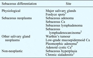
Approximately 149 primary sebaceous tumours of salivary gland origin have been reported in the English literature (see Table I).Reference Marx and Stern2–Reference Daley4, Reference Pilch6 However, only 10 cases of intraoral sebaceous adenoma have been reported in the English language literature, with common sites being the buccal mucosa and retromolar area.Reference Lezzi, Rubini, Fioroni and Piattelli7 No cases of sebaceous adenoma of the palate have previously been reported.
Distribution of sebaceous glands in oral cavity

Ca = carcinoma
The presence of sebaceous glands, or evidence of sebaceous differentiation, has been noted in submandibular and parotid salivary glands. Their presence in the oral cavity is thought to originate in the intralobular ducts of the salivary glands.Reference Batsakis and el-Naggar1 However, an opposing school of thought states that the presence of sebaceous lobules derives from the inclusion of ectoderm in the oral cavity at the time of fusion of the maxillary and mandibular processes, during embryonic development.Reference Dent, Hunter and Svirsky5
Histological examination of sebaceous adenoma shows an encapsulated tumour consisting of nests of sebaceous cells in a fibrous stroma. These are well demarcated lesions with a multilobular pattern. The tumour comprises three types of cells: undifferentiated germinal cells (at the periphery), mature sebocytes and intermediate cells.Reference Marx and Stern2
The secretory product of sebaceous glands is called sebum. Sebum comprises a mixture of lipids, including triglycerides, waxes, squalene, and cholesterol and its esters. Sebum may have weak antibacterial and antifungal properties.Reference Junqueira8
Sudan Black B stain demonstrates the presence of sebum within sebaceous adenomas.
Immunohistochemical analysis of sebaceous adenomas utilises Ki 67, a proliferative nuclear marker found to show higher expression in these tumours, compared with lower proliferation indices in normal glands and sebaceous hyperplasia.Reference Azevedo, Almeida, Netto, Miranda, Santos and Della Coletta9
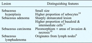
Sebaceous adenoma should be differentiated from other, sebaceous-related lesions (see Table II).Reference Pilch6, Reference Daley10, Reference Lever11
Oral cavity sebaceous lesions: differential diagnosis

Sebaceous adenomas do not recur after adequate excision, and the prognosis is excellent.Reference Marx and Stern2
• Sebaceous adenomas rarely occur intraorally; only 10 cases have previously been reported in the English language literature
• The first reported case of sebaceous adenoma of the palate is described
• Complete excision is curative
Sebaceous adenomas are also found in Muir–Torre syndrome. This is an autosomal dominant condition characterised by a combination of sebaceous tumours of the skin and one or more internal malignancies, most commonly colon cancer.Reference Schwartz and Torre12 An association with intraoral sebaceous adenomas has not been reported. However, as only a tiny number of intraoral sebaceous adenomas have been reported, an association of such tumours with Muir–Torre syndrome cannot be excluded at this stage.
Conclusion
Sebaceous adenomas occur very rarely in the palate. Investigation using a combination of clinical and histopathological analysis is essential. Sudan Black B special staining and Ki 67 immunohistochemical evaluation add valuable information when differentiating this tumour from other sebaceous lesions of the oral cavity. A possible association with Muir–Torre syndrome should also be kept in mind. Following adequate excision, recurrence does not occur.