Case details
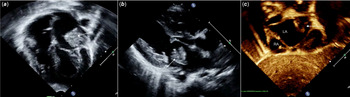
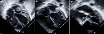
A 6-month-old female infant (patient 1), who underwent surgical closure of a sub-arterial ventricular septal defect, was found to have an echogenic mass in the left atrium (Fig 1a and b, Supplementary Videos 1 and 2) on post-operative transthoracic echocardiogram. The mass was arising from the lateral aspect of the left atrium and protruded towards the mitral orifice, without causing any obstruction to mitral inflow. Colour Doppler did not reveal any flow into the mass. Careful assessment of the lesion was performed in multiple echocardiographic views. Subcostal long-axis view clinched the diagnosis of left atrial appendage inversion (Fig 1c, Supplementary Video 3). The continuity of the left atrial wall with the inverted atrial appendage was clearly demonstrable in this view, which helped to differentiate this from left atrial thrombus. No active intervention was done. Though the lesion persisted on the echocardiogram performed at 3-month follow-up, there was complete disappearance of the lesion after 1 year. A similar image was noted in a 2-month-old male infant (patient 2) who underwent truncus arteriosus repair (Fig 2a). After confirming the diagnosis of left atrial appendage inversion, he was advised conservative management. Echocardiogram at 3-month follow-up revealed a significant decrease in the size of the lesion (Fig 2b). Subsequently, he was lost to follow-up and when seen after 2 years, the mass had disappeared suggesting complete resolution of the lesion (Fig 2c).
Echocardiographic images of patient 1. ( a ) Four-chamber view showing a large echogenic mass (white arrow) attached to the lateral wall of left atrium just above the mitral valve. ( b ) Parasternal long-axis view showing the echogenic mass in left atrium. ( c ) Subcostal long-axis view showing inverted left atrial appendage (white arrow). Note the continuity of the structure with left atrial wall. LA=left atrium, RA=right atrium.

Echocardiographic images of patient 2. ( a ) Four-chamber view showing a large echogenic mass (inverted appendage) in the left atrium attached to the lateral wall. ( b ) Echocardiogram at 3-month follow-up showing a significant reduction in the size of the mass. ( c ) Complete resolution noted at 2-year follow-up.

Discussion
Left atrial appendage inversion is a rare complication following cardiac surgery. Reference Allen, Ilbawi and Hartz1 This occurs due to the negative pressure created by the left atrial vent during surgery or during deairing procedures. Reference Cohen, Tamir and Yanai2,Reference Fujiwara, Naito and Noguchi3 There have been reports of left atrial appendage inversion in pericardial effusion, which could be due to the increased intrapericardial pressure. Reference Gecmen, Candan and Guler4 Spontaneous inversion of the left atrial appendage in the absence of above two situations is also reported. Reference Powell, Taylor and Cottrill5 If it is recognised during surgery in the post-operative transoesophageal echocardiogram, it may be corrected by digital manipulation. However, if it is recognised later, no intervention is required as spontaneous resolution occurs. Occasionally, if it causes haemodynamic compromise by obstructing the left ventricular inflow, intervention may be required. Reference Molaei, Tabib and Meraji6
In both our patients, transoesophageal echocardiogram was not performed intraoperatively as the weight of the infants was less than 5 kg. Thus, it went unnoticed in the immediate post-operative period. In both, left atrial vent was placed through the patent foramen ovale during surgery. The negative pressure caused by the vent was probably responsible for the inversion of the left atrial appendage. Thus, the appearance of a tubular mass protruding towards the mitral orifice with the absence of flow into it and having the same echodensity as the atrial wall is suggestive of left atrial appendage inversion. Though the lesion has a misleading appearance of a mass on the four-chamber and parasternal long-axis views; the subcostal views are diagnostic and clearly demonstrate the inversion of the appendage. Hence, we suggest a detailed subcostal echocardiographic examination when there is a new appearance of a left atrial mass following cardiac surgery. Cardiologists should be aware of this condition as recognition of this entity will avoid unnecessary investigations and unindicated treatment with anticoagulants.
Supplementary material
To view supplementary material for this article, please visit https://doi.org/10.1017/S1047951120003662
Acknowledgements
None.
Financial support
This research received no specific grant from any funding agency, commercial, or not-for-profit sectors.
Conflicts of interest
None.
Ethical standards
The authors assert that all procedures contributing to this work comply with the ethical standards of the relevant national guidelines on human experimentation and with the Helsinki declaration of 1975, as revised in 2008, and has been approved by the institutional committees.