Introduction
The retinal ganglion cell line (RGC-5) (derived by transforming postnatal day 1 rat retinal cells with Ψ 2 E1A virus) was originally established by Krishnamoorthy et al. (Reference Krishnamoorthy, Agarwal, Prasanna, Vopat, Lambert, Sheedlo, Pang, Shade, Wordinger, Yorio, Clark and Agarwal2001). This cell line displays some characteristics of retinal ganglion cells (RGCs), such as expressions of Thy-1, Brn3C, Neuritin, N-methyl-d-aspartate-R1 and GABA-B receptors, the sensitivity to glutamate excitotoxicity and neurotrophin withdrawal (Krishnamoorthy et al., Reference Krishnamoorthy, Agarwal, Prasanna, Vopat, Lambert, Sheedlo, Pang, Shade, Wordinger, Yorio, Clark and Agarwal2001; Nieto et al., Reference Nieto, Acosta-Rodríguez, Valdez and Guido2010), but no expression of glial fibrillary acidic protein (the marker of astrocyte) and HPC-1 or 8A1 (the markers of amacrine or horizontal cell) (Krishnamoorthy et al., Reference Krishnamoorthy, Agarwal, Prasanna, Vopat, Lambert, Sheedlo, Pang, Shade, Wordinger, Yorio, Clark and Agarwal2001). Thus, it may be used as a valuable model for in vitro (patho)physiological study of RGCs. For example, the characterization of cell death induced by growth factor withdrawal (Charles et al., Reference Charles, Khalyfa, Kumar, Krishnamoorthy, Roque, Cooper and Agarwal2005), hydrostatic pressure (Ju et al., Reference Ju, Liu, Kim, Crowston, Lindsey, Agarwal, Ellisman, Perkins and Weinreb2007, Reference Ju, Kim, Lindsey, Angert, Patel, Scott, Liu, Crowston, Ellisman, Perkins and Weinreb2009), endoplasmic reticulum stress (Shimazawa et al., Reference Shimazawa, Inokuchi, Ito, Murata, Aihara, Miura, Araie and Hara2007; Inokuchi et al., Reference Inokuchi, Nakajima, Shimazawa, Kurita, Kubo, Saito, Sajiki, Kudo, Aihara, Imaizumi, Araie and Hara2009), light exposure (Wood et al., Reference Wood, Lascaratos, Bron and Osborne2008), optineurin (Chalasani et al., Reference Chalasani, Radha, Gupta, Agarwal, Balasubramanian and Swarup2007), oxidative stress (Maher & Hanneken, Reference Maher and Hanneken2005a,Reference Maher and Hannekenb), calcium ionophore (Das et al., Reference Das, Garner, Del Re, Woodward, Kumar, Agarwal, Banik and Ray2006) has been carried out. The differentiation responses to various stimuli were also investigated (Frassetto et al., Reference Frassetto, Schlieve, Lieven, Utter, Jones, Agarwal and Levin2006; Nieto et al., Reference Nieto, Acosta-Rodríguez, Valdez and Guido2010; Wood et al., Reference Wood, Chidlow, Tran and Jonathan2010). In addition, the genes involved in the signal transduction pathways were determined (Khalyfa et al., Reference Khalyfa, Chlon, Qiang, Agarwal and Cooper2007). Furthermore, the cell line was also used to study biochemical and optic neuropathies involved in glaucoma in retinal cells (Khalyfa et al., Reference Khalyfa, Chlon, Qiang, Agarwal and Cooper2007).
Although this cell line has been extensively applied to study in vitro pathophysiology and physiology of RGCs since its establishment, only few data are available for its electrophysiological properties. Moorhouse et al. (Reference Moorhouse, Li, Vickery, Hill and Morley2004) reported that low levels of basal Cl− current, inwardly rectifying K+ current, and Gd3+-insensitive stretch-activated current were present in the RGC-5 cell line, but no voltage-dependent inward Na+ currents were detected. Frassetto et al. (Reference Frassetto, Schlieve, Lieven, Utter, Jones, Agarwal and Levin2006) found that treatment of broad-spectrum protein kinase inhibitor, staurosporine, induced voltage outward current in proliferating RGC-5 cell line. Tchedre et al. (Reference Tchedre, Huang, Dibas, Krishnamoorthy, Dillon and Yorio2008) investigated sigma-1 receptor regulation of a voltage-gated calcium channels in RGC-5 cell line.
Since disputes still remain in terms of its electrophysiological characteristics, it is necessary to further determine the existence and classification of ionic currents in the RGC-5 cell line. In the present study, we aim to identify the types of potassium and chlorine ionic currents in the RGC-5 cell line. We will provide basic documentary data, which will benefit further investigations in the RGC-5 cell line.
Materials and methods
Cell culture
RGC-5 cell line (gifted by Prof. Dingfeng Su in The Second Military Medical University, Shanghai, China) was cultured in 1 g/l glucose Dulbecco’s modified Eagle medium (DMEM) with l-glutamine, 10% fetal bovine serum (FBS), 100 U/ml penicillin, and 100 μg/ml streptomycin. Cells were incubated in humidified atmosphere of 95% O2 and 5% CO2 at 37°C. For electrophysiological recording, the cell line was subcultured in 35 × 10 mm dishes (treated polystyrene). Fresh medium was replaced 24 h before the experiments. DMEM, l-glutamine, FBS, penicillin, and streptomycin were from DingGuo ChangSheng Biotechnology Co., Ltd., Beijing, China.
Electrophysiological recording
Whole-cell currents were recorded by using the patch-clamp technique at room temperature (23–25°C), in which a Multiclamp700B amplifier, a Digidata 1440A digitizer, and pclamp 10.1 software (Molecular Devices Corporation, Union City, CA) were applied. The low-pass filter frequency and sampling frequency were set at 10 and 5 kHz, respectively. The patch pipette was pulled from 1.2 mm outer diameter and 0.5 mm inner diameter glass pipette using a horizontal puller (P-97; Sutter Instrument Co., Novato, CA). The resistance of the patch pipettes was 3–6 MΩ, when filled with the standard pipette solution. An Ag–AgCl pellet was used as the reference electrode in the bath. A little positive pressure was added in the patch pipette before immersing it into the bath solution. When the tip of the pipette got in touch with the cell surface, the positive pressure was withdrawn, and then, a little negative pressure was given, until a gigaseal (>1 GΩ) was formed. Whole-cell configuration was obtained by applying suitable suction to break the patch membrane, and then, the resting membrane potentials (RMPs) value could be obtained at the mode of I = 0 in Multiclamp700B and the whole-cell membrane capacitance could be obtained at the mode of cell in Clampex. The membrane capacitance value and RMPs of RGC-5 cell line were first detected in standard recording extracellular and pipette solution without any blockers as described in “Solutions” section. We then recorded the whole-cell membrane currents in the RGC-5 cell line and used different ion channel blockers to isolate and identify the types of the membrane currents. During whole-cell recording, series resistance (∼60%) and leakage compensations are conducted with MultiClamp Commander. The compositions of solutions used for current recordings are listed below. The liquid junction potentials were negligible.
Solutions
For whole-cell membrane currents recording, the extracellular solution contained (in mM) 140.0 NaCl, 5.0 KCl, 1.0 MgCl2, 1.5 CaCl2, 10.0 HEPES, and 10.0 glucose (pH 7.35 with NaOH); pipette solution contained (in mM) 135.0 KCl, 2.0 MgCl2, 10.0 HEPES, 10.0 EGTA, 1.0 Na2-ATP, and 10.0 glucose (pH 7.25 with KOH). For Cl− current recording, the extracellular solution contained (in mM) 95.0 NaCl, 25.0 CsCl, 2.0 CaCl2, 1.0 MgCl2, 10.0 HEPES, 5.0 glucose, 20.0 tetraethylammonium chloride (TEA-Cl) (pH 7.35 with NaOH); the pipette solution contained (in mM) 103.0 CsCl, 4.0 MgCl2, 10.0 D-glucose, 3.0 ATP disodium (Na2-ATP), 5.0 EGTA, 10.0 HEPES, and 10.0 TEA-Cl (pH 7.25 with CsOH). For inwardly rectifier K+ current recording, the extracellular solution contained (in mM) 40.0 NaCl, 105.0 KCl, 1.0 MgCl2, 1.5 CaCl2, 10.0 HEPES, and 10.0 glucose (pH 7.35 with NaOH); pipette solution contained (in mM) 135.0 KCl, 2.0 MgCl2, 10.0 HEPES, 10.0 EGTA, 1.0 Na2-ATP, and 10.0 glucose (pH 7.25 with KOH). For delay rectifier K+ current recording, the extracellular solution contained 40.0 NaCl, 105.0 KCl, 1.0 MgCl2, 1.5 CaCl2, 50 μM Zn2+, 10.0 HEPES, 10.0 glucose, and 600 μM disodium 4,4’-diisothiocyanatostilbene-2,2’-disulfonate (DIDS) (pH 7.35 with NaOH); pipette solution contained 135.0 KCl, 2.0 MgCl2, 10.0 HEPES, 10.0 EGTA, 50 μM Zn2+, 1.0 Na2-ATP, 600 μM DIDS, and 10.0 glucose (pH 7.25 with KOH). For voltage-gated sodium channels current (I Na) recording, the extracellular solution contained 125.0 NaCl, 25 NaHCO3, 1.25 KCl, 1.25 KH2PO4, 1.5 MgCl2, 1.5 CaCl2, 50 μM Zn2+, 10.0 HEPES, and 10.0 glucose (pH 7.35 with NaOH); pipette solution contained 135.0 CsCl, 2.0 MgCl2, 10.0 HEPES, 10.0 EGTA, 20.0 TEA-Cl, 50 μM Zn2+, 1.0 Na2-ATP, and 10.0 glucose (pH 7.25 with CsOH). TTX (tetrodotoxin) (2 μM) was used to determine if any TTX-sensitive I Na component was present. For transient outward potassium channels current (I A) recording, the extracellular solution contained 140.0 NaCl, 5.0 KCl, 1.0 MgCl2, 1.5 CaCl2, 600 μM DIDS, 10.0 HEPES, and 10.0 glucose (pH 7.35 with NaOH); pipette solution contained 135.0 KCl, 2.0 MgCl2, 10.0 HEPES, 10.0 EGTA, 600 μM DIDS, 1.0 Na2-ATP, and 10.0 glucose (pH 7.25 with KOH). DIDS, TEA-Cl, Na2-ATP, and CsOH were purchased from Sigma-Aldrich, Co., St. Louis, Missouri, Other reagents were analytical reagents (AR, Tianjin, China).
Results
The shape, RMP, and membrane capacitance of the cultured RGC-5 cell line
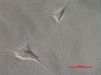
Fig. 1 shows the micrograph of proliferating RGC-5 cell line. The cultured RGC-5 cell line had projections, which was similar to some neuron. We measured the RMPs and the membrane capacitance value of RGC-5 cell line using the standard recording extracellular solution and pipette solution. The data indicated that the RMPs were between −30 and −6 mV (−17.63 ± 2.60 mV, n = 10). The average membrane capacitance value was 16.52 ± 1.08 pF (n = 10).
The micrograph of RGC-5 cells seeded on a dish for whole-cell patch-clamp recording, scale bar 50 μm.

Whole-cell membrane currents
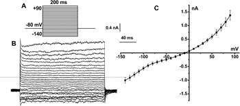
First, we recorded the total whole-cell currents by using standard recording extracellular and pipette solutions (as described in Materials and methods section) in the absence of any channel blockers. A representative family of current traces and the I-V relationship are shown in Fig. 2. The currents were elicited by a series of increasing voltages from −140 to +90 mV with 10-mV step for 200 ms from a holding potential of −80 mV (Fig. 2A). The membrane currents trace (Fig. 2B) exhibited both outward and inward currents. In addition, the I-V relationships (Fig. 2C) showed both inwardly rectifying (probably due to inwardly rectifying K+ currents) and outwardly rectifying (probably due to delayed rectifier K+ current or Cl−current).
Typical current traces and the I-V relationship of whole-cell currents recorded in RGC-5 cell line by a series of increasing voltages from −140 to +90 mV with a 10-mV step for 200 ms from a holding potential of −80 mV. (A) The voltage-clamp protocol. (B) The whole-cell current traces including inward and outward current recorded in RGC-5 cell line. The zero current level is indicated by the dashed line. (C) The current–voltage (I-V) relationships showed a reversal potential at about −10 mV (n = 6).

Inwardly rectifying currents including Cl− and K+ current
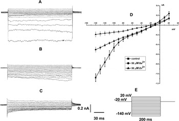
We observed an inwardly rectifying current, which was inhibited by Ba2+ and Zn2+ in RGC-5 cell line (Fig. 3). The inwardly rectifying currents were recorded by a series of increasing voltages from −140 to +20 mV with 10-mV step for 200 ms from a holding potential of −20 mV (Fig. 3E). As shown in Fig. 3A, an inwardly rectifying current was elicited at hyperpolarized membrane potentials. It was blocked by Ba2+, a blocker of inwardly rectifying K+ current (I k1). The peak inward current was inhibited by 50 μM Ba2+ from −1079.11 ± 81.37 pA to −502.16 ± 40.70 pA (n = 5; P < 0.01). Moreover, the peak inward current was further inhibited from −502.16 ± 40.70 pA to −190.98 ± 49.38 pA (n = 5; P < 0.01) after addition of 50 μM Zn2+, a blocker of inwardly rectifying Cl− current (Hartzell & Qu, Reference Hartzell and Qu2003) (Fig. 3C). I-V relationships of the inwardly rectifying currents under aforementioned conditions were shown in Fig. 3D. These results suggest that the inwardly rectifying currents may contain an I k1 and inwardly rectifying Cl−current component.
Whole-cell inwardly rectifying current was inhibited by Ba2+ and Zn2+ in RGC-5 cell line. (A) Current traces recorded in the absence of any blockers. (B) Current traces recorded in the presence of 50 μM Ba2+. (C) Current traces recorded in the presence of 50 μM Ba2+ and 50 μM Zn2+. (D) The I-V relationships obtained from the above three recording statuses (n = 5). (E) The voltage-clamp protocol: a series of increasing voltages from −140 to +20 mV with 10-mV step for 200 ms from a holding potential of −20 mV (n = 5).

Inwardly rectifying Cl− currents
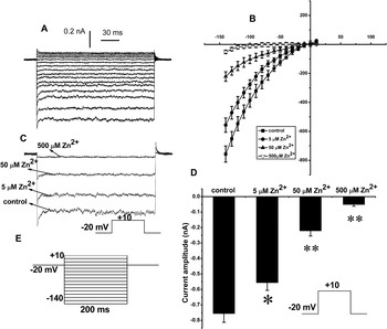
Fig. 4 showed the typical inwardly rectifying Cl− current traces and the I-V relationship recorded in RGC-5 cell line by a series of increasing voltages from −140 to 10 mV with 10-mV step for 200 ms from a holding potential of −20 mV in the presence of different concentration of Zn2+. In the present recording, the inwardly rectifying Cl− currents were recorded by applying K+-free extracellular and pipette solutions (replacing K+ with Cs+) and with 20 mM tetraethylammonium (TEA), a blocker of delayed outward K+ current. Zn2+ has been used as a blocker of cloned ClC-2 channels (Hartzell & Qu, Reference Hartzell and Qu2003). As shown in Fig. 4, a Zn2+-sensitive inwardly rectifying Cl− current was obtained in RGC-5 cell line. At voltage of 10 mV, 0, 5, 50 and 500 μM of Zn2+ treatment resulted in −757.00 ± 48.72 pA (n = 9), −556.68 ± 39.28 pA (n = 9), −221.79 ± 31.17 pA (n = 5), and −51.83 ±11.09 pA (n = 4) of I Cl,ir, respectively. Compared with 0 μM of Zn2+ treatment, 5, 50, and 500 μM of Zn2+ treatment, respectively, produced P < 0.05, P < 0.01, P < 0.01 significant difference. The treatments with 5, 50, and 500 μM Zn2+ significantly suppress I Cl,ir. According to Hill equation, the IC50 of Zn2+ for suppressing I Cl,ir was 17.6 μmol/l. These results further confirmed the existence of a Zn2+-sensitive I Cl,irin the RGC-5 cell line.
Typical inwardly rectifying Cl− current traces and the I-V relationship recorded in RGC-5 cell line by a series of increasing voltages from −140 to 10 mV with 10-mV step for 200 ms from a holding potential of −20 mV in presence of different concentration of Zn2+. (A) showed inwardly rectifying Cl− current traces in no presence of Zn2+. (B) showed the different I-V relationships of inwardly rectifying Cl− current recorded in presence of 0, 5, 50, and 500 μM of Zn2+, respectively (n = 9, 9, 5, and 4); * and ** respectively indicated P < 0.05 and P < 0.01 significant difference compared with 0 μM of Zn2+ treatment. (C) showed the typical inwardly rectifying Cl− amplitude current traces in presence of 0, 5, 50, and 500 μM of Zn2+. (D) showed the different inwardly rectifying Cl− current amplitude in presence of 0, 5, 50, and 500 μM of Zn2+, respectively (n = 9, 9, 5, and 4); * and ** respectively indicated P < 0.05 and P < 0.01 significant difference compared with 0 μM of Zn2+ treatment. (E) showed the voltage-clamp protocol applied for (A) and (B).

Ca2+-activated Cl− current
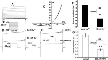
In the following experiments, we also observed a Ca2+-activated Cl− current in the RGC-5 cell line. In the present recording, the K+ current and inward Cl− current were eliminated by applying the extracellular and pipette solution without K+ (replacing K+ with Cs+) and with 20 mM TEA and 500 μM Zn2+. As shown in Fig. 5, a Ca2+-activated Cl− current was recorded from RGC-5 cell line. The current was strongly inhibited by DIDS, a blocker of I Cl. When the extracellular Ca2+ concentration was decreased from 2 to 0.2 mM, the magnitude of the current decreased by 52 ± 6% (n = 4, P < 0.05; Fig. 5D and 5F). The peak current was reduced 65 ± 7% by 600 μM DIDS (n = 5, P < 0.01; Fig. 5E and 5G).
The Ca2+-activated Cl− current elicited in RGC-5 cell line. (A) The typical I Cl,Ca traces elicited by a series of increasing voltages from −140 to +90 mV in a 10-mV increment for 200 ms from a holding potential of −20 mV. (B) The voltage-clamp protocol for (A) and (C). (C) The I-V relationship of I Cl,Ca in RGC-5 cell line (n = 6). (D, F) Comparison of the amplitudes of I Cl,Ca elicited in 2 mM Ca2+ and 0.2 mM Ca2+ of extracellular solution (n = 4, *P < 0.05). (E, G) showed I Cl,Ca inhibited by 600 μM DIDS (n = 5, **P < 0.01).

Delay rectifier K+ current
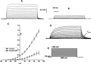
A delay rectifier K+ current (I K) was also elicited in the RGC-5 cell line (Fig. 6). In the present recording, the I Cl,Ca was eliminated by applying the extracellular and pipette solution with 600 μM DIDS. As shown in Fig. 6A and 6B, the peak value of I K was decreased from 467.22 ± 31.85 pA to 111.91 ± 6.58 pA (n = 5; P = 0.001) by 50 mM TEA added to extracellular solution. I K tail currents (I K,tail) were shown clearly in Fig. 6D, a recording from another cell. I-V relationship revealed that I K was activated at about −30 mV in RGC-5 cell line (Fig. 6C).
Delay rectifier K+ current in RGC-5 cells. (A) The typical outward current traces showing a delay rectifier K+ current. (B) The current traces were blocked by 50 mM TEA. (C) The I-V relationships recorded in presence of 0 and 50 mM of TEA, respectively (n = 5; P = 0.001). (D) I K tail currents are shown clearly in a recording from another RGC-5 cell. (E) The voltage-clamp protocol: a series of increasing voltages from −60 to +80 mV in 10 mV increments for 200 ms from a holding potential of −70 mV.

Absence of voltage-gated sodium channels current and transient outward/A-type potassium channels current
Last, we also tested whether voltage-gated sodium channels current (I Na) and transient outward/A-type potassium channels current (I A) existed in the cell line. A series of voltage pulses from −70 to +40 mV in 10 mV increments for 200 ms from a holding potential of −80 mV was used for I Na recording. As indicated in the Materials and methods section, we have included 135.0 mM CsCl and 20.0 TEA-Cl in the pipette solution for I Na and I Ca recordings. Even under this recording condition where the large opposing potassium currents were blocked, we observe neither I Na nor I Ca. In some experiments, we added TTX (2 μM) in the bath solution and compared the current traces in the absence and in the presence of 2 μM TTX. However, we did not observe any TTX-sensitive current component. A series of increasing voltages from −40 to +80 mV in 10 mV increments for 500 ms at a holding potential of −80 mV were used for I A recording. However, neither I Na nor I Awas observed in RGC-5 cell line (data not shown).
Discussion
The present experiments showed that the cultured RGC-5 cell line had projections, which was similar to some neuronal cells. Meanwhile, inwardly rectifying Cl− current (I Cl,ir), Ca2+-activated Cl− current (I Cl,Ca), K+ currents including inwardly rectifying K+ current (I K1), and delay rectifier K+ current (I K) are present in this cell line, whereas voltage-gated sodium channels current (I Na) and transient outward potassium channels current (I A) are absent. The RGC-5 cell line maintained at a RMP of −17.6 ± 2.6 mV and had an average membrane capacitance value of 16.52 ± 1.08 pF. Some results of present experiment were consistent with the results by Moorhouse et al. (Reference Moorhouse, Li, Vickery, Hill and Morley2004) such as the presence of Cl− current, inwardly rectifying K+ current, a low RMP, but no I Na in this cell line. However, in the present study, we applied Zn2+, Ba2+, DIDS, and TEA as the blocker of I Cl,ir, I K1, I Cl, and I K, respectively. We also classified two Cl− currents, that is, I Cl,ir and I Cl,Ca, and two K+ currents, that is, I K1, and I K. These results provided more information on electrophysiological properties in the RGC-5 cell line. It is beneficial for future (patho)physiological and pharmacological studies on the ionic channels of RGC-5 cell line.
In generally, the main function of inwardly rectifying potassium channels current (I K1) is to establish the negative RMP and to control cell signal (Sakmann & Trube, Reference Sakmann and Trube1984; Butt & Kalsi, Reference Butt and Kalsi2006; Jang et al., Reference Jang, Park, Hur, Hong, Hur, Chae, Kim, Kim, Kim and Kim2011). Furthermore, Kir channels also play a very important role in regulation of the action potential (AP) duration in excitable cells (Sakmann & Trube, Reference Sakmann and Trube1984). Some experiments reported that I K1 was also found in bovine retinal and choroidal microvascular endothelial cells and primary rat RGCs (Eschke et al., Reference Eschke, Richter, Brylla, Lewerenz, Spanel-Borowski and Nieber2002; Chen et al., Reference Chen, Yu, Zhao and Yang2004). Our experiment demonstrated that I K1 existed in RGC-5 cell line. And similar results have been reported by Moorhouse et al. (Reference Moorhouse, Li, Vickery, Hill and Morley2004). Henne et al. (Reference Henne, Pöttering and Jeserich2000), Chen et al. (Reference Chen, Yu, Zhao and Yang2004), and our results suggested that the electrophysiological properties of I K1in RGCs were similar to RGC-5 cell line.
The biophysical and pharmacological properties of I Cl,ir were first reported in mammalian heart (Duan et al., Reference Duan, Ye, Britton, Horowitz and Hume2000). Afterward, I Cl,ir was found in many kinds of cells, for example, intestinal pacemaker cells (Zhu et al., Reference Zhu, Mucci and Huizinga2005) and Xenopus retinal pigment epithelial cells (Hartzell & Qu, Reference Hartzell and Qu2003). The inwardly rectifying Cl− channel was assumed to play a potentially important role in the regulation of cardiac electrical activity and cell volume under physiological and pathophysiology conditions (Duan et al., Reference Duan, Ye, Britton, Horowitz and Hume2000). Moreover, the volume sensitivity, voltage sensitivity, anion selectivity, pharmacology, and extracellular pH sensitivity were considered as the physiological characterization of this current (Rutledge et al., Reference Rutledge, Bianchi, Christensen, Boehmer, Morrison, Broslat, Beld, George, Greenstein and Strange2001). I Cl,ir was consistent with currents carried by cloned ClC-2 channels (Duan et al., Reference Duan, Ye, Britton, Horowitz and Hume2000). ClC-2 channels have been mentioned to function in transepithelial salt, water transport, intracellular Cl− homeostasis, cell volume regulation, and intercellular signaling pathways (Siegfried & Jentsch, Reference Siegfried and Jentsch2000; Strange, Reference Strange2002). And ClC-2 was proved to express in the rat retina (Enz et al., Reference Enz, Ross and Cutting1999). A recent study by Yu et al. (Reference Yu, Han, Jiang, Zheng and Liu2011) suggested that the mRNA of ClC-2 channels expressed in RGC-5 cell line and ClC-2 channels correlated with glutamate-induced apoptosis. Our experiment unequivocally recorded the Zn2+-sensitive I Cl,ir in RGC-5 cell line. Based on the continuity of ClC-2 channels between the rat retina and RGC-5 cell line, and ClC-2 channels involving in glutamate-induced apoptosis, RGC-5 cell line might be used as a model in in vitro study of RGC apoptosis involving ClC-2 channels.
Ca2+-activated Cl− channels are widely expressed in all mammalian species. They control electrical excitability by regulating the Cl− equilibrium potential in various cells (Frings et al., Reference Frings, Reuter and Kleene2000; Fuller & Benos, Reference Fuller and Benos2000; McGahon et al., Reference McGahon, Needham, Scholfield, McGeown and Curtis2009). Ca2+-activated Cl− channels have been connected with important physiological functions, involving in control of AP waveform and firing pattern in neurons, membrane potential stabilization in photoreceptors, the regulation of neuronal excitability in animal (Frings et al., Reference Frings, Reuter and Kleene2000). I Cl,Ca has been reported in some types of retinal cells, for example, retinal arteriolar smooth muscle cells (McGahon et al., Reference McGahon, Needham, Scholfield, McGeown and Curtis2009) and Xenopus retinal pigment epithelial cells (Hartzell & Qu, Reference Hartzell and Qu2003). Our experiment recorded the outward Cl− currents, which were inhibited by DIDS and activated by Ca2+ in RGC-5 cell line. Some studies have found that high extracellular Ca2+ could increase intracellular Ca2+ concentration through the DAG (Diacylglycerol) lipase/lipoxygenase pathway, leading to the activation of I Cl,Ca in frog parathyroid cells (Okada et al., Reference Okada, Imendra, Miyazaki, Hotokezaka, Fujiyama and Toda2011). Recent experimental evidence indicates that the expression of I Cl,Ca is related to nerve injury (Boudes et al., Reference Boudes, Sar, Menigoz, Hilaire, Péquignot, Kozlenkov, Marmorstein, Carroll, Valmier and Scamps2009). And I Cl,Ca is not only a downstream element to the initial cation influx but also can greatly amplify the initial signal in vomeronasal sensory neurons (Yang & Delay, Reference Yang and Delay2010). Our results suggest that this cell line might provide a useful model in vitro for studying pathophysiology of cell injury involved in Ca2+-activated Cl−channels.
Previous studies have shown that cellular regulatory volume decrease (RVD) or regulatory volume increase (RVI) is involved in physiological and pathophysiological processes, such as cell differentiation and proliferation, apoptosis, and necrosis (Okada, Reference Okada2004; Okada et al., Reference Okada, Maeno, Shimizu, Manabe, Mori and Nabekura2004, Reference Okada, Sato and Numata2009). The RVD and RVI are mainly mediated by a number of anion channels, of which the volume-sensitive outwardly rectifying Cl− channel is the most prominently activated. Our present study observed two major anion channels, that is, the inwardly rectifying Cl− current (I Cl,ir) and Ca2+-activated Cl− current (I Cl,Ca). Further experiments are needed to test whether these currents are regulated by stimulus factors such as volume changes, apoptotic stimulation, and reagents affecting signaling molecules to elucidate the role of observed currents in such processes (Okada et al., Reference Okada, Sato and Numata2009).
I K has been demonstrated to determine the neuronal excitability and contribute to the repolarization of membrane potentials following APs in many kinds of neurons. In most species, I K could be separated into two different channel currents: the slowly activating delayed rectifier potassium channel current, I Ks, and the rapidly activating type, I Kr (Liu & Antzelevitch, Reference Liu and Antzelevitch1995; Heath & Terrar, Reference Heath and Terrar1996). Frassetto et al. (Reference Frassetto, Schlieve, Lieven, Utter, Jones, Agarwal and Levin2006) have detected a voltage-gated K+ channel (K V) conductance in RGC-5 cell line. Consistent with this finding, we also found a small amount of I K and I K,tail in this cell line. The kinetics of current activation and tail currents resemble those of I Ks, although it needs to be confirmed by applying specific channel blockers (e.g., HMR 1556). We have also clearly shown an inward rectifier potassium current (I K1) in RGC-5 cell according to its electrophysiological (inwardly rectifying) and pharmacological (Ba2+ blockage) properties (Fig. 3). To date, seven subtypes of inward rectifier potassium channels (Kir1–Kir7) and several subtypes of delayed rectifier potassium channels (Kv7, Kv11, mink, MiRP, etc.) have been identified in different types of body cells. We will determine the molecular identifications of these currents (channel subtypes) in RGC-5 cell line in our future study.
In summary, our present study showed the presence of I Cl,ir, I Cl,Ca, I K1, and I K in RGC-5 cell line, while I A and I Na were not observed. Moorhouse et al. (Reference Moorhouse, Li, Vickery, Hill and Morley2004) also reported that no voltage-dependent inward Na+ current was detected, which may reflect some differences in electrophysiology between the RGC-5 cell line and the acutely dissociated or in situ RGCs (Wang et al., Reference Wang, Ratto, Bisti and Chalupa1997; Guenther et al., Reference Guenther, Schmid, Reiff and Zrenner1999). However, accumulating evidence has indicated that I Cl,ir, I Cl,Ca, I K1, and I K are involved in physiology and pathophysiology of retina such as RGC apoptosis and injury. Especially, the chloride channels are involved not only in RGC apoptosis but also in other neuron (such as pheochromocytoma cells, PC12) apoptosis (Zuo et al., Reference Zuo, Zhu, Bai, Zhang, Mao, Chen and Wang2009; Chang et al., Reference Chang, Zhang, Cai and Yin2010). Based on our experiments, both I Cl,ir and I Cl,Ca were recorded in the RGC-5 line, suggesting that this cell line is useful for in vitro study of pathophysiology of neurons apoptosis and selection of chemical medicine for antiapoptosis or cell damage. However, the molecular identities of these channels in the RGC-5 cell line need to be determined in the future.
Limitation
It needs to be noted that most of our findings were each obtained from four to five cells. However, numerous morphologically and electrophysiologically distinct ganglion cell types have been found in vivo. The voltage-dependent Na+ current (I Na) was not detected in RGC-5 cell line under our experimental condition, which may reflect an immature stage of these cells compared to ganglion cells in vivo. Approaches promoting further differentiation toward “real” ganglion cells are needed to provide a more useful experimental model for ganglion cell studies.
Acknowledgments
We thank Prof. Dingfeng Su for kindly providing the RGC-5 cell line. The work was supported by grants by the Natural Science Foundation of China (No. 30470223). Conflict of Interest: The authors have declared that there is no conflict of interest.