Introduction
During the last decade, the small, long-inactive Torrecillas mine, in the northern Atacama Desert of Chile, has yielded a remarkable array of new mineral species, all but one of which are arsenic oxysalts. These include twelve new hydrogen arsenates and five new arsenites (see Kampf et al., Reference Kampf, Nash, Dini and Molina Donoso2019a, Reference Kampf, Chukanov, Möhn, Dini, Molina Donoso and Friis2019b, Reference Kampf, Nash and Molina Donoso2020). The new mineral species nafeasite, described herein, is the 13th new hydrogen arsenate to be discovered at Torrecillas. The majority of the new arsenic oxysalts from the Torrecillas mine are either analogues of known minerals or contain important structural components that are similar to those in known minerals; however, several of the new hydrogen arsenates, including nafeasite, have novel structure types.
The name nafeasite is based on the species-defining cations Na+, Fe3+ and As5+. The new mineral and the name have been approved by the International Mineralogical Association (IMA2021-103, Kampf et al., Reference Kampf, Schlüter, Malcherek, Paulenz, Pohl, Ma, Dini and Molina Donoso2022). The description is based upon one holotype specimen and two cotype specimens. The holotype (collected by JS) is deposited in the Mineralogisches Museum Hamburg, Leibniz-Institut zur Analyse des Biodiversitätswandels, Grindelallee 48, 20146 Hamburg, Germany, catalogue number MD760. The two cotypes, one of which is a piece from the holotype, are deposited in the collections of the Natural History Museum of Los Angeles County, 900 Exposition Boulevard, Los Angeles, CA 90007, USA, catalogue numbers 76193 (piece of the holotype) and 76194 (collected by AAMD).
Occurrence
The new mineral was found on two specimens collected in 2014 at the Torrecillas mine, Salar Grande, Iquique Province, Tarapacá Region, Chile (~20°58'13''S, 70°8'17''W). Torrecillas Hill, on which the Torrecillas mine is located, is composed of four different rock units. The Coastal Range Batholith (mainly gabbros) extends from the seashore to the Pan-American Road along the base of Torrecillas Hill. At the foot of Torrecillas Hill is a small area of contact metamorphic rocks in which garnet crystals occur in metamorphosed shales. Higher on the hill, the rocks are predominantly porphyritic andesitic lavas of the Jurassic La Negra Formation (García, Reference García1967; Buchelt and Tellez, Reference Buchelt, Tellez, Bahlburg, Breitkreuz and Giese1988). The Torrecillas deposit, in which the new minerals were found, consists of two main veins rich in secondary arsenic and copper minerals that intersect metamorphosed marine shales and lavas. These mineralised veins are genetically related to the aforementioned porphyritic andesitic lavas of the Jurassic La Negra Formation. More information on the geology and mineralogy of the area is provided by Gutiérrez (Reference Gutiérrez1975).
The rare secondary chlorides, arsenates and arsenites (and associated sulfates) have been found at three main sites on the hill: an upper pit measuring ~8 m long and 3 m deep, a lower pit ~100 m from the upper pit and measuring ~5 m long and 3 m deep, and a mine shaft adjacent to the lower pit and lower on the hill. Nafeasite was found in a small brecciated boulder at the base of the hillslope below the aforementioned workings by two of the authors (JS and MD) and in the upper pit by one of the authors (AAMD). The new mineral is a secondary alteration phase occurring in association with anhydrite, gypsum, halite, lavendulan, magnesiokoritnigite, natrojarosite and a phase probably equivalent to synthetic Na1.91Fe3+H1.09(AsO4)2⋅1.15H2O reported by D`Yvoire and Screpel (Reference D'Yvoire and Screpel1974). The new mineral is very rare, having been found only on a few specimens.
The secondary assemblages at the Torrecillas deposit are interpreted as principally having formed from the oxidation of native arsenic and other As-bearing primary phases, followed by later alteration by saline fluids derived from evaporating meteoric water under hyperarid conditions (cf. Cameron et al., Reference Cameron, Leybourne and Palacios2007); however, considering the proximity of the Torrecillas deposit to the Pacific Ocean, it seems possible that the frequent dense coastal camanchaca fogs have also played a role in the alteration of the veins and the formation of the secondary minerals, particularly in the recent past, since the exhumation of the deposit well above sea level on Torrecillas Hill.
Physical and optical properties
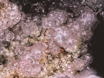
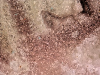
Nafeasite occurs in tightly intergrown aggregates and druses of equant crystals, up to 0.1 mm in diameter (Figs 1 and 2). The complete morphology could not be measured because the crystals are small, tightly intergrown and often twinned by threefold rotation about a* (or [301]). Observed crystal faces include (010), (001) and (1$\bar{1}$ 0). Crystals are light to medium pink and transparent, with vitreous lustre and white streak. The mineral does not fluoresce in long- or short-wave ultraviolet light. The Mohs hardness is ~2½ based on scratch tests. Crystals are brittle with irregular fracture and no cleavage. The density measured by flotation in a mixture of methylene iodide and toluene is 3.23 g⋅cm–3. The calculated density is 3.221 g⋅cm–3 based on the empirical formula of the holotype and 3.239 g⋅cm–3 for the ideal formula. The mineral dissolves slowly in dilute HCl at room temperature.
Pink nafeasite crystals with pale-yellow crystals of a Na–Fe-arsenate on cotype specimen 76193; the field of view is 1.14 mm across.

Pink nafeasite druse with white magnesiokoritnigite on cotype specimen 76194; the field of view is 0.84 mm across.

Optically, nafeasite is biaxial (+) with α = 1.679(3), β = 1.682(3) and γ = 1.730(5) measured in white light. The 2V determined from extinction data analysed with EXCALIBRW (Gunter et al., Reference Gunter, Bandli, Bloss, Evans, Su and Weaver2004) is 27(2)°; the calculated 2V is 28.7°. Dispersion is slight, r < v. The partially determined optical orientation is Y = b. No pleochroism was observed.
Raman spectroscopy
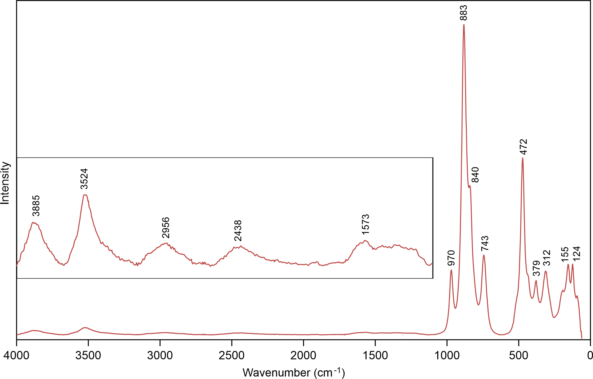
Raman spectroscopy was done on a Horiba XploRa+ micro-Raman spectrometer using an incident wavelength of 532 nm, laser slit of 100 μm, 1800 gr/mm diffraction grating and a 100× (0.9 NA) objective. The spectra for both the holotype and the cotype from the upper pit were recorded from 4000 to 60 cm–1. The spectra were almost identical, the principal difference being that the bands originating from framework vibrations (<1000 cm–1) in the cotype spectrum were shifted to slightly higher wavenumbers due to a lower Fe:Al ratio. Only the spectrum for the holotype is shown in Fig. 3.
Raman spectrum of nafeasite recorded with a 532 nm laser. Inset shows vertically exaggerated section of the spectrum from 4000 to 1100 cm–1.

In the 4000–1100 cm–1 region (vertically exaggerated in the inset in Fig. 3), several broad weak bands are observed. The band centred at 3524 cm–1 is attributable to weak hydrogen bonds corresponding to O⋅⋅⋅O distances around 2.90–2.95 Å; whereas the broad weak band centred at 2956 cm–1 is attributable to stronger hydrogen bonds corresponding to O⋅⋅⋅O distances around 2.63 Å (Libowitzky, Reference Libowitzky1999). The bands approximately centred at 3885 and 2438 cm–1 do not appear to fit any likely bonds in the structure and are presumed to be artefacts. The rather indistinct band between ~1700 and ~1500 cm–1 with a maximum at ~1573 cm–1 is probably attributable to the ν2 (δ) H–O–H bending mode of the H2O groups.
The bands between ~1000 and ~700 cm–1 are probably due to the ν3 and ν1 stretching modes of AsO3OH groups. The band at 472 cm–1, with shoulders to either side, and the band at 379 cm–1 are probably due to ν4 and ν2 bending modes of AsO3OH groups. The bands at lower wavenumbers are attributable to various lattice vibrations.
Composition
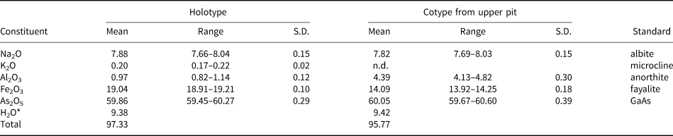
Analyses (holotype: 7 points; cotype: 4 points) were performed at Caltech on a JEOL 8200 electron microprobe in wavelength dispersive spectroscopy mode. Analytical conditions were 15 kV accelerating voltage, 2 nA beam current and 15 μm beam diameter. We attribute the low analytical totals to the irregular surfaces of the crystals. Insufficient material is available for the determination of H2O, so it is calculated based on the structure. Analytical data are given in Table 1.
Analytical data (in wt.%) for nafeasite.

* Based on the structure
S.D. – standard deviation
The empirical formulae of the holotype and cotype (based on 9 O atoms per formula unit) are Na0.98K0.02Fe0.92Al0.07As2.00O9H4.01 and Na0.97Fe0.68Al0.33As2.00O9H4.01, respectively. The simplified structural formula is (Na,K)(Fe3+,Al)(AsO3OH)2⋅H2O. The idealised formula is NaFe3+(AsO3OH)2⋅H2O, which requires Na2O 8.23, Fe2O3 21.20, As2O5 61.01, H2O 9.56, total 100 wt.%. The Gladstone–Dale compatibility 1 – (K P/K C) for the empirical formula is –0.021 in the range of excellent compatibility (Mandarino, Reference Mandarino2007); for the ideal formula, it is –0.007, in the range of superior compatibility.
X-ray crystallography and structure refinement
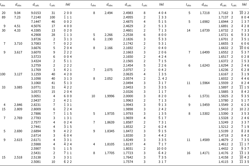
Powder X-ray diffraction data for the holotype were recorded using a Rigaku R-Axis Rapid II curved imaging plate microdiffractometer with monochromatised MoKα radiation. A Gandolfi-like motion on the φ and ω axes was used to randomise the sample. Observed d-values and intensities were derived by profile fitting using JADE Pro software (Materials Data, Inc.). Data (in Å for MoKα) are given in Table 2.
Powder X-ray diffraction data (d in Å) for nafeasite. Only calculated lines with intensities > 1.5 are listed.

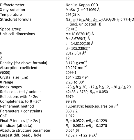
Single-crystal X-ray studies were carried out on 4-circle Kappa CCD diffractometer with monochromatized MoKα radiation. The Nonius Collect software package was used for the collection of the structure data. Eval15 (Schreurs et al., Reference Schreurs, Xian and Kroon-Batenburg2010) was used for processing the data from a twinned crystal consisting of three domains, related by rotation of ~120° about the a* direction. The major domain accounts for about 60% of the crystal volume. The application of an empirical absorption correction using the multi-scan method was done using SADABS (Sheldrick, Reference Sheldrick2008). The structure was solved by the charge-flipping method using Superflip (Palatinus and Chapuis, Reference Palatinus and Chapuis2007) as implemented in Jana2006 (Petříček et al., Reference Petříček, Dušek and Palatinus2014). Refinement proceeded by full-matrix least-squares on F 2 using SHELXL-2016 (Sheldrick, Reference Sheldrick2015).
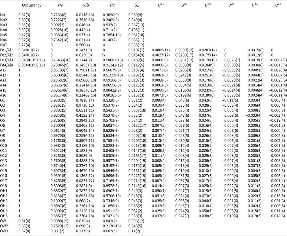
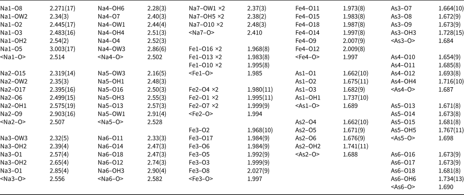
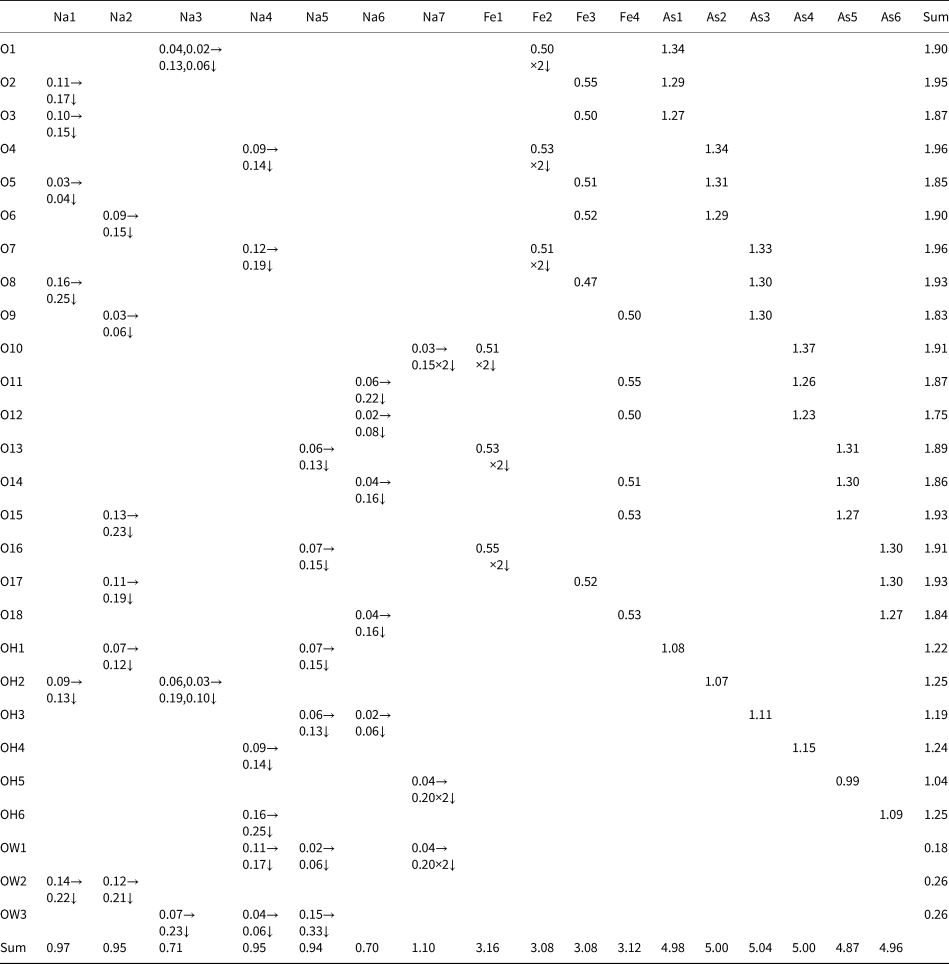
The framework portion of the structure consisting of AsO3OH tetrahedra and (Fe3+,Al)O6 octahedra (refined with joint Fe–Al occupancies) is well ordered and all framework sites were successfully refined with full occupancies and anisotropic displacement paraments; however, the channels in the framework are occupied by a disordered array of Na cations and H2O (OW) groups. We tried a variety of refinement strategies to model the channel constituents. The sites (seven Na and three OW) were identified on the basis of bond distances. All channel sites were found to be partially occupied. Although most could be refined successfully with anisotropic displacement paraments, the ellipsoids of most (particularly Na4 and Na5) were pronouncedly oblate or prolate. Attempts to split these sites created more issues in that the resulting sites either appeared to require further splitting or their displacement parameters became nonpositive definite. In addition, the resulting Na–O distances no longer defined sensible coordinations. Ultimately, we decided to refine all of the channel constituents with isotropic displacement parameters and without site splitting, with two exceptions: (1) the 0.38 occupied Na3 site is split across a two-fold axis, 1.07(6) Å from an equivalent Na3 site and (2) the Na6 and OW2 sites, which are 1.18(4) Å apart were refined with joint full occupancy. To further improve the modelling of the channel constituents, the total occupancy of the Na sites was constrained to one Na atom per formula unit. Difference Fourier failed to locate possible H atom sites. The data collection and refinement details are given in Table 3, atom coordinates and displacement parameters in Table 4, selected bond distances in Table 5 and a bond-valence analysis in Table 6. The crystallographic information file has been deposited with the Principal Editor of Mineralogical Magazine and is available as Supplementary material (see below).
Data collection and structure refinement details for nafeasite.

R int = Σ|F o2–F o2(mean)|/Σ[F o2]. GoF = S = {Σ[w(F o2–F c2)2]/(n–p)}½. R 1 = Σ||F o|–|F c||/Σ|F o|. wR 2 = {Σ[w(F o2–F c2)2]/Σ[w(F o2)2]}½; w = 1/[σ2(F o2)+(aP)2+bP] where a is 0.0451, b is 72.2 and P is [2F c2+Max(F o2,0)]/3.
Atom coordinates and displacement parameters (Å2) for nafeasite.

Selected bond distances (Å) for nafeasite.

Bond valence analysis for nafeasite. Values are expressed in valence units (vu).*

*Bond valences are based on refined site occupancies. Bond-valence parameters are from Gagné and Hawthorne (Reference Gagné and Hawthorne2015). Hydrogen-bond contributions are not included.
Description of the structure
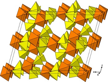
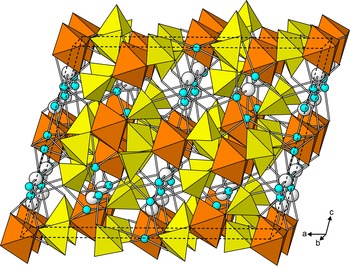
The structure of nafeasite is based on a loose 3D framework of alternating AsO3OH tetrahedra and Fe3+O6 octahedra (Fig. 4). Each of the four Fe3+O6 octahedra (centred by Fe1, Fe2, Fe3 and Fe4) shares all six of its vertices with an AsO3OH tetrahedron and each AsO3OH tetrahedron (centred by As1, As2, As3, As4, As5 and As6) shares its three O vertices with an Fe3+O6 octahedron, the remaining OH vertex being unshared. The resulting framework consists of layers of mutually unconnected AsO3OH tetrahedra and layers of mutually unconnected Fe3+O6 octahedra, alternating along [100]. Relatively large interstices within the framework accommodate seven partially occupied Na sites (Na1, Na2, Na3, Na4, Na5, Na6 and Na7) and three partially occupied H2O sites (OW1, OW2 and OW3) (Fig. 5). Hydrogen bonding clearly plays a significant role in the stability of the structure; however, the complexity of the disordered interstitial portion of the structure made it impossible to devise a comprehensive and unambiguous hydrogen-bonding scheme. Nevertheless, it is worth noting that the O12 site, with a bond-valence sum of 1.75 valence units (vu), is only 2.62 Å from the OH5 site and, based on the relation of Ferraris and Ivaldi (Reference Ferraris and Ivaldi1988), would receive a hydrogen bond contribution of 0.27 vu.
3D framework of corner-sharing octahedra and tetrahedra in the structure of nafeasite, viewed slightly canted along [010].

The same view of the structure of nafeasite as in Fig. 4 but with interstitial Na (light blue) and H2O (white) sites shown.

The structure of nafeasite is unique. Arsenate mineral structures based on simple frameworks of alternating octahedra and tetrahedra are uncommon. The only ones noted by Majzlan et al. (Reference Majzlan, Drahota, Filippi, Bowell, Alpers, Jamieson, Nordstrom and Majzlan2014) were parascorodite, Fe(H2O)2AsO4; the isostructural minerals scorodite, Fe(H2O)2AsO4, mansfieldite, Al(H2O)2AsO4, and yanomamite, In(H2O)2AsO4; and berzeliite, (NaCa2)Mg2(AsO4)3, and manganberzeliite, (NaCa2)Mn2(AsO4)3, which have the garnet structure. None of these structures is very similar to that of nafeasite.
Though the Al content of the holotype is rather small, Al3+ occupies approximately one third of the octahedral sites in the cotype crystal, which suggests the possible existence of a yet unknown Al analogue of nafeasite.
Acknowledgements
Two anonymous reviewers and Structural Editor Peter Leverett are thanked for their constructive comments on the manuscript. A portion of this study was funded by the John Jago Trelawney Endowment to the Mineral Sciences Department of the Natural History Museum of Los Angeles County.
Supplementary material
To view supplementary material for this article, please visit https://doi.org/10.1180/mgm.2022.58
Competing interests
The authors declare none.