Implications
Milk is a complex physiological liquid containing biological active components with various health-related effects. This review summarises some of the available cell-based models that can be used for testing milk-derived bioactives with focus on bovine milk. Cell-based assays have advantages and limitations that have to be considered before choosing an appropriate model. Also milk sample preparation and relevant endpoint parameters have to be considered carefully when milk-derived bioactives are to be tested. If the advantages of using cell-based models are combined with studies in in vivo models, it may be a durable way to produce valid documentation for claiming specific health-related effects of milk-derived bioactives in humans.
Introduction
The pharmaceutical, biotechnology and nutraceutical industries have a strong interest in screening for novel bioactives in complex mixtures such as milk and dairy products. These bioactives are not only important as nutritional supplements, but also have potential therapeutic applications for the treatment of a number of diseases. To identify and test the effect of bioactives such as hormones, growth factors, fatty acids, immunoglobulins, proteins, peptides, minerals or vitamins, robust screening assays are essential. More detailed overviews of milk-derived bioactives can be found elsewhere (Shah, Reference Shah2000; Michaelidou and Steijns, Reference Michaelidou and Steijns2006; Gibson, Reference Gibson2011). The present review summarises some of the available models for testing milk-derived bioactives with special focus on bovine milk. First, we discuss the advantages and limitations of cell-based models for testing bioactive components. Then elements to consider for choosing an appropriate cell-based model will be outlined followed by considerations on sample preparation when milk-derived bioactives are to be tested in vitro. Cell culture parameters or endpoints of interest for assessing the bioactivity of milk-derived compounds will be outlined, and examples of cell models of specific target tissues including the gastrointestinal tract and the mammary gland will be described. Finally, cell-based models of immune function will be described briefly.
Advantages and limitations of cell-based models
The use of in vitro cell-based models for screening and testing has the advantage that several concentrations of mixtures or specific compounds can be assayed at the same time in cells from specific tissues in a controlled environment. The hormesis concept that low doses of chemical or toxic substances are stimulatory while higher doses are inhibitory (Calabrese and Baldwin, Reference Calabrese and Baldwin1998; Calabrese, Reference Calabrese2010) makes it necessary to apply many concentrations of bioactive compounds to investigate the biological effect. Primary cell cultures from target organs or established cell lines can be used to identify the most sensitive cells. For each of the mixtures or specific compounds, different endpoints inherent to basal or more sophisticated cellular functions such as cell viability and proliferation, cell migration, regulation of gene expression, morphology or metabolic alterations can be investigated. This makes in vitro models an essential tool in mechanistic biological studies. Furthermore, the use of in vitro models have the advantage that they represent a possible alternative to animal experiments, thereby saving the lives of laboratory animals and reducing costs for expensive animal experimentation. Alternatives to animal studies are also included in recent legislation from the European Union, with the wording that: ‘authorities will have to assess the animal welfare implications of all scientific experiments involving animals with a view to promoting alternative testing methods where possible’ (http://www.euractiv.com/en/science/eu-wants-fewer-animals-used-research-news-497607).
However, cell-based models also have a number of limitations. Cell-based models are in vitro systems, which may not reflect the in vivo conditions of cells and tissues in their natural state in the organism, where signalling and interplay between organs play important roles. The cultural environment also lacks the several systemic components involved in the homeostatic regulation in vivo, principally those of the endocrine and nervous systems. Cellular metabolism therefore may be more constant in vitro than in vivo and may not be truly representative of the tissue from which the cells were derived. Another limitation is that in vitro studies do not incorporate the effects of biological processes such as bioavailability, pharmacokinetics, metabolism, distribution and interacting with binding and transport proteins that occurs in the intact organism, which may all influence the biological effects of intake of milk-derived bioactives. Consequently, it is necessary to extend the results obtained in vitro to animal or human studies to elaborate the biological effects of bioactive compounds. The way to improve the use of cell-based models is to carefully consider the choice of cell-based model (target organ), how the milk sample (bioactive) is prepared and added to the cell culture medium and not at least the endpoint measured.
Choosing an appropriate cell-based model
Overall, in vitro cell-based models to test the activity of milk-derived bioactives have to live up to at least two basal requirements: it should be easy to handle for high-throughput testing; and it should have good human predictive power, that is, provide data that support interpretation of results for the in vivo situation. Therefore, careful consideration should be given in choosing the specific target organ of interest and also in choosing the physiological stage of the cells (e.g. undifferentiated v. differentiated), and developmental stage of humans or animals from where cells are obtained. Another important thing to consider is whether to choose an established cell line or primary cells isolated from the intact organism before experimentation. For a number of cellular functions such as apoptosis and the regulation of gene expression, primary cultures or freshly isolated cells of human or animal tissue are often preferred as primary cells normally retain the majority of the in vivo functionality. However, primary cells often only survive for a few days in cell culture, and as they usually derive from different individuals in each assay, the reproducibility of results may significantly differ from one assay to another. When choosing established cell lines it is important to keep in mind that these cells relies on immortalised or transformed cells in an artificial environment, and they may therefore dedifferentiate and have lost some functional properties observed in vivo during passaging. Furthermore, the proliferation and behaviour under environmental stimuli of cell lines of cancer origin may be significantly altered as cancerous cells have a different glycosylation (Brooks et al., Reference Brooks, Carter, Royle, Harvey, Fry, Kinch, Dwek and Rudd2008). A number of cell-based models of the gastrointestinal tract and the mammary gland will be discussed in detail later in this review.
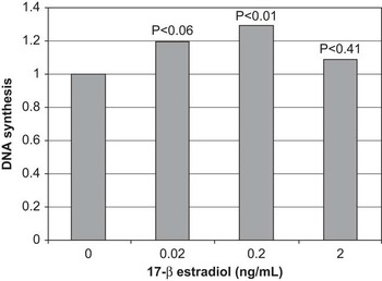
Historically, fibroblast cell lines and mink lung epithelial cells were some of the first cell lines to be used for test of biological activity in milk (Klagsbrun and Neumann, Reference Klagsbrun and Neumann1979; Klagsbrun and Shing, Reference Klagsbrun and Shing1984; Rogers et al., Reference Rogers, Belford, Francis and Ballard1995; Belford et al., Reference Belford, Rogers, Francis, Payne, Ballard and Goddard1997). Furthermore, there are a number of cell-based studies showing immunoregulatory and antibacterial effects of milk proteins and peptides (Meisel and Gunther, Reference Meisel and Gunther1998; Moller et al., Reference Moller, Scholz-Ahrens, Roos and Schrezenmeir2008). More recently, a number of mammary and intestinal cell lines (transformed or cancer origin) have been used (e.g. Sejrsen et al., Reference Sejrsen, Pedersen, Vestergaard and Purup2000; Purup et al., Reference Purup, Vestergaard, Pedersen and Sejrsen2007; Nielsen et al., Reference Nielsen, Norgaard, Purup, Frette and Bonefeld-Jorgensen2009). As mentioned above, the choice of model is essential for the outcome when milk bioactives are tested in cell cultures. To illustrate this point, it has proved difficult to demonstrate a direct mitogenic effect of 17β-estradiol on cell lines of bovine mammary epithelial cells in vitro (Akers et al., Reference Akers, McFadden, Purup, Vestergaard, Sejrsen and Capuco2000), although oestrogen overall is known to play an important role in the development of the bovine mammary gland (Purup et al., Reference Purup, Sejrsen, Foldager and Akers1993b; Capuco et al., Reference Capuco, Ellis, Wood, Akers and Garrett2002). As seen in Figure 1, we showed mitogenic effects of very low concentrations of 17β-oestradiol (E2) in heifer mammary gland explants. Mammary explants were treated with increasing concentrations of E2 and cell proliferation (DNA synthesis) measured as incorporation of tritiated thymidine was used as the endpoint. DNA synthesis was significantly increased at 0.02 and 0.2 ng/ml of E2 in both explants and organoids, whereas the higher concentration of 2 ng/ml had no effect on DNA synthesis.
Effect of 17β-oestradiol on DNA synthesis in mammary tissue explants from prepubertal Holstein heifers. Values are presented relative to DNA synthesis obtained in medium without 17β-oestradiol. P-values for the effect of the three concentrations of 17β-oestradiol are shown. A significant effect of 17β-oestradiol on DNA synthesis was seen in low concentrations (adapted from Purup et al., Reference Purup, Sejrsen and Akers1993a).

Sample preparation for test of milk bioactives
Milk is a complex biological matrix containing several bioactive components associated with different fractions of milk (Clare and Swaisgood, Reference Clare and Swaisgood2000; Shah, Reference Shah2000). The major challenges when testing milk bioactives are to develop and apply sample purification procedures so that cytotoxicity problems with the final extract applied into the cellular cultures are avoided; to obtain a certain level of selectivity in order to collect the bioactives of interest in the same fraction; and to limit the presence of undesired substances in the extract to be tested. Furthermore, many milk borne bioactives are latent, requiring proteolytic release of bioactive peptides from inactive native milk proteins. Therefore, to investigate the bioactivity of milk components in vitro, activation may be required.
The procedures applied for preparation of the milk fractions for cell-based assays depend on in what fraction of the milk the bioactive of interest is present. An initial step is typically to spin the raw milk at low gravity to obtain an upper fat fraction. The fat can be further sonicated and ultracentrifuged to obtain, for example, the milk fat globule membranes. The skim milk fraction can be further processed by high speed or ultra-centrifugation and the casein and whey fraction can be separated by acid precipitation (Purup et al., Reference Purup, Vestergaard, Pedersen and Sejrsen2007). If the bioactive of interest is related to the protein fraction of milk, extracts of the whey fraction can be obtained by microfiltration (Rusu et al., Reference Rusu, Drouin, Pouliot, Gauthier and Poubelle2010), whereas peptides of caseins can be obtained by enzymatic digestion followed by fractionation and purification by reverse phase chromatography (Kitazawa et al., Reference Kitazawa, Yonezawa, Tohno, Shimosato, Kawai, Saito and Wang2007). The challenge is thereafter to add the potential milk bioactive to the cell culture medium without creating a milieu for cells that are too different from the internal milieu in the intact organism/organ from which the cells derive so that the culture conditions become unfavourable. It is our experience that the proportion of complete whey that can be included in the cell culture media should not exceed 10%, otherwise whey itself has a cytotoxic effect on most cell types. However, the concentration and in what form the milk bioactive is added to the cell culture medium depends fully on what the bioactive is of interest and whether the bioactive is isolated directly from milk or purchased commercially (Pocock et al., Reference Pocock, Sales and Milligan2002; Nielsen et al., Reference Nielsen, Norgaard, Purup, Frette and Bonefeld-Jorgensen2009; Rusu et al., Reference Rusu, Drouin, Pouliot, Gauthier and Poubelle2010).
Milk contains relatively high levels of endogenous steroid hormones (Hartmann et al., Reference Hartmann, Lacorn and Steinhart1998; Malekinejad et al., Reference Malekinejad, Scherpenisse and Bergwerff2006; Courant et al., Reference Courant, Antignac, Maume, Monteau, Andre and Le2007; Farlow et al., Reference Farlow, Xu and Veenstra2009) and also feed-derived phytoestrogens (Hoikkala et al., Reference Hoikkala, Mustonen, Saastamoinen, Jokela, Taponen, Saloniemi and Wahala2007; Steinshamn et al., Reference Steinshamn, Purup, Thuen and Hansen-Moller2008; Nielsen et al., Reference Nielsen, Norgaard, Purup, Frette and Bonefeld-Jorgensen2009), the latter in concentrations reflecting the type and amount of legumes included in the feed ration. Both steroid hormones and phytoestrogens are mainly present in milk as biologically inactive glucuronide and sulphate conjugates (King et al., Reference King, Mano and Head1998; Courant et al., Reference Courant, Antignac, Maume, Monteau, Andre and Le2007; Farlow et al., Reference Farlow, Xu and Veenstra2009). In order to test the oestrogenic or anti-oestrogenic effects of these substances in bioassays, they have to be de-conjugated and extracted from milk. Pocock et al. (Reference Pocock, Sales and Milligan2002) described a simple method in which de-conjugation and precipitation of milk proteins was performed chemically with hydrochloric acid followed by extraction using diethyl ether. The final extract was then dissolved in ethanol before added to the cell culture media. However, this method does not allow a separation of endogenous steroid hormones from dietary-derived phytoestrogens, the two potentially interacting with each other. Antignac et al. (Reference Antignac, Gaudin-Hirret, Naegeli, Cariou, Elliott and Le Bizec2009) developed a more sophisticated protocol in which a preliminary extraction with acetone (precipitation of proteins) was followed by enzymatic hydrolysis (Helix pomatia) and a two purification steps onto reverse (C18) and normal (SiOH) phase solid phase extraction (SPE) cartridges. The latter step resulted in the removal of endogenous oestrogens from the extract. The extract was reconstituted in dimethyl sulfoxide (DMSO) to make it water soluble before tested in an oestrogen-receptor transactivation assay with T47D.luc cells (Dip et al., Reference Dip, Lenz, Antignac, Le Bizec, Gmuender and Naegeli2008).
When enzymatic hydrolysis is performed on milk, some interfering compounds will inevitably be introduced. It is our experience that cell lines such as the human breast cancer cells MCF-7 are sensitive to these residues showing signs of cytotoxicity (unpublished data) when exposed to extracts produced with enzymatic hydrolysis, and has to be considered when the sample preparation protocol is developed.
Choosing an endpoint to measure
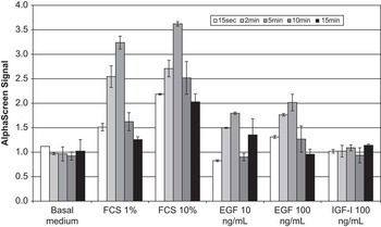
One of the first things to decide before choosing the cell-based assay format is the endpoint of interest. Assays are available to measure a variety of different markers that indicate the number of viable cells (viability assay), the proliferation of cells, the number of dead cells (cytotoxicity assays), the total number of cells or the mechanism of cell death (e.g. apoptosis). Other endpoint parameters include cell morphology, cell migration, inflammation and synthesis and secretion of proteins. Cell viability or cytotoxicity measurements usually are determined at the end of the exposure period. Assays that require only a few minutes to generate a measurable signal, such as ATP quantification or lactate dehydrogenase-release assays, provide information representing a snapshot in time and have an advantage over assays that may require several hours or days of incubation to develop a signal. Although the technological innovations in the fields of proteomics, genomics and metabolomics in recent years have provided lots of new types of data, in vitro models are still essential tools in biological mechanistic studies. Furthermore, in vitro models can provide material for proteomic and genomic studies. The proteomic, genomic or metabolomic profiling in association with a systems biology approach are very important endpoints in this context. The effect of milk-derived bioactives on intracellular signalling proteins represents a fast way to get a response in cells representing a specific tissue. Instead of the endpoint response a signalling event is measured after a short treatment of cells with the bioactives to study. The Amplified Luminescence Proximity Homogenous Assay (ALPHA) screen (AlphaScreen®; PerkinElmer Danmark A/S, Skovlunde, Denmark) technology is a very sensitive way of measuring intracellular signalling proteins by a luminescence proximity assay. At present, more than 60 different ALPHA screen kits has been developed within many different areas such as immunity, cancer, diabetes, inflammation and neurodegeneration (PerkinElmer, Skovlunde, Denmark). In Figure 2, the phosphorylated extracellular signal-regulated kinase is measured in intestinal cells. Cells were treated with foetal calf serum, epidermal growth factor or insulin-like growth factor-I (IGF-I) for 15 s, 2, 5, 10 and 15 min. The results show that the maximal response was obtained after only 5 min of treatments. We believe that the ALPHA screen technology will be an important technology in intracellular signalling studies in cell-based models.
Measurement of phosphorylated extracellular signal-regulated kinase in intestinal cells by the use of Amplified Luminescence Proximity Homogenous Assay (ALPHA) screen technology. The results show that the maximal response to foetal calf serum and epidermal growth factor (EGF) is obtained after only 5 min of treatment of cells (Purup S., unpublished data). FCS = foetal calf serum.

Cell-based models of the mammary gland
The mammary gland is a target organ of considerable interest when it comes to milk-derived bioactives, as it has been estimated that up to 50% of all newly diagnosed breast cancers may be related to dietary factors (Doll, Reference Doll1992; Hilakivi-Clarke et al., Reference Hilakivi-Clarke, Cho, deAssis, Olivo, Ealley, Bouker, Welch, Khan, Clarke and Cabanes2001) The relation between breast cancer and diet is complex, but the fact is that milk contains hormones and growth factors such as oestrogens, phytoestrogens, progesterone and IGF-1 that are involved in the aetiology of breast cancer. A number of bovine mammary cell lines have been established, among these the MAC-T (Huynh et al., Reference Huynh, Robitaille and Turner1991) and the BME-UV1 (Zavizion et al., Reference Zavizion, van Duffelen, Schaeffer and Politis1996) cell lines, both of which were immortalised by transfection with the SV40 T-antigen. MAC-T cells have been used for studies of many bioactives, including fatty acids, growth factors, steroids, retinoids, cytokines and mammary extracts, and parameters such as viability, proliferation, apoptosis, gene expression, epithelial transport, cell signalling and lipogenesis (Woodward et al., Reference Woodward, Turner, Hung and Zhao1996; Cohick and Turner, Reference Cohick and Turner1998; Berry et al., Reference Berry, Weber Nielsen, Sejrsen, Pearson, Boyle and Akers2003; Peterson et al., Reference Peterson, Matitashvili and Bauman2004; Zarzynska et al., Reference Zarzynska, Gajewska and Motyl2005; Thorn et al., Reference Thorn, Purup, Cohick, Vestergaard, Sejrsen and Boisclair2006 and Reference Thorn, Purup, Vestergaard, Sejrsen, Meyer, Van Amburgh and Boisclair2008; Bruzelius et al., Reference Bruzelius, Purup, James, Onning and Akesson2008; Keating et al., Reference Keating, Zhao, Finucane, Glimm and Kennelly2008; Sorensen et al., Reference Sorensen, Chris, Murdoch, Keating, Cruz-Hernandez, Wegner, Kennelly, Okine and Weselake2008; Zhou et al., Reference Zhou, Capuco and Jiang2008). MAC-T cells also represent a more general model of the mammary gland, as these cells retain a number of biochemical and morphological characteristics typical of mammary epithelial cells in vivo. In addition the immortalized bovine mammary epithelial cell line, BME-UV, have been used for elaborating effects of a number of bioactives such as growth factors, mycotoxins, steroids, lactoferrin proteins and retinoids, and parameters such as viability, proliferation, apoptosis and gene expression (Weber et al., Reference Weber, Purup, Vestergaard, Ellis, Scndergard-Andersen, Akers and Sejrsen1999; Cheli et al., Reference Cheli, Politis, Rossi, Fusi and Baldi2003; Baldi et al., Reference Baldi, Losio, Cheli, Rebucci, Sangalli, Fusi, Bertasi, Pavoni, Carli and Politis2004; Fusi et al., Reference Fusi, Rebucci, Pecorini, Rossi, D'Ambrosio and Baldi2008; Kozlowski et al., Reference Kozlowski, Gajewska, Majewska, Jank and Motyl2009; Pecorini et al., Reference Pecorini, Sassera, Rebucci, Saccone, Bandi and Baldi2010). Monolayer culture models of mammary cells are easy and convenient in vitro systems. However, they do not recapitulate the glandular structure of epithelium in vivo and therefore cannot provide the optimal system for studying the regulation of proliferation, polarisation and differentiation of the glandular epithelium. The development of three-dimensional cell culture models has allowed investigators to make significant progress towards characterisation of factors of importance for establishment and maintenance of epithelial architecture. Of importance is also the exposure of primary mammary cells or established cell lines to physiological exogenous matrices that mimic the composition of the normal basement membrane. In a recent study (Kozlowski et al., Reference Kozlowski, Gajewska, Majewska, Jank and Motyl2009), the growth and transcriptomic profile was studied in BME-UV1 mammary cells cultured in traditional monolayers on a plastic surface, and on extracellular matrix, Matrigel™ (BD Biosciences, Erembodegem, Belgium). It was shown that BME-UV1 cells grown on Matrigel™ formed polarised acinar structures during 16 days of culture and microarray analysis showed that the difference in spatial architecture between mammary epithelial cells cultured in monolayer and a three-dimensional structure was reflected by differences in transcriptomic profile. This study shows the importance of developing models with polarised mammary epithelial cells and that BME-UV1 may be a good model for studying, for example, developmental processes of the bovine mammary gland in vitro.
Primary cells of the bovine mammary gland have also been used as models including both organoids, explants and epithelial cells for studies of effects of growth factors, steroids, lactoferrin, retinoids, cytokines, biogenic amines, mammary extracts, and endpoint parameters such as mitogenic activity and gene expression have been measured (Matitashvili and Bauman, Reference Matitashvili and Bauman1999 and Reference Matitashvili and Bauman2001; Weber et al., Reference Weber, Purup, Vestergaard, Ellis, Scndergard-Andersen, Akers and Sejrsen1999; Akers et al., Reference Akers, McFadden, Purup, Vestergaard, Sejrsen and Capuco2000; Ellis et al., Reference Ellis, Purup, Sejrsen and Akers2000; Purup et al., Reference Purup, Vestergaard, Weber, Plaut, Akers and Sejrsen2000 and Reference Purup, Jensen and Sejrsen2001; Weng et al., Reference Weng, Beyenbach and Quaroni2005; Thorn et al., Reference Thorn, Purup, Cohick, Vestergaard, Sejrsen and Boisclair2006 and Reference Thorn, Purup, Vestergaard, Sejrsen, Meyer, Van Amburgh and Boisclair2008; Ernens et al., Reference Ernens, Clegg, Schneider and Larondelle2007; Fusi et al., Reference Fusi, Rebucci, Pecorini, Rossi, D'Ambrosio and Baldi2008; Swanson et al., Reference Swanson, Stelwagen, Dobson, Henderson, Davis, Farr and Singh2009; Wang and Baumrucker, Reference Wang and Baumrucker2010). Both primary mammary cells and cell lines of human and rodent origin have been used to study effects of milk-derived bioactives such as CLA and phytoestrogens. Cells in these models include MCF-7, MDA-MB-231, MCF-10a, MVLN, TM4T, as well as primary mammary epithelial and stromal cells (Ip et al., Reference Ip, Masso-Welch, Shoemaker, Shea-Eaton and Ip1999; Park et al., Reference Park, Allen and Shultz2000; Strange et al., Reference Strange, Wilkinson and Emerman2002; Zhang et al., Reference Zhang, Warren, Shoemaker and Ip2007; Ou et al., Reference Ou, Wu, Ip, Meng, Hsu and Ip2008; Wang et al., Reference Wang, Huang, Liu, Yan and Lin2008; Nielsen et al.,Reference Nielsen, Norgaard, Purup, Frette and Bonefeld-Jorgensen2009; Bernard-Gallon et al., Reference Bernard-Gallon, Satih, Chalabi, Rabiau, Bosviel, Fontana and Bignon2010).
We developed a model with mammary cells isolated from parenchyma of the prepubertal heifer mammary gland. Several studies have shown that this this model is a very sensitive screening model for milk-derived bioactives (Norup et al., Reference Norup, Purup and Sejrsen1997; Weber et al., Reference Weber, Purup, Vestergaard, Akers and Sejrsen1997 and Reference Weber, Purup, Vestergaard, Akers and Sejrsen2000; Purup et al., Reference Purup, Jensen and Sejrsen1999, Reference Purup, Vestergaard, Weber, Plaut, Akers and Sejrsen2000 and Reference Purup, Vestergaard, Weisbjerg, Hvelplund and Sejrsen2002; Ellis et al., Reference Ellis, Purup, Sejrsen and Akers2000; Lametsch et al., Reference Lametsch, Rasmussen, Johnsen, Purup, Sejrsen, Petersen and Heegaard2000; Sejrsen et al., Reference Sejrsen, Pedersen, Vestergaard and Purup2000; Hansen et al., Reference Hansen, Purup and Christensen2003; Thorn et al., Reference Thorn, Purup, Cohick, Vestergaard, Sejrsen and Boisclair2006; Fusi et al., Reference Fusi, Rebucci, Pecorini, Rossi, D'Ambrosio and Baldi2008). Groups of mammary epithelial cells are isolated from parenchymal tissue by enzymatic treatment and precipitation and these isolated organoids are then cultured in collagen gels, allowing the cells to proliferate in three dimensions. This model has been used to elucidate the effect of colostrum and milk (whey samples) during lactation (Sejrsen et al., Reference Sejrsen, Pedersen, Vestergaard and Purup2000), and when the bioactivity of the same whey samples were studied in human small intestinal cells, differences between the tissues were evident (Purup et al., Reference Purup, Vestergaard, Pedersen and Sejrsen2007). For both target tissues, there was a decline in the mitogenic activity of milk obtained on post-partum day 1 until 1 week after calving. However, mature milk and milk from dry cows had significant mitogenic activity in intestinal cells but not in mammary cells despite the fact that high concentrations of the IGF-I present in milk from dry cows and IGF-I is known to stimulate mammary epithelial cell proliferation (Purup et al., Reference Purup, Sandowski and Sejrsen1995 and Reference Purup, Vestergaard, Weber, Plaut, Akers and Sejrsen2000; Sejrsen et al., Reference Sejrsen, Pedersen, Vestergaard and Purup2000).
In a recent study, primary mammary cells were also used as a model system in which responses to inflammation-related proteins TNF-α, IL-1β and IL-6, MCP-1 and PAI-1 were measured (Thorn et al., Reference Thorn, Purup, Vestergaard, Sejrsen, Meyer, Van Amburgh and Boisclair2008). The responses to these proteins were also measured in MAC-T cells at Cornell University (Thorn et al., Reference Thorn, Purup, Vestergaard, Sejrsen, Meyer, Van Amburgh and Boisclair2008), and the results showed that the responses in MAC-T cells were very similar to primary mammary epithelial cells.
Cow's milk contains endogenous hormones such as oestrogens and progesterone, and also various phytoestrogens (Purup et al., Reference Purup, Hansen-Møller, Sejrsen, Christensen, Lykkesfeld, Leffers and Skakkebaek2005; Steinshamn et al., Reference Steinshamn, Purup, Thuen and Hansen-Moller2008; Andersen et al., Reference Andersen, Nielsen, Purup, Kristensen, Eriksen, Søegaard, Sørensen and Fretté2009), a diverse group of non-steroidal plant-derived compounds that show structural and functional similarities with 17-β oestradiol. Phytoestrogens have the potential to exert oestrogenic and anti-oestrogenic activities by interacting with numerous molecules, carrier proteins, enzymes and membrane and nuclear receptors directly or indirectly involved in oestrogen signals (Benassayag et al., Reference Benassayag, Perrot-Applanat and Ferre2002; Committee on Toxicity, 2003; Martin et al., Reference Martin, Crotty and Nelson2007). As high levels of leguminous plants, such as lupine, clover, horse beans and peas, are used in organic milk production, and as leguminous plants have a naturally high content of phytoestrogens, these plants are a potential source of phytoestrogens for the cow and in turn for consumers of milk and dairy products. To elucidate the oestrogenic effect of phytoestrogens in bovine milk, an oestrogen-receptor reporter gene assay based on the stably transfected human mammary epithelial MVLN cells has been used (Nielsen et al., Reference Nielsen, Norgaard, Purup, Frette and Bonefeld-Jorgensen2009). MVLN cells derived from oestrogen receptor-positive MCF-7 breast cancer cells and comprise a very sensitive assay for studying activation of the oestrogen receptor by potential oestrogenic compounds. We showed that extracts of high-phytoestrogen milk (especially high in equol) had a higher oestrogenicity than low-phytoestrogen milk in MVLN cells Nielsen et al., Reference Nielsen, Norgaard, Purup, Frette and Bonefeld-Jorgensen2009).
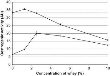
The MCF-7 breast cancer cell line has also been used as a model system to test the proliferative activity of milk from cows fed different forages, including milk from cows fed rations with fresh clover grass, fresh herb mixture or concentrate (control) (Purup S. and Jensen S.K., in preparation). In another study, the proliferative effect of milk from cows in mid-lactation was measured in cells when whey was added to media with or without oestrogen (Nielsen T.S., Andersen C., Purup S. and Sejrsen K., in preparation). Interestingly, addition of milk clearly inhibited cell proliferation of cells simultaneously treated with oestrogen (Figure 3). Although there is a long step from in vitro measurements to in vivo effects, these results indicate that milk may inhibit the effect of high levels of endogenous oestrogen.
Effect of whey prepared from milk from 16 cows in mid lactation on mammary cancer cells in culture. Whey was added to control media with (upper curve) or without (lower curve) oestradiol-17β in a concentration of 1 nM, and oestrogenic activity was measured as proliferation of cells after 6 days of treatment (Andersen C., Purup S. and Sejrsen K., in preparation)

Cell-based models of the gastrointestinal tract
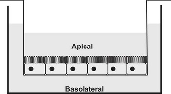
The gastrointestinal tract is an obvious target for bioactive molecules delivered through milk and dairy products, because it lies at the interface between dietary components in the lumen and the internal processes of the host. Therefore, intestinal models are of great interest to the food and pharmaceutical industry, providing tests of newly developed food ingredients and drugs before bringing products to the market. The gastrointestinal tract, however, is a complex system with many interacting cell types and the microbiota, so in cell models as many as possible of these factors should be taken into account. As with mammary epithelial cells, monolayer culture models are convenient in vitro systems, but are often not optimal systems for studying regulation of biological parameters in intestinal cells. Tight junctions are necessary for the formation of intestinal epithelial barrier, integrity and polarity (Shin et al., Reference Shin, Fogg and Margolis2006). Tight junctions are developed in vitro when epithelial cells are cultured on microporous membranes, enabling polarisation of the cells and development of transepithelial resistance. Below the microporous membrane is the basolateral side corresponding to the internal side of the intestine and above the microporous membrane is the apical side corresponding to the lumen side (Figure 4). Although, to our knowledge, there are no publications available, this two-compartment system could potentially be applied to mammary cells as well, studying transport or metabolism of blood borne substances across the epithelium. However, interesting applications for mammary cells of the two-compartment system have been described previously in studies of polymorphonuclear neutrophil migration (Smits et al., Reference Smits, Cifrian, Guidry, Rainard, Burvenich and Paape1996 and Reference Smits, Burvenich, Guidry, Heyneman and Massart-Leën1999) and protein and plasminogen activator production in mammary epithelial cells after extracellular calcium treatment (Cheli et al., Reference Cheli, Baldi, Dell'Orto, Zavizion and Politis2001).
Schematic presentation of two-compartment model with polarised intestinal epithelial cells growing on a microporous membrane (figure obtained from M. Fredborg, Faculty of Science and Technology, Aarhus University, Denmark).

Such two-compartment models also provide the possibility of applying epithelial cells in combination with other cell types such as immune cells like macrophages or dendritic cells that attach on the bottom of the culture wells. When microbiota is added to the apical side, this three-component model (epithelial cells, immune cells and microbiota) is close to the in vivo situation. The available cell-based models of the intestine and their potential uses especially in studies of interactions between the host, pathogens and the intestinal microflora have been reviewed by Cencic and Langerholc (Reference Cencic and Langerholc2010) and Chopra et al. (Reference Chopra, Dombkowski, Stemmer and Parker2010). As it has been difficult to propagate cell lines of normal intestinal epithelium, cell lines of cancer origin (transformed cell lines) are often used as in vitro models for the intestinal tract. For mechanistic studies and attachment assays, the human colon tumorigenic cell lines Caco-2, T84 and HT-29 have been widely used, while the most widely used rodent cell lines are the rat IEC-6 and IEC-18 (Cencic and Langerholc, Reference Cencic and Langerholc2010). Caco-2 cells are often used as these cells differentiate spontaneously, and when differentiated they display morphological and functional characteristics of small intestinal cells, that is, form tight junctions, develop apical microvilli, express brush border enzymes and express growth factor receptors (Delie and Rubas, Reference Delie and Rubas1997). For studies of human milk-derived bioactives, the small intestinal FHs 74 Int. cell line has been widely used as a model (Ichiba et al., Reference Ichiba, Kusuda, Itagane, Fujita and Issiki1992; Kawamura et al., Reference Kawamura, Miki, Kurokawa and Kojima1994; Wagner et al., Reference Wagner, Forsythe and Wagner1998; Hirai et al., Reference Hirai, Ichiba, Saito, Shintaku, Yamano and Kusuda2002; Takeda et al., Reference Takeda, Sakata, Minekawa, Yamamoto, Hayashi, Tasaka and Murata2004). We have shown that these small intestinal cells represent a very sensitive model for milk-derived bioactives (Purup et al., Reference Purup, Vestergaard, Pedersen and Sejrsen2007). Colonic cells of cancer origin have also been used to test the effects of milk-derived bioactives. Of our cell-based models with Caco-2, LoVo, HCT-116, Sw-480, Colo-320, CCK-81 and HT-29, we selected models with Caco-2 and HT-29 cells for measuring bioactivity in milk samples (whey) from the Nordic countries to study the effect of feeding different feed stuffs on the milk content of bioactives stimulating or inhibiting cell proliferation (Purup S. et al., unpublished data).
The exact choice of the right cell-based models for a particular study can be difficult. We recently compared the proteomic signatures of intestinal cell lines of different origin including human colonic cancer cell lines, a normal human colon cell line, human and porcine small intestinal cell lines and porcine intestinal tissue. On the basis of the non-redundant proteins a dendrogram was performed showing clusters of colonic cancer cell lines and small intestinal cell lines together, while the porcine intestinal tissue clustered with the normal human colon cell line (Fredborg M. et al., in preparation). This study indicates that the results obtained are dependent on the origin of the intestinal cell line.
Various intestinal epithelial cell lines have been widely exploited in wound-healing assays as in vitro models that allow simple manipulation of the external environment. A variety of factors that affect the rate of intestinal epithelial cells to heal a wound have been identified using these models, including growth factors, cytokines and extracellular matrix (ECM) components (Seltana et al., Reference Seltana, Basora and Beaulieu2010). Although some ECM components may be synthesised and secreted from intestinal epithelial cells such as cellular fibronectin (Vachon et al., Reference Vachon, Simoneau, Herring-Gillam and Beaulieu1995), it is a limitation of these cell models that cell monolayers are grown on plastic or on an artificial coating rich in ECM factors. Seltana et al. (Reference Seltana, Basora and Beaulieu2010) recently established co-cultures with intestinal epithelial (Caco-2/15) cells on top of a myofibroblasts network as a model to investigate the mechanisms that regulate wound healing in the intestinal epithelium. Using this model, they showed that addition of type I collagen or human fibronectin to the culture medium accelerated wound closure on epithelial cell co-cultures (Seltana et al., Reference Seltana, Basora and Beaulieu2010).
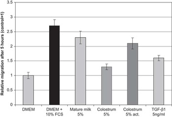
The re-establishment of the epithelial surface can in a simplified way be divided in three distinct mechanisms after the ulceration: cell migration cell, proliferation and cell maturation and differentiation. In collaboration with TGR BioSciences Pty Ltd, Adelaide, Australia, we have established a wound-healing model to identify bioactive peptides in milk or whey that affects migration of intestinal epithelial cells in culture. Rat intestinal epithelial IEC-6 cells, originally derived from the rat crypt, are cultured to confluence and monolayers are wounded by scraping a disposable pipette tip across the dishes. Then media-containing whey samples are added at various concentrations to the cells. The distance migrated by the cells at the anterior edges of the wounded monolayer is determined before and after 5 to 6 h of treatment. Figure 5 shows an example of results obtained by this model when cells are treated with whey fraction of mature milk (5% in culture medium), colostrum (5% in culture medium), acid-activated colostrum (5% in culture medium) and TGF-β1 (5 ng/ml). The distance migrated by the cells was increased when colostrum was acid-activated compared with non- acid-activated colostrum. TGF-β1 plays a role in the re-epithelialisation process (Barrientos et al., Reference Barrientos, Stojadinovic, Golinko, Brem and Tomic-Canic2008; Sturm and Dignass, Reference Sturm and Dignass2008), and therefore, this growth factor was included as a treatment. However, the concentration of TGF-β1 (5 ng/ml), corresponding to the concentration found in colostrum at post-partum day 1 and higher than the concentration found in mature milk (Purup et al., Reference Purup, Vestergaard, Pedersen and Sejrsen2007), could not alone explain the migration caused by the mature milk sample or the acid-activated colostrum sample assayed (Figure 5).
Effect of pools of bovine whey prepared from mature milk, colostrum, acid-activated colostrum and transforming growth factor-β1 (TGF-β1) on migration of rat intestinal epithelial cells (IEC-6; DSMZ, Braunschweig, Germany) in culture. Whey was added to control media (Dulbecco's Minimal Essential Medium) and distance migrated was measured after 5 h treatment. Values are given as means ± s.e.m. relative to distance migrated in control medium. As a positive control, foetal calf serum (FCS) was added in a concentration of 10% in culture medium (Purup S., unpublished data).

Cell-based models of immune function
Milk proteins has been identified as a rich source of biologically active peptides with immunoregulatory functions (Kawahara et al., Reference Kawahara, Katayama and Otani2004; Kitazawa et al., Reference Kitazawa, Yonezawa, Tohno, Shimosato, Kawai, Saito and Wang2007; Rusu et al., Reference Rusu, Drouin, Pouliot, Gauthier and Poubelle2010; Fiedorowicz et al., Reference Fiedorowicz, Jarmolowska, Iwan, Kostyra, Obuchowicz and Obuchowicz2011). The proliferative effect of milk protein β-casein (1 to 28) was measured in human T, B and monocyte cell lines (T-cell lines HUT-78, Jurkat Clone E6-1, MOLT-4; B-cell lines BALL, KHM-1B, U266B1; monocyte cell lines U937 HL-60) by a colorimetric assay supplemented with measurements of immunoglobulin A levels in supernatants and mRNA expression of interleukin-6 (IL-6) in some of the cell lines (Kawahara et al., Reference Kawahara, Katayama and Otani2004). In another study, the chemotactic effect of peptides derived from enzymatic digestion of β-casein was studied in a murine macrophage-like cell line (J774-1) and in human neutrophils and T lymphocytes (Kitazawa et al., Reference Kitazawa, Yonezawa, Tohno, Shimosato, Kawai, Saito and Wang2007). Fiedorowicz et al. (Reference Fiedorowicz, Jarmolowska, Iwan, Kostyra, Obuchowicz and Obuchowicz2011) used freshly isolated human blood mononuclear cells (monocytes + lymphocytes, BMCs) to study the influence of opioid peptides released from β-casein during digestion on BMC proliferation and cytokine secretion, whereas Rusu et al. (Reference Rusu, Drouin, Pouliot, Gauthier and Poubelle2010) isolated human neutrophils and studied effects of whey extracts (β-lactalbumin, α-lactalbumin, glycomacropeptide, lactoferrin, immunoglobulin and bovine serum albumin) on cytokine production in vitro. Primary cultures of mouse spleen and rabbit Peyer's patch cells have also previously been used to study the effect of β-casein digests on proliferation and immunoglobulin production (Hata et al., Reference Hata, Higashiyama and Otani1998).
The specific results from these studies are beyond the scope of this review to summarise, but it illustrates the variety of cell-based models that can be applied in studying the immuno-modulatory effects of milk-derived bioactives, primarily of protein origin.
Summary and conclusion
The use of in vitro cell-based models for screening and testing of milk-derived bioactives represent a potential alternative to the use of a large number of experimental animals. Although the list of currently validated in vitro alternative models is relatively short, there are many new models in the process of validation that can be introduced in the near future. Numerous effects of milk-derived bioactives in bovine milk have been described in vitro, and there certainly is potential for the application of these bioactive compounds as functional foods or pharmaceutical products. However, data from in vitro studies alone are insufficient in claiming an effect on human health, as in vitro models do not take into account the bioavailability, pharmacokinetics, metabolism and distribution and interaction with binding and transport proteins that occur in the intact organism, which may all influence the biological effects of intake of milk-derived bioactives. Therefore, results from studies of milk-derived bioactives in in vitro cell-based models have to be interpreted as such and need further studies in animal or human models. Guidelines on what specific cell-based models are most promising to apply in order to develop, standardise and validate assay platforms with robust performance and reproducibility are not possible to give, as it depends completely on the bioactive and the target organ of interest.
Acknowledgements
The authors thank Annette K. Nielsen, Kasper B. Poulsen and Marlene Fredborg for their technical assistance. The financial support from ERA-net CORE organic, the Danish Research Agency projects ‘Identification of bioactives in complex natural mixtures’ (contract no. 2056-030024), ‘Wound healing effects of bioactive components from milk in relation to gastro-intestinal diseases’ (contract no. 2101-05-0060) and ‘Infectobesity’ (contract no. 2101-07-0111) is greatly acknowledged.