Case report
An 81-year old woman presented at the Department of Geriatric Medicine complaining of dizziness which was preventing her from taking part in daily activities and causing falls. The first symptoms of dizziness had occurred five weeks previously and were accompanied by a short period of dysarthria, which resolved after three weeks. Concomitantly, she experienced short-term memory problems, in combination with difficulty in word-finding and mild depressive symptoms.
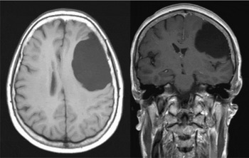
On examination she had no orthostatic hypotension and no dysarthria. On neurological examination she had asymmetrical reflexes, dysdiadochokinesis and a positive Barré test, with the right side being affected. Mild disorientation, short-term memory problems, bradyphrenia, diminished attention and depressive symptoms were observed during the psychiatric assessment. She scored 23 out of 30 points on the Mini-mental State Examination (MMSE), and abnormalities were also seen in her executive functions. Laboratory tests were normal, including thyroid function, vitamin B12 and folate levels. Electrocardiography showed a normal sinus rhythm. The diagnosis of depression was made and, given the presence of neurological signs, a magnetic resonance imaging (MRI) scan of the brain was performed. This showed a large (6.2 cm × 4.3 cm) arachnoid cyst frontotemporally on the left side of the brain. The cyst had induced a midline shift, and probably a compression of the hippocampus on the left side (Figure 1).
Magnetic imaging resonance of the brain showing a large cystic lesion and compression of cerebral parenchyma.

The patient underwent surgery by means of fenestration of the skull and cyst. A Rickham reservoir was left behind. The peri-operative period was uncomplicated and the patient recovered well. Post-operatively the patient was seen again at our outpatient clinic. Her depressive symptoms had disappeared, the short-term memory problems had resolved and she no longer experienced any dizziness. The MMSE score improved, being 28 out 30 points. The mild asymmetrical neurological problems also resolved completely after fenestration. This patient appears to be one of the oldest patients with an arachnoid cyst described in the literature.
The patient gave written informed consent to publication of this case report in this journal.
Discussion
Arachnoid cysts, first described in 1831 by Bright, are benign intra-arachnoid fluid-filled compartments (Seizeur et al., Reference Seizeur, Forlodou, Coustons and Dam-Hieu2007; Cincu et al., Reference Cincu, Agrawal and Eiras2007). In recent years there has been an increasing incidence of these cysts, mostly because of frequent routine use of brain imaging. Arachnoid cysts can be found in 4% of the population (Flaherty, Reference Flaherty2000). The pathophysiology of arachnoid cysts is not completely understood, but genetic predisposition may play a role in their development. There is a distinction between primary and secondary cysts. Primary cysts are considered a maldevelopmental anomaly, in which splitting or duplicating of the primitive arachnoid membrane in early embryonic life leads to a collection of cerebro-spinal fluid (CSF). The cysts may enlarge because of changes in CSF flow, ball valve mechanisms and fluid production of the cell lining of the cyst wall (Seizeur et al., Reference Seizeur, Forlodou, Coustons and Dam-Hieu2007). Secondary cysts may develop following trauma or inflammation. Most cysts remain asymptomatic during life and are located in the middle cranial and posterior fossa conservatively (Yamasaki et al., Reference Yamasaki2003). Symptomatic cysts tend to occur at an early age. Elderly people with cysts may experience headaches, vomiting, hemiparesis, dizziness and gait disturbances (Strojnik, Reference Strojnik2006; Seizeur et al., Reference Seizeur, Forlodou, Coustons and Dam-Hieu2007). These symptoms are non-specific and may also indicate ischemic brain events, hydrocephalus and other intracranial pathology. Our patient had problems which were likely to be due to the arachnoid cyst, which was causing depressive symptoms. The symptoms resolved after surgery, which only takes place if the arachnoid cysts are symptomatic. The most common neurosurgical approach is fenestration of the skull and cyst. Asymptomatic cysts are managed conservatively (Yamasaki et al., Reference Yamasaki2003).
In describing this case we want to emphasize that depressive symptoms associated with neurological symptoms and signs might be due to brain lesions.
Conflict of interest
None.
Description of authors’ roles
Hester R. Boomkens, Tischa J. M. van der Cammen and Francesco U.S. Mattace-Raso observed the patient described in this case report and were responsible for the preparation of the paper.