The reproductive tracts in mammals
In mammalian embryos, the reproductive tracts develop from the Wolffian (in the male) and Mullerian (in the female) ducts. In males, the reproductive tract is composed of the efferent ducts, the epididymis and the vas deferens. In females, the tract is composed of the oviduct, the uterus and the vagina. These tracts, together with the gonads and accessory glands, are the fundamental reproductive organs (Cooper, Reference Cooper1986; Brenner & Maslar, Reference Brenner, Maslar, Knobil and Neil1988).
Both tracts are principally composed of a muscular layer and a mucosa. The muscular layer appears enlarged in some regions, such as the vas deferens, in which muscular activity is important for ejaculation. The same is seen in the uterus, where the strong muscular layer plays an important role in gestation and parturition. The luminal surface of each tract is composed of a mucosal layer, in which the epithelium is the most important physiological component. In the male tract, the epithelium of the epididymis is responsible for the maturation of the spermatozoon during its passage through this organ (Bedford, Reference Bedford, Fawcett and Bedford1979; Cooper, Reference Cooper1986; Kirchhoff et al., Reference Kirchhoff, Pera, Derr, Yeung and Cooper1997). Several proteins secreted by the epithelium of the epididymis become associated with the spermatozoon plasma membrane and have probable functions during sperm capacitation, acrosomal reaction and/or sperm/egg interaction (Jones, Reference Jones1998; Lye & Hinton, Reference Lye and Hinton2004). In the female tract, the analogous oviduct epithelium participates in the transport of gametes, creates the necessary medium for fertilization and recognizes the embryo during its implantation (Hunter, Reference Hunter1988; Brenner & Maslar, Reference Brenner, Maslar, Knobil and Neil1988). Therefore, an important component of reproduction is associated with the physiology of these epithelia. From this perspective, the possibility of modifying the characteristics and functions of these epithelial cells is an important goal for controlling and manipulating reproduction.
Methods of transfection and transfection efficiency
Gene transfer or transfection consists of introducing a foreign gene or genes into cells, tissues or complete animals (Ledley, Reference Ledley1995; Mulligan, Reference Mulligan1993). Transfection procedures have been widely applied in recent years to understand the function of different genes or proteins (Ledley, Reference Ledley1995; Mulligan, Reference Mulligan1993), and these methods have been the basis for gene therapy (Mulligan, Reference Mulligan1993; Verma & Weitzman, Reference Verma and Weitzman2005). Three types of transfection techniques have been used: (i) In vitro methods, which consist of the transfection of tissues or cells maintained in culture media. (ii) In vivo methods, in which the gene constructs are introduced into live organisms. (iii) Ex vivo methods, in which a fresh organ or cells recently extracted from the animal are transfected. Alternatively, cultured cells can be transfected and then introduced into a living animal. This procedure and the in vivo method have been principally employed for gene therapy (Verma & Weitzman, Reference Verma and Weitzman2005).
Gene constructs used for transfections sometimes consist of naked DNA (Wolff & Budker, Reference Wolff and Budker2005). However, gene vectors are usually employed. Gene vectors are molecules to which the DNA (or RNA) is attached; these vectors help transfer exogenous genes into cells. The most frequently employed vectors have been viruses (adenovirus or retrovirus). Viral vectors are the best vehicles for gene transfer because they easily recognize the cell, reach the nucleus and induce a high level of expression. A drawback is that they have the ability to replicate inside the host, thereby causing disease (Daftary & Taylor, Reference Daftary and Taylor2003). Non-viral methods include the use of lipid molecules such as liposomes, neutral lipids, polymeric matrixes and nanoparticles (Mulligan, Reference Mulligan1993; Hart, Reference Hart2005; Vasir & Labhasetwar, Reference Vasir and Labhasentwar2006). Liposomes can easily associate with nucleic acid molecules and do not show a deleterious effect on cells, but their transfection efficiency is sometimes low (Ledley, Reference Ledley1995). Other procedures, such as in vivo electroporation (Trezise et al., Reference Trezise, Palazon, Davies and Colledge2003) or the bombardment of cells with high-velocity microprojectiles (Zelenin et al., Reference Zelenin, Alimov, Zelenina, Semenova, Rodova, Chernov and Kolesnikov1993), have also been successfully employed.
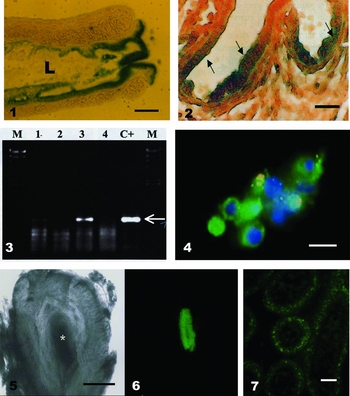
Several gene constructs used for genital tract transfection express the β-galactosidase gene, a commonly used reporter. β-Galactosidase is a bacterial enzyme that is not present in mammalian tissues and can be easily identified by a simple histochemical procedure (Figs. 1 and 2). Green fluorescent protein (GFP) is another extensively used reporter gene. GFP is naturally present in a jellyfish species, and its expression can be observed in fixed or live cells (Kendall & Badminton, Reference Kendall and Badminton1998). GFP expression can be easily observed under a fluorescence microscope in the cytoplasm of transfected cells (Figs. 4, 6 and 7).
Longitudinal section of a mouse uterus during metaestrus transfected with the β-galactosidase gene construct. The green colour indicates the presence of the enzyme. A positive reaction is observed in the epithelium that surrounds the lumen (L) of the uterus and presents as patches along the epithelium. Scale bar = 250μm. Figure 2 Mouse oviduct during metaestrus transfected with the β-galactosidase gene construct. Arrows indicate the positive areas of β-galactosidase expression in the lower region of the glands. Reproduced from Relloso & Esponda (Reference Relloso and Esponda2000). Scale bar = 50μm. Figure 3 PCR of episomal DNA fractions collected from the oviduct (lanes 1 and 2) or uterus (lanes 3 and 4) treated with the β-galactosidase gene construct (lanes 1 and 3) or untreated (lines 2 and 4). The pCMV-β plasmid was employed as a control (C+). Lane M, molecular weight marker. The reaction yields a major product of 580 bp (arrow). Reproduced from Relloso & Esponda (Reference Relloso and Esponda2000). Figure 4 Human uterine cells transfected ex vivo with a gene construct expressing GFP. Cells were collected and then rapidly transfected in vitro. Some cells express GFP, as evidenced by the green fluorescence of the cytoplasm. Blue fluorescence indicates DAPI-stained nuclei. Scale bar = 10μm. Figures 5 and 6 Thick section of about 1 mm from human vas deferens tissue transfected with the GFP gene construct. In 5 the section is observed under light microscopy (* indicates the epithelium). In 6 the same section under fluorescence microscopy, showing fluorescence exclusively in the epithelium. Reproduced from Esponda et al. (Reference Esponda, Goldstein and Witkin2004). Figure 7In vivo transfection of the mouse cauda epididymis using the GFP gene construct. A cross-section shows four epididymal tubules in which several epithelial cells show the green fluorescence of GFP. Reproduced from Esponda & Carballada (Reference Esponda and Carballada2009). Scale bar = 25μm.

Cells in culture can be easily transfected using a plasmid vector, but in vivo gene transfer, which is usually achieved by gene injection into a tissue or an animal, is a more difficult technology, and its efficiency is often low.
When in vivo transfections are developed and gene constructions are administered inside of tubular organs, the most transfected region is usually the epithelium. This result has been observed for different tissues, such as trachea (Fortunati et al., Reference Fortunati, Bout, Zanta, Valerio and Scarpa1996), alveolar epithelium (Katkin et al., Reference Katkin, Gilbert, Langston, French and Baudet1995), endothelial cells (Cuevas et al., Reference Cuevas, Garcia-Calvo, Carceller, Reimers, Zazo, Cuevas, Muñoz-Willery, Martinez-Coso, Lamas and Gimenez-Gallego1996) and in the male and female genital tracts, as we describe in this review.
Both the number of cells that express the foreign gene(s) and the time of expression are related to the efficiency of transfection (Mulligan, Reference Mulligan1993; Anderson, Reference Anderson1998). Transfection efficiency is low when the transgene is expressed in a few cells or during short periods of time. In other cases, the efficiency is higher when the transgene is expressed in a large number of cells and/or lasts for long time periods. This situation is the case for in vivo transfection conducted in muscle, in which gene expression can last for several months (Danko et al., Reference Danko, Williams, Herweijer, Zang, Latendresse and Bock1997). The efficiency of transfection is also related to other factors, such as the distribution and extracellular half-life of DNA, the efficiency of DNA uptake into the cells, the rate of degradation of DNA within the cell, the rate of transcription, the stability of mRNA and the rate of translation (Ledley & Ledley, Reference Ledley and Ledley1994). Additionally, cell death affects the transfection efficiency when the reporter protein is toxic or the immune system recognizes and eliminates the foreign gene products together with the cells expressing them (Anderson, Reference Anderson1998). The extent and efficiency of transfection depend on several factors, and the transgenes must avoid several defenses before arriving at the nucleus. To reach target cells, transgenes must avoid recognition by phagocytes and bind to the surface of target cells. Then, they must be internalized and avoid cytoplasmic degradation. The entrapment and degradation of plasmid DNA in endolysosomes and the nuclear envelope are the major impediments to efficient gene transfer (Nishikawa & Huang, Reference Nishikawa and Huang2001; Dean et al., Reference Dean, and Zimmer2005). Other factors include the physiological, mitotic and secretory activity of the host cell. As a result of these barriers, only a small fraction of plasmid DNA penetrates the nucleus (Lechardeur & Lukacs, Reference Lechardeur and Lukacs2002). Characteristics of plasmid DNA, the half life of DNA and the rate of RNA transcription, translation and stability are important factors that regulate transfection efficiency. Furthermore, gene vectors are also an important factor for efficiency, as mentioned above.
Gene transfer to the female genital tract
Several authors have described methods for in vitro gene delivery into the epithelial cells of the female reproductive tract. These methods have been applied to different uterine cell lines in culture (Telgmann & Gellersen, Reference Telgmann and Gellersen1998; Boggess et al., Reference Boggess, Zhou, Bae-Jump, Gehrig and Whang2006; Wadehra et al., Reference Wadehra, Dayali, Maimigi, Ord, Ier, Braun and Williams2006). The in vitro transfection of human endometrial cells has shown that liposomes are good gene vectors and that 80% of cells expressed the transgene (Daftary & Taylor, Reference Daftary and Taylor2003). This result is common when cell lines are transfected using novel gene vectors and transfection procedures. In vitro methods have been applied to human uterine cancer and have also been used as potential treatments for benign diseases such as leiomyoma (which is one of the leading indications for hysterectomy). Recently, a vaccine against human papillomavirus has been developed using gene therapy as the basis of technology. This therapy may have an effect on cervical neoplasia (Stribey et al., Reference Stribey, Rehman, Niu and Christman2002).
In the human uterus, ex vivo gene transfer was developed using tissue collected during a hysterectomy and the injection of DNA–liposome complexes into the uterine lumen (Daftary & Taylor, Reference Daftary and Taylor2001). In that experiment, β-galactosidase complexed to liposomes was used. Transgene expression was observed in 50% of the endometrial epithelium, in 25% of endometrial stromal cells and in the superficial myometrium. Furthermore, freshly isolated human uterine cells were also successfully transfected with β-galactosidase and luciferase (Charnock-Jones et al., Reference Charnock-Jones, Sharkey, Jaggers, Yoo, Heap and Smith1997) or with the GFP gene construct (Zizi et al., Reference Zizi, Minardi, Ciavattini, Giantomassi, Montironi, Muzzonigro, Di Primio and Lucarini2010; Esponda, unpublished). In all these cases, good expression was observed (Fig. 4).
The first in vivo transfection of the uterus was performed by Charnock-Jones et al. (Reference Charnock-Jones, Sharkey, Jaggers, Yoo, Heap and Smith1997) using a mouse model (Table 1). These authors used a β-galactosidase gene construct complexed with liposomes that was injected into the base of the uterine horn. Transgene expression appeared to be principally located in the glandular cells of the uterine epithelium, and 70% of cells expressed the construct. On the other hand, when transfection was done on day 2 of pregnancy, the pregnancy continued, and normal pups were delivered. Similar experiments for transfection of the mouse uterus were developed by Relloso & Esponda (Reference Relloso and Esponda1998, Reference Relloso and Esponda2000). In these experiments, the β-galactosidase gene construct complexed with liposomes was injected into the proximal end of the oviduct, and transgene expression was observed in the uterine glands for 14 days (Fig. 1). Furthermore, a similar experiment conducted in immature females showing a similar degree of transfection.
Gene delivery into the female or male genital tract
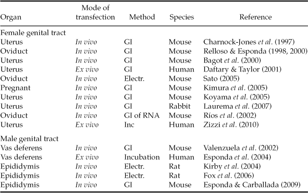
Electr: electroporation; GI: gene injection; Inc: incubation.
Gene transfer experiments have also been undertaken in the pregnant mouse uterus (Bagot et al., Reference Bagot, Troy and Taylor2000; Kimura et al., Reference Kimura, Nakamura, Koyama, Ogita, Tabata, Tsutsui, Shimoya, Koyama, Kaneda and Murata2005; Koyama et al., Reference Koyama, Kimura, Ogita, Nakamura, Tabata, Abu Haddi Noor, Temma-Asano, Shimoya, Tsutsui, Koyama, Kaneda and Murata2006). These experiments showed that the uterine mucosa can be successfully transfected by applying the hemagglutinating virus of Japan envelope vector instead of plasmids driven by a cytomegalovirus promoter (Nakamura et al., 2003). The use of this vector is 120-fold more efficient than cationic liposomes, and this transfection does not produce changes in the pregnancy rate, litter size, fetal growth or parturition (Nakamura et al., 2003). Other experiments were conducted using in vivo transfections in the mouse uterus to analyse the function of the homeobox gene HOXA10 (Daftary & Taylor, Reference Daftary and Taylor2003). Experiments using HOXA10 gene constructs and HOXA10 antisense sequences have revealed interesting details about the role of this gene during implantation in the mouse (Bagot et al., Reference Bagot, Troy and Taylor2000; Daftary & Taylor, Reference Daftary and Taylor2003).
In vivo transfections have also been performed in the oviduct (Relloso & Esponda Reference Relloso and Esponda1998, Reference Relloso and Esponda2000) (Fig. 2; Table 1). A method similar to that described for uterine transfection was employed to transfect gene constructs expressing β-galactosidase or GFP complexed with cationic liposomes. The epithelium was transfected, and the highest transgene expression occurred in the isthmus and juncture regions. A maximum of 6% of the cells were transfected, and the expression of the transgene lasted for 7 days (Relloso & Esponda, Reference Relloso and Esponda2000). Using an in vivo electroporation procedure, the entire oviduct of fertilized mouse females has been transfected with GFP, and the oviduct epithelium expressed the transgene (Sato, Reference Sato2005). This experiment showed that embryos contained in the oviduct also expressed the GFP. Thus, these results could help us better understand the physiology and development of preimplantation embryos (Sato, Reference Sato2005). On the other hand, in vivo transfections using mRNA have been done in the rat oviduct (Rios et al., Reference Rios, Venegas and Croxatto2002). These experiments showed that the translation of the exogenous mRNA did occur after the transfection of an estradiol-inducible mRNA or β-galactosidase mRNA.
In all cases, for both the uterus and oviduct, different molecular procedures have been used to better detect the presence of the transgene or assess transgene expression in the transfected tissue. PCR, RT-PCR, Southern blot, gene sequence and other methods have been applied, and the results of these experiments have supported the utility and feasibility of transfection (Fig. 3).
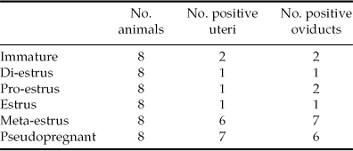
An important observation was that the efficiency of transfection in the uterus and oviduct is directly related to the cycle of uterine epithelial development, which is directly associated with the hormonal stage of the animal (Relloso & Esponda, Reference Relloso and Esponda2000). Only a few positive transfections occurred in immature, di-estrus, pro-estrus and estrus groups, whereas the highest numbers were observed during the meta-estrus and pseudopregnancy stages. During the latter two stages, the epithelial conditions are very similar, and there are high concentrations of progesterone and lower concentrations of estradiol (Relloso & Esponda, Reference Relloso and Esponda2000) (Table 2). Recently, studies of transfection in the rabbit uterus have shown that the transduction pattern of uterine tissues varies significantly with the reproductive cycle (Laurema et al., Reference Laurema, Lumme, Heinonen, Heinonen and Yla-Hertuala2007).
Influence of hormonal stage on β-galactosidase expression in the mouse uterus and oviduct
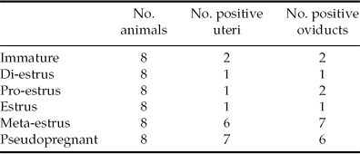
Reproduced from Relloso & Esponda (Reference Relloso and Esponda2000).
It should be noted that the transfection efficiency of the uterine glands and oviductal epithelium do not seem to be solely a consequence of the hormonal cycle, and apparently, the efficiency is also likely related to other cellular factors. During meta-estrus and pseudopregnancy, the uterine epithelium is well developed and the glands have their largest diameters. On the contrary, in young animals or during di-estrus, the epithelium is inactive. During pro-estrus and estrus, the epithelium is actively growing (Brenner & Maslar, Reference Brenner, Maslar, Knobil and Neil1988). The relative volume of mitochondria and the smooth and rough endoplasmic reticulum diameters are also larger during metaestrus. Alternatively, in stages correlated with low transfection efficiency, such as diestrus, the epithelium is thin and inactive (Eddy & Pauerstein, Reference Eddy and Pauerstein1980; Uhrin, Reference Uhrin1984). Secretory activity seems to be another important factor in the modulation of transfection during metaestrus and pseudopregnancy because at those times, this activity is low in both the uterus and oviduct. Alternatively, in other stages associated with low transfection efficiency (estrus, proestrus), epithelial cells are highly secretory (Brenner & Maslar, Reference Brenner, Maslar, Knobil and Neil1988). On the other hand, the deciliation phenomenon that occurs in the oviductal epithelium during metaestrus (Eddy & Pauerstein, Reference Eddy and Pauerstein1980) could facilitate the entrance of DNA–liposome complexes and concurrently increase transfection efficiency.
Gene transfer to the male genital tract
In the testis, sophisticated procedures using spermatogonial transplantation have been employed to collect transfected spermatozoa (Brinster & Avarbock, Reference Brinster and Avarbock1994, Yonezawa et al., Reference Yonezawa, Furuhata, Hirabayashi, Susuki, Takahayashi and Nishihara2001). In the male tract, experiments employing in vitro gene transfer have been scarce because epididymal cultured cell lines have only recently been available (Kirby et al., Reference Kirby, Yang, Labus, Lye, Hsia, Day, Cornwall and Hinton2004). Several epididymal cell lines have been used for in vitro transfections to analyse the expression of different genes and/or the roles of some proteins (Kirchoff et al., Reference Kirchhoff, Araki, Huhtaniemi, Matusik, Osterhoff, Poutanen, Samalecos, Sipila, Susuki and Orgebin-Crist2004; Dufresne et al., 2006; Yu et al., Reference Yu, Suzuki, Wang, Gupta, Jin, Orgebin-Crist and Matusik2006).
Human cultures of vas deferens cells have been collected from vasectomies and then transfected ex vivo using GFP as a reporter gene and liposomes or neutral lipids as gene vectors (Esponda et al., Reference Esponda, Goldstein and Witkin2004). GFP gene expression appeared exclusively in the vas epithelium and occupied 9–10% of the epithelial area (Figs. 5 and 6).
The mouse vas deferens was transfected in vivo using GFP or β-galactosidase complexed with liposomes (Valenzuela et al., Reference Valenzuela, Relloso and Esponda2002). Foreign gene expression occurred in 13.3% of cells after transfection with the GFP gene construct and persisted for three months in some areas of the vas. Modified cells appeared as patches along the epithelium. Furthermore, two gene constructs that express secretory proteins have been used to transfect the vas. These constructs were pSEAP–control, which expresses a secretory form of alkaline phosphatase, and pGFP–Ctk-37, which expresses a secreted form of GFP. In both cases, the vas secretions of the treated animals showed a marked increase of both proteins (Valenzuela et al., Reference Valenzuela, Relloso and Esponda2002).
The rat epididymis was recently transfected in vivo using an electroporation procedure to analyse the expression and function of some genes in the initial segment of the epididymis (Kirby et al., Reference Kirby, Yang, Labus, Lye, Hsia, Day, Cornwall and Hinton2004). In a mouse model, DNA–lipid complexes have also been used to transfect the cauda epididymis (Esponda & Carballada, Reference Esponda and Carballada2009). After a microinjection of the cauda tubules with the GFP gene construct, 39.7% of transfected cells expressed the transgene, accounting for 32.24% of the epithelial area. Transgene expression was observed for a maximum of 15 days after gene injection (Fig. 7).
The pSEAP-control (secreted alkaline phosphatase) gene construct was also employed to transfect the mouse cauda epididymis. A significant increase of the enzyme appeared in the fluid secreted by the cauda and in the cytosol of epithelial cells 7 days after gene injection (Esponda & Carballada, Reference Esponda and Carballada2009)
Conclusions
We can conclude the following: (i) The reported results demonstrate that the male and female mammalian reproductive tracts can be successfully transfected using different in vitro and in vivo methods. Furthermore, different gene constructs, reporter genes and procedures for transfection (gene injection, electroporation) can be employed to achieve transfection. (ii) We can also conclude that in vivo or ex vivo transfection induces good transgene expression in the epithelial region of the tracts. This result has been commonly observed when transfections were performed in other organs. (iii) In vitro transfection has been undertaken in different uterine cell lines and, more recently, in epididymal cell lines. It is important to evaluate the effectiveness of this approach in other uterine cancer cell lines and to understand gene expression and the function of other proteins. (iv) Considerable attention has been given to gene transfer in the female tract, particularly the uterus. This fact is obviously related to the prevalence of important pathologies that occur in the human uterus, and the uterus is currently considered a possible organ for gene therapies. (v) Transfection efficiency is closely related to the mitotic index and to the hormonal stage of the epithelium. The female tract is transfected most efficiently during the pseudopregnant or meta-estrus stages, which coincide with high progesterone and lower estradiol concentrations. The male tract seems to be more suitable for gene transfection because it is not subject to drastic cyclical hormonal changes. Furthermore, the male epithelium does not show a high mitotic index, does not have intermittent periods of variable secretory activity and inactivity and is not constantly renewed, as in the female tract due to the estrus cycle. (vi) In the male, it is feasible to modify the characteristics of secreted proteins. Transfection methods promote the expression of new proteins or modify those normally secreted by the epithelium. Therefore, the methods described herein could contribute to the development of useful tools to further investigate the control of the fertilizing ability of spermatozoa. Similar procedures could be applied to the female tract to modify uterine or oviduct secretions.
Perspectives
Gene transfer procedures applied to the male and female genital tracts will be employed with two principal purposes. One will be to better understand important details of the physiology of these tracts, including gene expression and protein functions in different stages, ages and pathological situations. The use of different genes, RNAs, siRNAs and overexpressed sense or antisense DNAs will allow for the study of unknown details of the reproductive tracts. In addition, these procedures can also be used as gene therapies. Today, gene therapies are a promising goal for biomedical sciences (Verma & Weitzman, Reference Verma and Weitzman2005). The use of transfection as a gene therapy modality in the reproductive tract could treat different human fertility problems. In the past, gene therapy was thought of as a possibility of the distant future; however, based on promising recent findings, the feasible use of gene therapy is closer than ever (Subramanian & Simon, Reference Subramanian and Simon2010).
It is important to recognize that gene therapy procedures induce several problems in cells and tissues, including the stimulation of immune responses that could lead to only transient expression of the transgene or inefficient re-administration of the vector (Zhou et al., Reference Zhou, Liu and Liang2004). On the other hand, the random integration of transgenes into the host genome can induce several important risks, such as oncogene activation and tumor suppressor gene inactivation (Hackett et al., Reference Hackett, Geurts and Hackett2007). The solution to these problems is to select a preferential site for the integration of the transgene. However, none of the vectors currently employed in preclinical or clinical trials prevents these hazardous side effects (Voigt et al., Reference Voigt, Izvák and Ivics2008). For this reason, strategies for targeted gene delivery are currently being developed (Sinn et al., Reference Sinn, Sauter and McCray2005; Daniel & Smith, Reference Daniel and Smith2008). The future use of these strategies and the development of new gene vectors could improve the efficiency and safety of gene therapies.
Acknowledgements
This work was partially supported by Grant BFU2008–02947-CO2–02/BMC from DGICYT (Spain).