It is well appreciated that there are defined windows in early life when neural circuitry can be robustly restructured in response to experience. These time-limited critical periods have been demonstrated for many brain functions across many brain regions and are thought to allow developing neural circuits to establish an individualized, optimal neural representation of a highly variable environment. The enhanced plasticity corresponds to the peak phases of physical growth and may, therefore, allow for constant perception during expansion of the body surface. For example, visual receptive fields must repeatedly remap as the distance between the two eyes increases. Indeed, experience-dependent matching of stimulus selectivity of the visual input from the two eyes occurs during the critical period (Wang et al., Reference Wang, Sarnaik and Cang2010).
The relative stability that follows the critical period may be advantageous in adult circuits, and also allow for conservation of energy/resources. However, the enhanced stability with age also inhibits large-scale adaptations to changes in the input during adulthood. Nonetheless the adult cortex retains the ability to express some forms of synaptic plasticity, but the mechanisms for the induction and expression of plasticity differ from those utilized during the critical period. In this context, it is important to bear in mind that many measures are in current use to study ‘ocular dominance (OD) plasticity.’ Originally defined as a change in the eye preference of the spiking output of V1 neurons (Wiesel & Hubel, Reference Wiesel and Hubel1963), current methods include visually evoked synaptic potentials, intrinsic hemodynamic signals, immediate early gene activation, thalamocortical axon or dendritic spine morphology and motility, and calcium responses in genetically identified cell types. Each of these methods yields different resolutions and may be variably sensitive to subthreshold inputs (Morishita & Hensch, Reference Morishita and Hensch2008), which are important considerations when informing therapies for recovery of the visual function in both amblyopic children and adults.
During the critical period, an asymmetry in the quality of the visual input across the two eyes leads to reduced visual acuity and a visually evoked spiking response through the affected eye with no obvious pathology in the eye, thalamus, or cortex. The severity of amblyopia depends on the age at initiation and the type of asymmetry, which can be caused by unequal alignment (strabismus), unequal refractive error (anisometropia), or form deprivation (e.g., cataract). The critical period for developing amblyopia in children extends to 8 years and is relatively easy to correct until that age by improving the quality of visual input in the affected eye (reviewed in Daw, Reference Daw1998; Mitchell & MacKinnon, Reference Mitchell and MacKinnon2002; Simons, Reference Simons2005) but becomes increasingly resistant to reversal with age.
In animal models, amblyopia is most often induced by monocular deprivation (MD)—an eyelid suture which significantly occludes the patterned visual input to one eye. Across various species, MD unleashes a sequence of functional and structural changes in V1 that shifts the OD of binocular neurons away from the deprived eye and toward the open eye, resulting in a reduction in deprived-eye acuity (Wiesel & Hubel, Reference Wiesel and Hubel1963, Reference Wiesel and Hubel1970; Olson & Freeman, Reference Olson and Freeman1975; Hubel et al., Reference Hubel, Wiesel and LeVay1977; Movshon & Dürsteler, Reference Movshon and Dürsteler1977; Blakemore et al., Reference Blakemore, Garey and Vital-Durand1978; LeVay et al., Reference LeVay, Stryker and Shatz1978; Shatz & Stryker, Reference Shatz and Stryker1978; Antonini & Stryker, Reference Antonini and Stryker1993; Fagiolini et al., Reference Fagiolini, Pizzorusso, Berardi, Domenici and Maffei1994; Gordon & Stryker, Reference Gordon and Stryker1996; Hensch et al., Reference Hensch, Fagiolini, Mataga, Stryker, Baekkeskov and Kash1998; Trachtenberg & Stryker, Reference Trachtenberg and Stryker2001; Mataga et al., Reference Mataga, Nagai and Hensch2002; Taha & Stryker, Reference Taha and Stryker2002; Prusky & Douglas, Reference Prusky and Douglas2003; Frenkel & Bear, Reference Frenkel and Bear2004; Sato & Stryker, Reference Sato and Stryker2008).
While OD plasticity peaks during the postnatal critical period, it (Lehmann and Lowel, Reference Lehmann and Löwel2008) persists beyond sexual maturity at some level in many species, including rats, mice and cats. The age at initiation and duration of MD then strongly impacts the severity of the subsequent amblyopia, as well as the potential for recovery (Liao et al., Reference Liao, Krahe, Prusky, Medina and Ramoa2004; Eaton et al., Reference Eaton, Sheehan and Quinlan2016). Accordingly, short durations of MD within the critical period induce a shift in OD and a reduction in the deprived eye visual acuity that are rapidly corrected by restoring normal vision (Schwarzkopf et al., Reference Schwarzkopf, Vorobyov, Mitchell and Sengpiel2007). In contrast, long-term MD initiated early and persisting through the end of the critical period induces a shift in OD and a loss of visual acuity that is highly resistant to reversal.
The initial response to MD during the critical period is a reduction in functional strength and selectivity of the deprived eye visual responses (Gordon & Stryker, Reference Gordon and Stryker1996; Hensch et al., Reference Hensch, Fagiolini, Mataga, Stryker, Baekkeskov and Kash1998; Trachtenberg et al., Reference Trachtenberg, Trepel and Stryker2000; Frenkel & Bear, Reference Frenkel and Bear2004) and disrupts the balance of excitatory ad inhibitory inputs to individual binocular neurons (Saiepour et al., Reference Saiepour, Rajendran, Omrani, Ma, Tao, Heimel and Levelt2015). Depression of deprived-eye responses may occur by synaptic depression at both thalamocortical and intracortical connections. Notably, rapid shifts in the visual response of parvalbumin (PV)-expressing inhibitory interneurons may enable these first functional changes within V1 (Yazaki-Sugiyama et al., Reference Yazaki-Sugiyama, Kang, Cåteau, Fukai and Hensch2009; Aton, Broussard et al., Reference Aton, Broussard, Dumoulin, Seibt, Watson, Coleman and Frank2013; Kuhlman et al., Reference Kuhlman, Olivas, Tring, Ikrar, Xu and Trachtenberg2013). Depression is then followed by a relatively slow, strengthening of open eye responses (Sawtell et al., Reference Sawtell, Frenkel, Philpot, Nakazawa, Tonegawa and Bear2003; Frenkel & Bear, Reference Frenkel and Bear2004; Kaneko et al., Reference Kaneko, Stellwagen, Malenka and Stryker2008).
Robust morphological plasticity is also induced by MD during the critical period. An initial degradation of the extracellular matrix by the upregulation of proteases occurs within the first 2 days after MD in the mouse and may elevate spine motility (Mataga et al., Reference Mataga, Mizaguchi and Hensch2004; Oray et al., Reference Oray, Majewska and Sur2004). Studies in cats, monkeys, and humans suggest that structural plasticity is facilitated by a reduction in the neurofilament-light protein within V1, which may de-stabilize the cytoskeleton and promote plasticity (Duffy & Livingstone, Reference Duffy and Livingstone2005; Duffy et al., Reference Duffy, Murphy, Frosch and Livingstone2007; Duffy & Mitchell, Reference Duffy and Mitchell2013). Brief MD during the critical period alters the spine density on pyramidal neurons (Mataga et al., Reference Mataga, Mizaguchi and Hensch2004; Tropea et al., Reference Tropea, Majewska, Garcia and Sur2010; Yu et al., Reference Yu, Majewska and Sur2011; Djurisic et al., Reference Djurisic, Vidal, Mann, Aharon, Kim, Ferrao Santos, Zuo, Hubener and Shatz2013) and induces a transient decrease in the density of synapses formed by thalamocortical axons originating from the lateral geniculate nucleus (LGN) (Coleman et al., Reference Coleman, Nahmani, Gavornik, Haslinger, Heynen, Erisir and Bear2010). Long-term MD yields enduring alterations in the length and extent of thalamocortical arbors serving the two eyes (Hubel et al., Reference Hubel, Wiesel and LeVay1977; Shatz & Stryker, Reference Shatz and Stryker1978; Antonini et al., Reference Antonini, Fagiolini and Stryker1999) and a significant reduction in dendritic spine density (Montey & Quinlan, Reference Montey and Quinlan2011).
Studies from humans and nonhuman primates suggest a protracted decline in visual plasticity that extends into adulthood rather than an abrupt closure of the critical period. However, the residual plasticity that persists in the adult visual cortex appears to differ from the plasticity during the critical period in several important ways: (1) the shift in OD in adults is slower and smaller and may require a longer duration of deprivation to engage; (2) it may not require depression of deprived eye responses for the subsequent strengthening of responses to the nondeprived eye; (3) it may be restricted to synapses in the supragranular and infragranular lamina, as plasticity in layer 4 has been shown to be constrained early in postnatal development; (4) it may be restricted by saturated synapses, setting limits on the amount of recovery of visual function that can be accomplished using this pathway. Additionally, MD in adults does not elicit the robust structural alterations that accompany OD plasticity during the critical period such as increased spine motility and pruning (Mataga et al., Reference Mataga, Mizaguchi and Hensch2004; Oray et al., Reference Oray, Majewska and Sur2004; Lee et al., Reference Lee, Huang, Feng, Sanes, Brown, So and Nedivi2006). Indeed, a general decline in structural plasticity is one of the hallmarks of the termination of the critical period. However, residual increases in the rate of formation and stability of dendritic spines may persist in the adult layer I after MD (Hofer et al., Reference Hofer, Mrsic-Flogel, Bonhoeffer and Hübener2009).
Inhibition and critical period induction
Powerful new tools in neuroscience, especially the molecular genetic control available in mice, are beginning to elucidate the cellular and molecular mechanisms that initiate and terminate critical periods. Ocular dominance plasticity peaks during the third postnatal week in rodents, demonstrating that elevated plasticity is not the initial state of immature circuits. Indeed, the maturation of specific inhibitory circuitry is necessary to initiate the critical period, which can be accelerated by activating inhibitory GABAA receptors with allosteric modulators such as benzodiazepines (Hensch et al., Reference Hensch, Fagiolini, Mataga, Stryker, Baekkeskov and Kash1998; Fagiolini & Hensch, Reference Fagiolini and Hensch2000; Iwai et al., Reference Iwai, Fagiolini, Obata and Hensch2003; Fagiolini et al., Reference Fagiolini, Fritschy, Löw, Möhler, Rudolph and Hensch2004). Promoting early maturation of a specific class of inhibitory interneurons that express the calcium binding protein parvalbumin (PV) by increasing the levels of growth factors (Huang et al., Reference Huang, Kirkwood, Pizzorusso, Porcialtti, Morales, Bear and Maffei1999; Hanover et al., Reference Hanover, Huang, Tonegawa and Stryker1999; Otx2: Sugiyama et al., Reference Sugiyama, Di Nardo, Aizawa, Matsuo, Volovitch, Prochiantz and Hensch2008; Spatazza et al., Reference Spatazza, Lee, Di Nardo, Tibaldi, Joliot, Hensch and Prochiantz2013) or by removing cell adhesion (PSA: Di Cristo et al., Reference Di Cristo, Chattopadhyaya, Kuhlman, Fu, Bélanger, Wu, Rutishauser, Maffei and Huang2007) or DNA binding proteins (MeCP2: Durand et al., Reference Durand, Patrizi, Quast, Hachigian, Pavlyuk, Saxena, Carninci, Hensch and Fagiolini2012; Krishnan et al., Reference Krishnan, Wang, Lu, Wang, Maffei, Cang and Huang2015) can induce premature initiation of the critical period.
The perisomatic inhibition mediated by these fast-spiking PV interneurons exerts powerful control over the excitability and plasticity of downstream pyramidal neurons, regulating many forms of downstream synaptic plasticity (Katagiri et al., Reference Katagiri, Fagiolini and Hensch2007; Kuhlman et al., Reference Kuhlman, Olivas, Tring, Ikrar, Xu and Trachtenberg2013; Toyoizumi et al., Reference Toyoizumi, Miyamoto, Yazaki-Sugiyama, Atapour, Hensch and Miller2013). Several proteins that regulate synaptic strength and/or number are highly enriched at excitatory synapses onto PV interneurons and impact the timing of the critical period and NRG1 (NARP: Chang et al., Reference Chang, Park, Pelkey, Grabenstatter, Xu, Linden, Sutula, McBain and Worley2010; Gu et al., Reference Gu, Huang, Chang, Worley, Kirkwood and Quinlan2013, Pelkey et al., Reference Pelkey, Barksdale, Craig, Yuan, Sukumaran, Vargish, Mitchell, Wyeth, Petralia, Chittajallu, Karlsson, Cameron, Murata, Colonnese, Worley and McBain2015; Gu et al., Reference Gu, Tran, Murase, Borrell, Kirkwood and Quinlan2016; kaplan et al., Reference Kaplan, Cooke, Komorowski, Chubykin, Thomazeau, Khibnik, Gavornik and Bear2016; Sun et al., Reference Sun, Ikrar, Davis, Gong, Zheng, Luo, Lai, Mei, Holmes, Gandhi and Xu2016). Accordingly, NARP-deficient mice fail to initiate a critical period unless rescued by enhancing the strength of the inhibitory output or excitatory drive onto PV interneurons (Gu et al., Reference Gu, Huang, Chang, Worley, Kirkwood and Quinlan2013; Gu et al., Reference Gu, Tran, Murase, Borrell, Kirkwood and Quinlan2016).
A further increase in perisomatic inhibition is thought to terminate the critical period. Hence, the critical period can be reopened in adulthood by pharmacological reduction of inhibition (Harazouv et al., 2010) or by the knockdown of Otx2 (Beurdeley et al., Reference Beurdeley, Spatazza, Lee, Sugiyama, Bernard, Di Nardo, Hensch and Prochiantz2012; Spatazza et al., Reference Spatazza, Lee, Di Nardo, Tibaldi, Joliot, Hensch and Prochiantz2013). Treatment with an NRG1 peptide induces a precocious termination of the critical period, while inhibition of the activity of the NRG receptor (ErbB) reactivates the critical period in adults (Gu et al., Reference Gu, Tran, Murase, Borrell, Kirkwood and Quinlan2016). Indeed, a developmental reduction of plasticity at excitatory synapses onto FS interneurons may explain the requirement for longer durations of MD with age (Kameyama et al., Reference Kameyama, Sohya, Ebina, Fukuda, Yanagawa and Tsumoto2010). Together, these studies indicate that PV inhibitory neurons exert bidirectional control over OD plasticity (van Versendaal & Levelt, Reference van Versendaal and Levelt2016).
Other classes of inhibitory neurons may influence the expression of plasticity, either independently or through the regulation of PV neurons. Interestingly, inhibitory neurons in layer 1 (L1) of the visual cortex and those expressing vasoactive intestinal peptide (VIP) are strongly activated during certain behavioral states and exert cortical effects by disinhibition of pyramidal neurons (Letzkus et al., Reference Letzkus, Wolff, Meyer, Tovote, Courtin, Herry and Lüthi2011; Donato et al., Reference Donato, Rompani and Caroni2013; Pfeffer et al., Reference Pfeffer, Xue, He, Huang and Scanziani2013; Pi et al., Reference Pi, Hangya, Kvitsiani, Sanders, Huang and Kepecs2013; Fu et al., Reference Fu, Kaneko, Tang, Alvarez-Buylla and Stryker2015). Locomotion activates VIP interneurons, which enhances neural activity in V1 (Niell & Stryker, Reference Niell and Stryker2010) and promotes adult plasticity by increasing inhibition onto other interneuron subtypes that target pyramidal neurons (Fu et al., Reference Fu, Tucciarone, Espinosa, Sheng, Daracy, Nicoll, Huang and Stryker2014; Fu et al., Reference Fu, Kaneko, Tang, Alvarez-Buylla and Stryker2015). Similarly, reinforcement signals (reward and punishment) during the performance of an auditory discrimination task activate VIP neurons in the auditory cortex, which increase the gain of a functional subpopulation of pyramidal neurons by disinhibition (Pi et al., Reference Pi, Hangya, Kvitsiani, Sanders, Huang and Kepecs2013). Thus, disinhibitory circuits which transiently suppress other inhibitory interneurons may be a general mechanism for enabling plasticity in the adult cortex.
Molecular reactivation of critical period in adulthood
Increasing evidence demonstrates that removing molecular ‘brakes’ in adulthood can enhance plasticity and promote recovery from amblyopia. For example, epigenetic mechanisms, such as histone deacetylase (HDAC) activity, may down regulate the expression of genes that promote plasticity over development. Accordingly, HDAC inhibition enhances plasticity in adult V1, allowing for recovery from amblyopia (Putignano et al., Reference Putignano, Lonetti, Cancedda, Ratto, Costa, Maffei and Pizzorusso2007; Silingardi et al., Reference Silingardi, Scali, Belluomini and Pizzorusso2010; Baroncelli et al., Reference Baroncelli, Scali, Sansevero, Olimpico, Manno, Costa and Sale2016). However, the downstream targets of histone acetylation at specific stages of development remain to be identified.
Alternatively, increased expression of specific genes over development can actively limit rewiring. The expression of Lynx1, an endogenous inhibitor of nicotinic acetylcholine receptors, emerges in V1 coincident with critical period closure, which would dampen the neuromodulatory actions of acetylcholine (Miwa et al., Reference Miwa, Ibanez-Tallon, Crabtree, Sanchez, Sali, Role and Heintz1999; Morishita et al., Reference Morishita, Miwa, Heintz and Hensch2010). Both genetic deletion of lynx1 and administration of acetylcholinesterase inhibitors enhance spine motility and the morphological plasticity induced by MD (Sajo et al., Reference Sajo, Ellis-Davies and Morishita2016) and enable recovery of visual acuity following MD throughout life (Morishita et al., Reference Morishita, Miwa, Heintz and Hensch2010). The major histocompatibility complex class I (MHCI) receptor, PirB, is another molecular brake. Disruption of PirB signaling enhances OD plasticity throughout life and facilitates recovery from amblyopia in adults (Syken et al., Reference Syken, Grandpre, Kanold and Shatz2006; Bochner et al., Reference Bochner, Sapp, Adelson, Zhang, Lee, Djurisi, Syken, Dan and Shatz2014). Another immune system molecule, Stat1, restricts the increase of open eye responses following monocular deprivation, while genetic deletion enhances this component of plasticity (Nagakura et al., Reference Nagakura, Van Wart, Petravicz, Tropea and Sur2014). The identification of specific molecules that actively suppress plasticity in the adult visual cortex may inform strategies for pharmacological interventions to reopen the critical period.
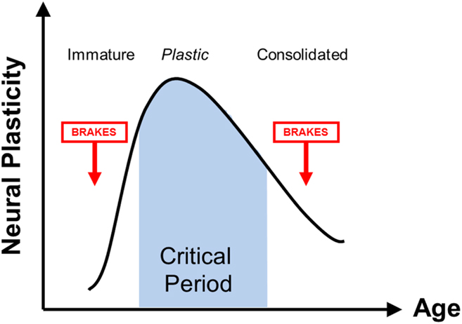
Molecular brakes can also present physical barriers to morphological plasticity. Perineuronal nets are highly enriched around PV neurons and reach maturity at the end of the critical period. Disrupting the molecular latticework of this extracellular matrix (Pizzorusso et al., Reference Pizzorusso, Medini, Berardi, Chierzi, Fawcett and Maffei2002; Pizzorusso et al., Reference Pizzorusso, Medini, Landi, Baldini, Berardi and Maffei2006; Carulli et al., Reference Carulli, Pizzorusso, Kwok, Putignano, Poli, Forostyak, Andrews, Deepa, Glant and Fawcett2010) or the molecules which bind to it (i.e., Otx2: Beurdeley et al., Reference Beurdeley, Spatazza, Lee, Sugiyama, Bernard, Di Nardo, Hensch and Prochiantz2012) enables OD plasticity and recovery from amblyopia in adults. Consistent with this, mice lacking the Nogo receptor (Ngr1), a bimodal receptor for chondroitin sulfate proteoglycans and myelin-derived inhibitory factors (Dickendesher et al., Reference Dickendesher, Baldwin, Mironova, Koriyama, Raiker, Askew, Wood, Geoffroy, Zheng, Liepmann, Katagiri, Benowitz, Geller and Giger2012), also retain critical period plasticity into adulthood and spontaneously recover visual acuity following long-term MD (McGee et al., Reference McGee, Yang, Fischer, Daw and Strittmatter2005; Stephany et al., Reference Stephany, Chan, Parivash, Dorton, Piechowicz, Qiu and McGee2014). Interestingly, PirB may act in concert with Ngr1 (Atwal et al., Reference Atwal, Pinkston-Gosse, Syken, Stawicki, Wu, Shatz and Tessier-Lavigne2008) to dampen the morphological plasticity of dendritic spines on layer 5 pyramidal neurons in adults (Bochner et al., Reference Bochner, Sapp, Adelson, Zhang, Lee, Djurisi, Syken, Dan and Shatz2014) (Fig. 1).
Critical period plasticity as a function of age. Initially, immature brain circuits are dominated by excitatory inputs and fail to express plasticity. As inhibitory circuits mature, a highly plastic critical period is induced. Plasticity then declines with age as inhibitory circuits and brake-like factors dominate, harboring the potential for plasticity throughout life. Dynamic changes in the excitatory/inhibitory balance across age are shown below the graph. Figure courtesy of Takao Hensch (Harvard University).

Interestingly, one recently discovered molecular brake may lie within the dendritic spine itself. Postsynaptic density protein 95 (PSD-95), an intracellular scaffold highly enriched at excitatory synapses, is thought to accelerate maturation of excitatory synapses. PSD-95 promotes the incorporation of AMPA-type glutamate receptors into synapses containing only NMDA receptors, which are normally functionally “silent” at the resting membrane potential. In contrast, the immediate early gene Arc promotes removal of AMPA receptors from cortical synapses and deletion precludes visual plasticity (McCurry et al., Reference McCurry, Shepherd, Tropea, Wang, Bear and Sur2010). Genetic reduction of PSD-95 in adulthood increases the number of silent synapses and reactivates the juvenile form of OD plasticity, characterized by a rapid and robust deprived-eye depression (Huang et al., Reference Huang, Stodieck, Goetze, Cui, Wong, Wenzel, Hosand, Dong, Löwel and Schlüter2015). Notably, no changes in GABAergic or NMDA receptor currents are observed, suggesting that the reactivation of plasticity by PSD-95 deletion lies downstream of the regulation of inhibitory circuitry. A conversion of ‘silent’ to functional synapses has been proposed as a general mechanism to constrain plasticity across brain regions (Greifzu et al., Reference Greifzu, Pielecka-Fortuna, Kalogeraki, Kremplar, Favaro, Schlüter and Löwel2014; Huang et al., Reference Huang, Stodieck, Goetze, Cui, Wong, Wenzel, Hosand, Dong, Löwel and Schlüter2015).
Environmental reactivation of critical period in adulthood
Characteristics of the physical or sensory environment strongly impact the function and plasticity of cortical circuits. Remarkably, adding social, sensory, or motor enrichment to the typically impoverished environment of the laboratory rodent influences the expression and time course of OD plasticity. Robust OD plasticity persists into adulthood when mice are raised in large complex cages with multisensory and motor enrichment (Sale et al., Reference Sale, Maya Vetencourt, Medini, Cenni, Baroncelli, De Pasquale and Maffei2007; Scali et al., Reference Scali, Baroncelli, Cenni, Sale and Maffei2012; Greifzu et al., Reference Greifzu, Pielecka-Fortuna, Kalogeraki, Kremplar, Favaro, Schlüter and Löwel2014). In fact, enriched rearing may better reflect the sensorimotor environment of primates including humans. At a molecular level, exposure to enriched environments in adulthood increases H3 acetylation (Baroncelli et al., Reference Baroncelli, Scali, Sansevero, Olimpico, Manno, Costa and Sale2016), reduces the expression of PV and GAD67 within inhibitory neurons of the visual cortex, weakens GABA signaling, and fosters plasticity in both the cortex and hippocampus (Sale et al., Reference Sale, Maya Vetencourt, Medini, Cenni, Baroncelli, De Pasquale and Maffei2007; Donato et al., Reference Donato, Rompani and Caroni2013; Greifzu et al., Reference Greifzu, Pielecka-Fortuna, Kalogeraki, Kremplar, Favaro, Schlüter and Löwel2014).
In this regard, it is intriguing that total visual deprivation also reactivates robust plasticity in adult V1 and promotes recovery from chronic MD (He et al., Reference He, Ray, Dennis and Quinlan2007; Montey & Quinlan, Reference Montey and Quinlan2011; Duffy & Mitchell, Reference Duffy and Mitchell2013; Stodieck et al., Reference Stodieck, Greifzu, Goetze, Schmidt and Löwel2014; Eaton et al., Reference Eaton, Sheehan and Quinlan2016; Mitchell et al., Reference Mitchell, MacNeill, Crowder, Holman and Duffy2016). The BCM theory of a sliding synaptic modification threshold forecasted that dark exposure would enhance plasticity and indeed several mechanisms are engaged by the dark exposure that are predicted to lower the threshold for synaptic plasticity in pyramidal neurons (Cooper & Bear, Reference Cooper and Bear2012). For example, the composition of the NMDA type glutamate receptors is reset to a “juvenile” form (containing the NR2B subunit; Quinlan et al., Reference Quinlan, Philpot, Huganir and Bear1999) which exhibit enhanced temporal summation (Yashiro et al., Reference Yashiro, Corlew and Philpot2005; He et al., Reference He, Hodos and Quinlan2006). In addition, forms of synaptic plasticity typically limited to juveniles are re-expressed (Huang et al., Reference Huang, Gu, Quinlan and Kirkwood2010; Montey & Quinlan, Reference Montey and Quinlan2011), spines on pyramidal neurons are shifted toward an immature structure and dynamics (Tropea et al., Reference Tropea, Majewska, Garcia and Sur2010), and immature excitatory synapses on pyramidal neurons are strengthened, thereby increasing excitability and expanding the integration window for spike-timing dependent plasticity (Goel & Lee, Reference Goel and Lee2007; He et al., Reference He, Ray, Dennis and Quinlan2007; Guo et al., Reference Guo, Huang, de Pasquale, McGehrin, Lee, Zhao and Kirkwood2012).
In contrast, dark exposure decreases the excitability of PV interneurons, and the reactivated plasticity can be reversed by increasing the strength of their excitatory synaptic inputs (Gu et al., Reference Gu, Tran, Murase, Borrell, Kirkwood and Quinlan2016). A loss of specific neurofilament protein associated with cytoskeletal stability is observed in the LGN following dark exposure, which may further contribute to the reactivation of structural OD plasticity (O’Leary et al., Reference O’Leary, Kutcher, Mitchell and Duffy2012; Duffy et al., Reference Duffy, Lingley, Holman and Mitchell2016). Importantly, dark exposure restores a period of susceptibility to MD and also promotes the recovery of visual function in adults with amblyopia, as has been demonstrated in rats, mice, and cats (He et al., Reference He, Hodos and Quinlan2006; He et al., Reference He, Ray, Dennis and Quinlan2007; Duffy & Mitchell, Reference Duffy and Mitchell2013; Stodieck et al., Reference Stodieck, Greifzu, Goetze, Schmidt and Löwel2014; Duffy et al., Reference Duffy, Lingley, Holman and Mitchell2016). The reactivation of plasticity by dark exposure has also been shown to strengthen the thalamic input to the cortex (Montey & Quinlan, Reference Montey and Quinlan2011). Thus, the seemingly opposite interventions of environmental enrichment and dark exposure may both enhance cellular plasticity by the removal of functional and structural constraints that normally accumulate over development to stabilize the V1 circuitry.
It is important to note that dark exposure alone does not impact visual acuity or neuronal stimulus selectivity, which is regained only after repetitive visual experience (Montey et al., Reference Montey, Eaton and Quinlan2013; Eaton et al., Reference Eaton, Sheehan and Quinlan2016). Likewise, enrichment or locomotion alone does not strengthen visual performance (Kaneko & Stryker, Reference Kaneko and Stryker2014; Greifzu et al., Reference Greifzu, Kalogeraki and Löwel2016). This suggests that the recovery from amblyopia in adulthood is a two-stage process that requires (1) the reactivation of plasticity in the adult amblyopic cortex (permissive step) and (2) focused visual experience to stimulate perceptual learning (instructive step). One of the challenges, therefore, is to identify the optimal visual stimulation to drive recovery of the function, which may differ based on age and depth of amblyopia (Montey & Quinlan, Reference Montey and Quinlan2011; Eaton et al., Reference Eaton, Sheehan and Quinlan2016). However, prolonged plasticity by environmental enrichment makes it unclear if complex environments better mimic those of primates including humans. At a minimum, these experiments provides a valuable condition with which to better understand the biological basis of critical period closure.
Reactivating plasticity to enhance recovery
The reactivation of plasticity in the primary visual cortex has revised the idea that critical periods are strictly limited to early postnatal development (Bavelier et al., Reference Bavelier, Levi, Li, Dan and Hensch2010; Takesian & Hensch, Reference Takesian and Hensch2013; Sengpiel, Reference Sengpiel2014). However, the mechanisms that engage the cortical plasticity necessary to treat amblyopia may be very different from the plasticity that enables the cortex to regain sensitivity to MD. Indeed, it has long been known that the critical period for plasticity in response to MD differs from the critical period for recovery of the binocularity and orientation selectivity by removing the MD (Liao et al., Reference Liao, Krahe, Prusky, Medina and Ramoa2004). Although initially assumed to overlap with the critical period for susceptibility to amblyopia, it is now clear that the treatment window for the reversal of amblyopia in humans may extend beyond early life (reviewed in Daw, Reference Daw1998). It is, therefore, important that key molecular effectors be tested in their ability to recover (not induce) amblyopia in adults.
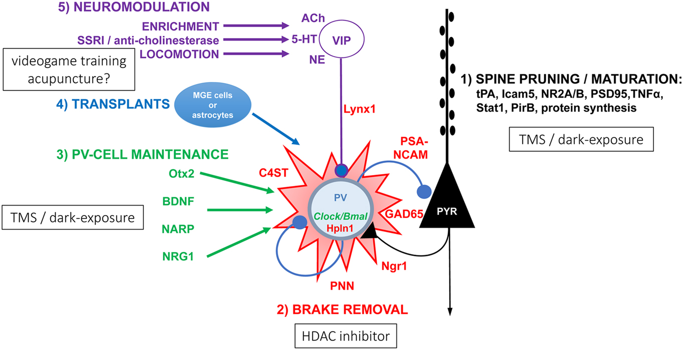
Mechanistic studies performed primarily in mice, have identified novel therapies with translational potential to reverse the developmental constraints on recovery from amblyopia. Several commonly prescribed drugs, such as cholinesterase inhibitors (Morishita et al., Reference Morishita, Miwa, Heintz and Hensch2010), valproate (Gervain et al., Reference Gervain, Vines, Chen, Seo, Hensch, Werker and Young2013; Lennartsson et al., Reference Lennartsson, Arner, Fagiolini, Saxena, Andersson, Takahashi, Noro, Sng, Sandelin, Hensch and Carninci2015), or selective serotonin reuptake inhibitors (Maya Vetencourt et al., Reference Maya Vetencourt, Sale, Viegi, Baroncelli, De Pasquale, O’Leary, Castrén and Maffei2008), could be repurposed to reactivate plasticity in adult amblyopic patients. Interestingly, a reduction in PV interneuron function may be a mechanism common to several of these interventions. The SSRI antidepressant fluoxetine reduces the basal levels of extracellular GABA (Maya Vetencourt et al., Reference Maya Vetencourt, Sale, Viegi, Baroncelli, De Pasquale, O’Leary, Castrén and Maffei2008) and the number of PV interneurons surrounded by dense perineuronal nets (Guirado et al., Reference Guirado, Perez-Rando, Sanchez-Matarredona, Castrén and Nacher2014). Similarly, dark exposure may rejuvenate intracortical inhibition by reducing the excitatory drive onto PV neurons (Gu et al., Reference Gu, Tran, Murase, Borrell, Kirkwood and Quinlan2016) (Fig. 2).
Pathways known to enhance plasticity in the adult visual cortex.

Future work that explores noninvasive ways to tap into these mechanisms to trigger plasticity may generate novel amblyopia treatments for adults. It will also be important to learn if the success of behavioral manipulations, such as dark rearing, environmental enrichment and exercise, promotes the recovery from amblyopia in rodents by reversing molecular brakes or engaging residual plasticity mechanisms that are normally expressed in the adult cortex (Sale et al., Reference Sale, Maya Vetencourt, Medini, Cenni, Baroncelli, De Pasquale and Maffei2007; He et al., Reference He, Ray, Dennis and Quinlan2007; Duffy & Mitchell, Reference Duffy and Mitchell2013; Eaton et al., Reference Eaton, Sheehan and Quinlan2016). Such biological insights gleaned from animal model systems have provided the foundation for a number of promising ongoing clinical trials aimed at improving vision in amblyopic patients (Stryker & Löwel, this volume).
In addition, it is important to keep in mind that while robust OD plasticity is lost with age, the adult visual cortex does retain the ability to learn. This is reflected in the success of visual training (repetitive visual task performance, visual perceptual learning) to promote enhancement of acuity and recovery of stereoscopic vision in both amblyopic humans and experimental animals (Levi & Li, Reference Levi and Li2009; Bonaccorsi et al., Reference Bonaccorsi, Berardi and Sale2014; Kawato et al., Reference Kawato, Lu, Sagi, Sasaki, Yu and Watanabe2014; Sengpiel, Reference Sengpiel2014). Recent approaches to visual training include dichoptic visual stimulation (in humans and cats) to normalize the quality of visual input across the strong and weak eye, and the use of action video games, to recruit neuromodulatory pathways that engage attention and motivation (Stryker & Löwel, this volume; Mitchell & Duffy, Reference Mitchell and Duffy2014; Hess & Thompson, Reference Hess and Thompson2015; Murphy et al., Reference Murphy, Roumeliotis, Williams, Beston and Jones2015; Levi et al., Reference Levi, Knill and Bavelier2015). However, the improvements in visual acuity achieved with these methods in humans have been relatively modest to date (Tsirlin et al., Reference Tsirlin, Colpa, Goltz and Wong2015, but see; Hess & Thompson, Reference Hess and Thompson2015).
Expanding the focus beyond ocular dominance
The primary aspects of visual system function assessed in animal studies of amblyopia are OD and spatial acuity. However, amblyopia is associated with a range of visual deficits, including loss of stereoscopic depth perception, crowding, impairments in shape discrimination, deficits in motion and direction perception, and object tracking (reviewed in Daw, Reference Daw2013). Some of these impairments, such as the loss of stereoscopic depth perception and visual crowding, may greatly impact the quality of the life of the amblyopic patient (Levi et al., Reference Levi, Knill and Bavelier2015). Expanding the focus of animal studies of amblyopia beyond the recovery of OD will broaden the ability of this work to inform clinical strategies.
Furthermore, separable neuronal response properties of individual V1 neurons have distinct, overlapping critical periods (reviewed in Kiorpes, Reference Kiorpes2015). For example, it has long been known that the critical period for direction selectivity in kittens precedes the critical period for OD (Daw & Wyatt, Reference Daw and Wyatt1976). In the primate visual system, critical periods for basic spectral sensitivities end relatively early (6 months), whereas those for complex representations such as contrast sensitivity and binocularity extend much later (25 months; Harwerth et al., Reference Harwerth, Smith, Duncan, Crawford and von Noorden1986). There is also evidence that some manipulations may globally reinstate V1 plasticity across these distinct visual functions. For example, dark exposure in adulthood, which reactivates plasticity for the recovery of normal OD in amblyopic rats, mice, and kittens (He et al., Reference He, Ray, Dennis and Quinlan2007; Montey & Quinlan, Reference Montey and Quinlan2011; Duffy & Mitchell, Reference Duffy and Mitchell2013; Stodieck et al., Reference Stodieck, Greifzu, Goetze, Schmidt and Löwel2014; Eaton et al., Reference Eaton, Sheehan and Quinlan2016; Mitchell et al., Reference Mitchell, MacNeill, Crowder, Holman and Duffy2016) promotes the recovery of stimulus selectivity and visual response strength (Montey et al., Reference Montey, Eaton and Quinlan2013). As critical periods for different visual functions may depend on separate underlying mechanisms, some manipulations may restore only selective features of V1 responses. For example, a genetic deletion of PSD-95 disrupts the development of orientation preference in the mouse visual cortex, without impacting the development or plasticity of OD in juveniles (Fagiolini et al., Reference Fagiolini, Katagiri, Miyamoto, Mori, Grant, Mishina and Hensch2003).
It is particularly important to ask if the interventions that promote the recovery of OD and/or visual acuity also promote the visual functions that underlie stereopsis, such as retinal disparity tuning and/or binocular matching of stimulus preference. Binocular integration in the primary visual cortex is an important first step in the perception of depth from retinal disparity (Scholl et al., Reference Scholl, Burge and Priebe2013). It has been demonstrated that shortly after eye opening, V1 neurons exhibit orientation tuning and respond to visual stimulation of either eye; however, the orientation preference through each eye, which is initially mismatched, becomes tuned to similar orientations during the critical period (Wang et al., Reference Wang, Sarnaik and Cang2010). Indeed, manipulations that prolong V1 plasticity, such as environmental enrichment, accelerate binocular matching of the stimulus selectivity in the developing mouse primary visual cortex (Gu et al., Reference Gu, Tran, Murase, Borrell, Kirkwood and Quinlan2016). In contrast, manipulations that accelerate plasticity in the cortex, such as heterozygous loss of Mecp2, prevent the acquisition of matched stimulus selectivity (Krishnan et al., Reference Krishnan, Wang, Lu, Wang, Maffei, Cang and Huang2015). Recovery of stereopsis in rodents can be assessed through behavioral measures such as visual cliff or SLAG performance (Gil-Pagés et al., Reference Gil-Pagés, Stiles, Parks, Neier, Radulovic, Oliveros, Ferrer, Reed, Wilton and Schrum2013). Incorporation of physiological and psychophysical assessments that examine contrast sensitivity, direction selectivity, and stereoscopy would greatly improve assessement of the treatment efficacy across multiple aspects of vision in patients with amblyopia.
Expanding the focus beyond primary visual cortex
The majority of animal work on amblyopia has focused on regions early in the visual pathway, as MD induces significant structural re-arrangements in V1, including pruning of thalamocortical inputs that serve the deprived eye (Wiesel & Hubel, Reference Wiesel and Hubel1963; Hubel et al., Reference Hubel, Wiesel and LeVay1977; Shatz & Stryker, Reference Shatz and Stryker1978). Long-term MD induces a near complete loss of stimulus selectivity for input coming in through the chronically deprived eye (Montey & Quinlan, Reference Montey and Quinlan2011). Given these severe structural and functional deficits in V1, it is even more remarkable that full recovery of visual acuity has been demonstrated with some interventions.
However, the magnitude of compromised vision observed in psychophysical experiments is often not mirrored by changes in the function of V1 neurons, suggesting that physiological changes may be propagated and amplified in higher cortical areas (Shooner et al., Reference Shooner, Hallum, Kumbhani, Ziemba, Garcia-Marin, Kelly, Majaj, Movshon and Kiorpes2015). Indeed, psychophysical and neural recording data suggest that amblyopia is also associated with abnormalities in extrastriate regions (reviewed in Kiorpes, Reference Kiorpes2015). For example, deficits in higher order visual functions, such as motion perception, which have been described in amblyopic monkeys (Kiorpes et al., Reference Kiorpes, Tang and Movshon2006) may be partly explained by aberrant development of the extrastriate area MT/V5. Here neurons driven by the amblyopic eye exhibit reduced sensitivity to coherent motion and reduced ability to integrate motion information over time (El-Shamayleh et al., 2010).
Higher brain areas and neuromodulatory pathways are also potential targets to facilitate visual responses and plasticity within V1 of amblyopic adults. For example, children with macular degeneration show large regions of V1 that are unresponsive during passive viewing of the visual stimuli, but can be activated by engaging the subjects in a stimulus-related task, suggesting a powerful role of top-down influences. Remarkably, the same visual task-related responses are not observed in simulated lesion zones in normal binocular subjects, suggesting that macular degeneration may potentiate or unmask feedback signals (Masuda et al., Reference Masuda, Dumoulin, Nakadomari and Wandell2008).
Regions outside of the primary sensory cortices are thought to express late, prolonged windows of plasticity that extend well beyond that of V1. Thus, devising treatments to target these regions may be an effective strategy for recovery of visual function in adulthood that does not require the reactivation of plasticity in V1. Advanced tools enabling the monitoring, activation, or silencing of specific neural circuits in mice or higher species will contribute to our understanding of the top-down influences on plasticity within V1. Future primate studies will also be essential to examine plasticity within higher order visual regions.
Path forward
Rapidly evolving genetic, imaging, and physiological tools have allowed mechanistic insights into how critical period plasticity is regulated, including the identification of molecular ‘triggers’ and ‘brakes’ that control the initiation and termination in V1. Much of this knowledge has been gleaned recently from mouse models, which offer unprecedented experimental control of specific neuronal and synaptic populations, including optogenetic, chemogenetic, and magnetogenetic approaches. However, to better inform amblyopia treatment, mechanistic work should be expanded to additional species, especially those with a columnar organization of ocular preference and neurons tuned for small retinal disparities. The increasing availability of molecular genetic techniques such as CRISPR makes this a likely goal.
In addition, examination of the incidence and expression of amblyopia across human populations may elucidate the impact of environmental and genetic factors on individual differences in visual plasticity. An assessment of OD plasticity across a large number of recombinant inbred mouse strains revealed striking variability in response to MD. Interestingly, there was no correlation between the weakening of deprived eye responses and the strengthening of nondeprived eye responses, suggesting that these two pathways may be regulated by separate genetic factors (Heimel et al., Reference Heimel, Hermans, Sommeijer and Levelt2008). In addition, several molecules implicated in regulating the timing of the critical period, including the constraints on adult plasticity, are the known risk factors for neurodevelopmental disorders such as schizophrenia. These include HDAC and NRG1 (Rico & Marín, Reference Rico and Marín2011; Penzes et al., Reference Penzes, Buonanno, Passafaro, Sala and Sweet2013). Interestingly, male schizophrenics are two times less likely to have refractive errors (Caspi et al., Reference Caspi, Vishne, Reichenberg, Weiser, Dishon, Lubin, Shmushkevitz, Mandel, Noy and Davidson2009), raising the possibility that common genetic risk factors contribute generally to the maturation of neuronal circuitry, including the normal development of binocular vision. Further work is necessary to identify human populations that may be at a greater or at a lesser risk for the development of amblyopia.
Capitalizing on these biological insights, one goal is to develop targeted strategies to guide clinical trials by enhancing plasticity in the postcritical period visual cortex in humans. In addition, such a critical period regulation could also be extended to strabismus, eye movement control disorders, and the restoration of the optimal neural function after damage from a stroke or other traumatic brain injuries.
Recommendations
• During developmental ‘critical periods,’ neural circuitry can be potently shaped by experience. Although the brain retains the capacity to re-wire beyond early life, adult forms of plasticity may utilize mechanisms distinct from those available to juveniles. Understanding the differences between developmental and adult plasticity, including differences in how they are typically measured, will provide key insights into novel therapies for recovery of the visual function from amblyopia in both children and adults.
• Evolving tools in neuroscience have shed new light on the ‘triggers’ and ‘brakes’ that determine the onset and offset of critical periods. Strikingly, the brain’s intrinsic potential for plasticity is not lost with age but instead is actively constrained beyond early critical periods. Indeed, lifting molecular ‘brakes’ unmasks potent plasticity in adulthood. Ongoing work to determine how the various ‘brakes’ act within common cellular and circuit networks will lead to targeted therapeutic strategies to promote plasticity and biologically-inspired clinical studies for amblyopia recovery.
• Most animal studies have focused on reinstating a period of susceptibility to MD in adulthood. Yet, the plasticity necessary to recover from amblyopia may be different from that required to recover sensitivity to MD. Thus, future work should emphasize animal studies that specifically examine recovery of the visual function in amblyopic brains.
• Amblyopia is associated with a range of visual impairments beyond acuity, but the majority of studies in mouse models of amblyopia have focused exclusively on OD shifts. Future work should identify other physiological measures and behavioral paradigms to examine widespread visual functions beyond OD, such as contrast sensitivity and stereopsis that can be applied across species.
• Some of the deficits associated with amblyopia may result from abnormalities in regions beyond the primary visual cortex (V1). Moreover, signals from higher brain areas may facilitate visual responses and plasticity within V1. Thus, understanding developmental trajectories and critical period mechanisms throughout the visual system may identify new treatments for the recovery from amblyopia in adulthood.
• Future work should include the development of better models for amblyopia across animal species and humans. Identifying biochemical correlates of plasticity will allow us to compare developmental trajectories more readily across species. Capitalizing on genetic diversity in mice and humans will provide insights into the individual variability that influences the etiology or recovery from amblyopia.
Acknowledgments
The authors would like to thank Anne Takesian,* who served as the scribe for this discussion section. We would also like to thank all discussion group participants: Mark Bear, Dennis Dacey, Kevin Duffy, Elizabeth Engle, Donald Mitchell, Hirofumi Morishita, Kathryn Murphy, Siegrid Löwel, Anu Sharma, Michael Steinmetz, and Jan Wensveen.
*FM Kirby Neurobiology Center, Boston Children’s Hospital, Harvard Medical School, Boston, MA, USA.