Introduction
Cochlear implantation in a chronically diseased ear presents an uncommon but challenging situation, with a high risk of infection, meningitis, recurrent cholesteatoma and device extrusion. Implantation in this setting was previously considered a contraindication. More recently, varying surgical techniques have been described, with resultant successful implantation in this patient population.Reference Leung and Briggs 1 – Reference Free, Falcioni, Di Trapani, Giannuzzi, Russo and Sanna 6 The aim is to provide a closed, clean, infection-free environment, which is essential in patients about to receive a cochlear implant.Reference Hamzavi, Baumgartner, Franz and Plenk 7 Subtotal petrosectomy, cavity obliteration and blind sac closure of the external auditory canal is the technique most commonly described, to eradicate disease, with resultant safe cochlear implantation.Reference Vincenti, Pasanisi, Bacciu and Bacciu 8 This technique is also performed to gain access to the cochlea in the presence of challenging temporal bone anatomy.
There is significant heterogeneity in the literature regarding the obliterative materials used and the number of surgical stages required. Thus, no consensus exists on the single most successful technique for cochlear implantation in the setting of chronic ear disease.
We present our data on subtotal petrosectomy with cochlear implantation, and review the indications, outcomes and complications from a single cochlear implant centre. We performed a comparative review of the literature and suggest a management algorithm for patients with chronic ear disease undergoing cochlear implantation.
Materials and methods
A retrospective review of cases over a five-year period was performed using the Ear Science Institute Australia database. All patients who underwent subtotal petrosectomy and blind sac closure of the external auditory canal with cochlear implantation, performed by the senior author (MDA), were included. Clinical information was recorded, including the aetiology of hearing loss, the indication for subtotal petrosectomy, and pre- and post-operative hearing thresholds. Pre-operative evaluation included clinical examination, high-resolution computed tomography (CT) for all patients, and magnetic resonance imaging (MRI) in selected patients. The surgical details recorded included the number of surgical stages, the length of time between procedures, and post-operative complications.
Subtotal petrosectomy was performed as described by Fisch and Mattox.Reference Fisch and Mattox 9 Cochlear implantation was performed simultaneously, as a single-stage procedure, or after a minimum of three months, as a two-stage procedure. All patients underwent a complex hearing assessment and multidisciplinary team review to determine their suitability for cochlear implantation. Pre- and post-implantation audiological assessments were conducted, including pure tone audiometry and speech discrimination testing using City University of New York sentences in quiet conditions. Mean follow-up duration was 37 months (range, 8–84 months).
Results
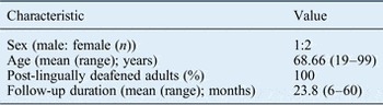
From January 2008 to March 2013, 355 patients received a cochlear implant at the Ear Science Institute Australia. Sixteen patients underwent combined subtotal petrosectomy and cochlear implant surgery. The patient demographics are outlined in Table I. All patients were post-lingually deafened adults.
Demographic data

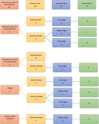
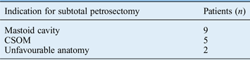
The indications for subtotal petrosectomy are outlined in Table II and Figure 1. Nine patients had an existing mastoid cavity. Six of these were modified radical mastoidectomy cavities for cholesteatoma and three were fenestration cavities for otosclerosis. Only two cavities, both cholesteatoma cavities, had signs of infection (persistent otorrhoea not responsive to conservative treatment such as aural toilet and topical medication).
Decision ladder and implant-related complications in patients who underwent subtotal petrosectomy and cochlear implantation. CSOM = chronic suppurative otitis media

Indications for surgery
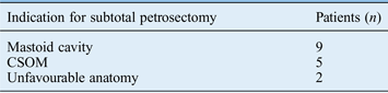
CSOM = chronic suppurative otitis media
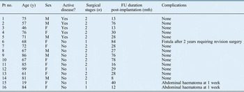
All patients with infection, with or without a mastoid cavity, underwent a two-stage procedure. The two patients with unfavourable anatomy had no prior history of ear disease and underwent a single-stage procedure. One patient with no infection in a dry, stable mastoid cavity underwent a single-stage procedure. This patient developed a fistula of the blind sac two years after the initial implant surgery and required subtotal petrosectomy revision. The implant was preserved. One patient with inactive chronic suppurative otitis media (CSOM) (dry tympanic membrane perforation, with no history of recurrent otorrhoea), underwent a single-stage procedure because of advanced age (99 years). There were no other implant-related complications recorded at the time of writing (Table III).
Follow-up duration and complications

Pt no. = patient number; y = years; FU = follow-up; mth = months; M = male; F = female
Electrode insertion was achieved via a round window approach extended into an anterior-inferior round window cochleostomy. Two patients required a drill-out of the basal turn of the cochlea because of extensive cochlear sclerosis. All patients had full electrode insertion, with good electrode impedance recorded in all electrodes.
Cavity obliteration was carried out with vascularised musculoperiosteal flaps in 10 patients and with abdominal fat in 5 patients. In general, infected cavities or less pneumatised mastoids could be effectively obliterated with vascular muscle flaps. There were no technical problems recorded during the second stage of surgery in cavities obliterated with fat (n = 3) or musculoperiosteal flaps (n = 10). Two patients with fat obliteration developed a post-operative abdominal haematoma in the first post-operative week. Twelve months' audiological data were available for 15 patients.
All patients fulfilled the pre-operative audiological candidacy for cochlear implantation. Specifically, all had bilateral hearing loss, ranging from moderately severe to profound, with unaided, pure tone audiometry scores of: less than 65 per cent for open-set sentences in quiet conditions in the worse ear and less than 85 per cent in the better ear, and less than 45 per cent for open-set phonemes in quiet conditions in the worse ear and less than 65 per cent in the better ear. Post-operative aided testing indicated thresholds of 20–35 dB in 14 patients and 40–50 dB in 1 patient. Aided speech perception in quiet conditions demonstrated a mean score of 95 per cent (range, 87–100 per cent).
Discussion
Cochlear implantation in the setting of chronic ear disease, with or without infection, is complex and challenging. It can be associated with potentially devastating complications, including recurrent cholesteatoma, electrode extrusion and spread of infection via the otic capsule. In the setting of chronic ear disease, the goals of cochlear implant surgery are to eradicate disease, and to create a closed and sterile cavity. This will minimise infection and close off the cochlear implant from the external environment.Reference Free, Falcioni, Di Trapani, Giannuzzi, Russo and Sanna 6 In a previous mastoid cavity, it is also essential to create a strong protective layer of tissue with which to cover the implant and electrodes to prevent extrusion. For patients with a history of CSOM, subtotal petrosectomy is recommended to avoid the complications of extrusion, infection and cholesteatoma formation.Reference Issing, Schonermark, Winkelmann, Kempf and Ernst 10
Donnelly et al. were one of the earlier groups to conclude that if implantation was to be contemplated in the presence of CSOM, a radical mastoidectomy with obliteration should be considered.Reference Donnelly, Pyman and Clark 11 They described the implantation of three patients with CSOM without cholesteatoma, all of whom developed recurrence of middle-ear disease, necessitating further surgery. One patient developed infection within the otic capsule via the cochleostomy, with resultant explantation of their cochlear implant.
Although no clear consensus exists regarding the optimal surgical technique, it is generally accepted that in the presence of infection, a two-stage procedure is warranted.Reference Issing, Schonermark, Winkelmann, Kempf and Ernst 10 , Reference Postelmans, Stokroos, Linmans and Kremer 12 , Reference Incesulu, Kocaturk and Vural 13
Issing et al. recommended a two-stage procedure to diminish post-operative complications in the presence of existing inflammatory conditions in the cavity, as 2 of their 14 cases were complicated by a retroauricular fistula following a single-stage procedure on infected mastoid cavities.Reference Issing, Schonermark, Winkelmann, Kempf and Ernst 10 Leung and Briggs reported 2 of 10 patients who underwent a two-stage procedure for active infection that required revision of the obliteration before cochlear implantation.Reference Leung and Briggs 1
The debate continues as to whether patients with inactive disease should undergo a single- or two-stage procedure. Advocates of a single-stage procedure include Hamzavi et al., who described no complications in seven patients who underwent single-stage subtotal petrosectomy and cochlear implantation for stable mastoid cavities.Reference Hamzavi, Baumgartner, Franz and Plenk 7 However, one significant complication of single-stage surgery is implantation cholesteatoma, which may not be detected after single-stage surgery. Several groups that described a single-stage procedure for inactive disease have also described patients requiring revision surgery and with device extrusion.Reference Free, Falcioni, Di Trapani, Giannuzzi, Russo and Sanna 6 , Reference Vincenti, Pasanisi, Bacciu and Bacciu 8 , Reference Postelmans, Stokroos, Linmans and Kremer 12 This mirrors our own experience with a post-operative wound infection that required revision surgery in a patient with seemingly inactive disease, who underwent a single-stage procedure.
It has been stated that a simple, dry perforation can be easily repaired and cochlear implantation can be performed using either a simultaneous or staged procedure.Reference Axon, Mawman, Upile and Ramsden 2 , Reference Postelmans, Stokroos, Linmans and Kremer 12 However, it is difficult to determine clinically if the underlying eustachian tube dysfunction will cause a reactivation of otitis media when the tympanic membrane is repaired – a significant risk in the setting of cochlear implantation. Postelmans et al. advocate a single-stage procedure in patients with no active infection, defined as no discharge in the six-month period before surgery.Reference Postelmans, Stokroos, Linmans and Kremer 12 The single-stage procedure either involved tympanoplasty and cochlear implantation for simple dry perforations, or subtotal petrosectomy and cochlear implantation for an existing mastoid cavity. They described one major complication requiring explantation in one of three patients who underwent single-stage tympanoplasty and cochlear implantation. Axon et al. also reported a patient who developed reactivation of acute otitis media following two-stage tympanoplasty and cochlear implantation. Their case was successfully managed with parenteral antibiotic therapy and the implant was preserved. We believe that in patients with uninfected single dry perforations, single-stage subtotal petrosectomy and cochlear implantation is the safest method. Meticulous technique should prevent inclusion cholesteatoma.
Two patients in our series underwent subtotal petrosectomy in combination with cochlear implantation in order to attain better access to the round window during surgery. Neither of these patients had a prior history of ear disease. One patient had a very sclerotic mastoid bone on the pre-operative CT scan, with an anteriorly placed sigmoid sinus, and access to the round window via a facial recess approach was not possible. The second patient had a moderately pneumatised mastoid bone, but a very narrow facial recess which could not provide adequate visualisation of the round window niche. Converting to subtotal petrosectomy in these patients improved access and facilitated safe cochlear implant insertion.
In our series, two patients who underwent subtotal petrosectomy in previous mastoid cavities also required a drill-out of the basal turn of the cochlea because of cochlear obliteration. Cochlear ossification and obliteration is also often seen as a result of meningitis, otosclerosis, chronic ear disease or trauma. Extensive drill-out procedures may be required, with their attendant risks to the internal carotid artery, and subtotal petrosectomy is advocated in this setting to identify the at-risk structures.
Performing subtotal petrosectomy and cochlear implantation has also been described in patients with inner-ear malformations. Twenty-five per cent of inner-ear malformations are associated with an abnormally positioned facial nerve, or cerebrospinal fluid (CSF) or perilymph ‘gusher’.Reference Papsin 14 Free et al. highlighted three specific reasons to consider subtotal petrosectomy with cochlear implantation in this group with complex temporal bone anatomy: in order to identify anatomical landmarks in the presence of anomalous middle-ear anatomy, in preparation for a CSF or perilymph leak, and to reduce the risk of meningitis in patients with severe inner-ear malformations.Reference Free, Falcioni, Di Trapani, Giannuzzi, Russo and Sanna 6
Vascularised musculoperiosteal flapsReference O'Sullivan and Atlas 15 and abdominal fatReference Free, Falcioni, Di Trapani, Giannuzzi, Russo and Sanna 6 have both been described as suitable tissue for cavity obliteration, and one does not seem to confer a particular advantage over the other. We favour the use of vascularised flaps where possible, especially in the presence of pre-operative infection.
Long-term follow up of these patients is recommended, as late complications can occur. In our series, one patient required revision surgery two years after their initial implant surgery. The length of follow up in our series ranged from 8 to 84 months. Although no other implant-related complications have been observed to date, ongoing clinical follow up over a longer time period may reveal future complications. However, the detection of recurrent cholesteatoma is limited clinically and radiologically by the blind sac closure of the external auditory canal and the artefact caused by the cochlear implant making MRI redundant.
Computed tomography scanning cannot reliably distinguish between soft tissue and cholesteatoma. Some authors advocate sequential CT scanning to look for soft tissue expansion or bony erosion as an indicator of recurrent cholesteatoma.Reference Vincenti, Pasanisi, Bacciu and Bacciu 8
Two papers have described their results with long-term radiological follow up using CT.Reference Xenellis, Nikolopoulos, Marangoudakis, Vlastarakos, Tsangaroulakis and Ferekidis 4 , Reference Vincenti, Pasanisi, Bacciu and Bacciu 8 Xenellis et al. followed nine patients over a mean of seven years, and performed a CT scan one year post-operatively and subsequently every three to four years.Reference Xenellis, Nikolopoulos, Marangoudakis, Vlastarakos, Tsangaroulakis and Ferekidis 4 Vincenti et al. followed 12 patients for a mean of 12 years and routinely performed a high-resolution CT scan 12–18 months post-operatively.Reference Vincenti, Pasanisi, Bacciu and Bacciu 8 Neither series detected cholesteatoma post-operatively on routine radiological investigation. Our protocol is different; in line with Leung and Briggs,Reference Leung and Briggs 1 routine radiological investigations are not undertaken. In our series, high-resolution CT scanning was performed only in the presence of suspicious signs or symptoms, avoiding unnecessary radiation exposure for patients.
-
• Cochlear implant surgery in chronic ear disease aims to eradicate disease, avoid recurrence, and prevent implant infection or extrusion
-
• Subtotal petrosectomy and blind sac closure of the external ear canal facilitates a clean surgical field by eliminating disease and disconnecting from the external environment
-
• This technique is also performed for cochlear access in cases of challenging anatomy or in drill-out procedures
-
• Subtotal petrosectomy in cochlear implantation is safe and effective when surgical steps are carried out meticulously
-
• Detection of recurrent cholesteatoma is limited clinically and radiologically by the blind sac closure and cochlear implant artefact
-
• Efficacy of routine post-operative computed tomography and optimal timing of implantation after subtotal petrosectomy are debatable
Subtotal petrosectomy with blind sac closure of the external auditory canal has made cochlear implant surgery feasible in patients with chronic ear disease. The success of the procedure depends on a meticulous surgical approach, including: removal of all mucosal disease, complete and secure closure of the external auditory canal skin into a blind sac, and adequate securing and covering of the implant to prevent rotation or extrusion. This series indicates that infection and successful implantation can be achieved by single surgery in particular circumstances, but that staged surgery should be utilised in infected ears, with the consideration of vascularised flaps for obliteration. Our series adds to the growing body of evidence proposing subtotal petrosectomy as a safe and effective procedure for facilitating cochlear implantation in patients with chronic ear disease or unfavourable anatomy. In the setting of active or inactive CSOM, with or without cholesteatoma, we recommend a two-stage procedure to diminish post-operative complications in this patient population.