Significant outcomes
-
Patients with schizophrenia in remission have significantly lower sub-foveal choroidal thickness as compared to other groups.
-
Antipsychotic-naïve patients with first-episode schizophrenia have significantly lower macular volume in the left eye compared to the healthy controls.
-
After controlling for various potential confounders, patients with psychotic illnesses have lower sub-foveal choroidal thickness, compared to healthy controls.
Limitations
-
The study involved cross-sectional assessment.
-
The study did not evaluate the association of retinal changes with the brain changes by using neuroimaging.
-
The study did not evaluate the association of retinal changes with cumulative antipsychotic dose exposure in the lifetime.
Introduction
Among various plausible current theories of schizophrenia, the neurodegenerative theory of schizophrenia is a prominent one with much support (Zarogianni et al., Reference Zarogianni, Moorhead and Lawrie2013). Due to different clinical presentations in patients with schizophrenia, and considerable overlap of symptoms with other disorders, for diagnosing schizophrenia and related conditions, there is a need for having objective markers (Ascaso et al., Reference Ascaso, Rodriguez-Jimenez, Cabezón, López-Antón, Santabárbara, De la Cámara, Modrego, Quintanilla, Bagney, Gutierrez, Cruz, Cristóbal and Lobo2015; Zarogianni et al., Reference Zarogianni, Moorhead and Lawrie2013). Retina is considered to be an extension of the brain. Due to this, it has been evaluated as an indicator of brain changes seen in different diseases (hypertension, migraine, dementia and diabetes mellitus) and disorders, including schizophrenia (Bowd et al., Reference Bowd, Weinreb, Williams and Zangwill2000; Lin et al., Reference Lin, Yi, Zhang, Liu, Cai, Chen, Zhang, Zhao and Pan2020; Dhasmana et al., Reference Dhasmana, Sah and Gupta2016). Retina develops from the anterior part of the neural tube in an early phase of development (Schönfeldt-Lecuona et al., Reference Schönfeldt-Lecuona, Kregel, Schmidt, Pinkhardt, Lauda, Kassubek, Connemann, Freudenmann and Gahr2016). Due to this, the retina and cortical part of brain are similar in terms of their structure, neurotransmitters, and functions (Gordon-Lipkin et al., Reference Gordon-Lipkin, Chodkowski, Reich, Smith, Pulicken, Balcer, Frohman, Cutter and Calabresi2007; Pulicken et al., Reference Pulicken, Gordon-Lipkin, Balcer, Frohman, Cutter and Calabresi2007). The retinal nerve fibre layer (RNFL) is composed of axons of retinal ganglion cells and can be considered an extension of the brain (Pan et al., Reference Pan, Zhou, Xiang and Yu2018). It is expected that the changes occurring in the brain must reflect in the retina as it also has unmyelinated axons which can be visualised by recent techniques like optical coherence tomography (OCT) which is easy to perform (Silverstein et al., Reference Silverstein, Paterno, Cherneski and Green2018). OCT is a non-invasive and quick imaging method, which can be used for in vivo imaging of the retinal layers (Chu et al., Reference Chu, Kolappan, Barnes, Joyce and Ron2012). There are no known contraindications to OCT, and it opens a ‘window into the brain’ which allows measurement of retinal layers (Chu et al., Reference Chu, Kolappan, Barnes, Joyce and Ron2012).
Some of the studies have evaluated retinal layer changes in patients with schizophrenia. These studies have compared patients with schizophrenia with age-matched controls, and the sample size in these studies has varied from 10 to 81 patients with schizophrenia with all the studies except 2 including 40 or fewer patients with schizophrenia. In general, although not consistent, most of these studies suggest that there is a reduction in the RNFL thickness (Ascaso et al., Reference Ascaso, Rodriguez-Jimenez, Cabezón, López-Antón, Santabárbara, De la Cámara, Modrego, Quintanilla, Bagney, Gutierrez, Cruz, Cristóbal and Lobo2015, Reference Ascaso, Laura, Quintanilla, Gutiérrez Galve, López-Antón, Cristóbal and Lobo2010; Cabezon et al., Reference Cabezon, Ascaso, Ramiro, Quintanilla, Gutierrez, Lobo and Cristobal2012; Celik et al., Reference Celik, Kalenderoglu, Sevgi Karadag, Bekir Egilmez, Han-Almis and Şimşek2016; Lee et al., Reference Lee, Tajunisah, Sharmilla, Peyman and Subrayan2013; Samani et al., Reference Samani, Proudlock, Siram, Suraweera, Hutchinson, Nelson, Al-Uzri and Gottlob2018; Yılmaz et al., Reference Yılmaz, Küçük, Ülgen, Özköse, Demircan, Ulusoy and Zararsız2016) that represents a reduction in ganglion cell axons (Silverstein et al., Reference Silverstein, Fradkin and Demmin2019) and macular thinning (Ascaso et al., Reference Ascaso, Rodriguez-Jimenez, Cabezón, López-Antón, Santabárbara, De la Cámara, Modrego, Quintanilla, Bagney, Gutierrez, Cruz, Cristóbal and Lobo2015; Lee et al., Reference Lee, Tajunisah, Sharmilla, Peyman and Subrayan2013; Yılmaz et al., Reference Yılmaz, Küçük, Ülgen, Özköse, Demircan, Ulusoy and Zararsız2016; Silverstein et al., Reference Silverstein, Fradkin and Demmin2019) among patients with schizophrenia, compared to healthy controls. Other findings which are reported in one or another study include the increased ratio of cup and disc along with the volume of the cup (Cabezon et al., Reference Cabezon, Ascaso, Ramiro, Quintanilla, Gutierrez, Lobo and Cristobal2012). Overall studies of OCT in patients with schizophrenia consistently show structural retinal pathology; however, sites of abnormality vary across studies. A recent study evaluated the relationship between structural retinal metrics in schizophrenia and co-morbid medical conditions. No differences were seen in RNFL or macula measurements between patients with schizophrenia and controls. However, across patient and control groups, RNFL, macula and Ganglion cell-Inner Plexiform Layer (GCL-IPL) thinning were associated with the presence of co-morbid diabetes mellitus and hypertension. Even after controlling for diabetes mellitus and hypertension, patients with schizophrenia demonstrated enlarged optic cup volumes and cup-to-disc ratios, suggestive of tissue loss in the surrounding regions (Silverstein et al., Reference Silverstein, Fradkin and Demmin2019).
However, most of the existing studies are marked by certain limitations. For example, some of the studies have not excluded patients with co-morbid conditions such as diabetes mellitus and hypertension (Ascaso et al., Reference Ascaso, Rodriguez-Jimenez, Cabezón, López-Antón, Santabárbara, De la Cámara, Modrego, Quintanilla, Bagney, Gutierrez, Cruz, Cristóbal and Lobo2015, Reference Ascaso, Laura, Quintanilla, Gutiérrez Galve, López-Antón, Cristóbal and Lobo2010; Cabezon et al., Reference Cabezon, Ascaso, Ramiro, Quintanilla, Gutierrez, Lobo and Cristobal2012; Celik et al., Reference Celik, Kalenderoglu, Sevgi Karadag, Bekir Egilmez, Han-Almis and Şimşek2016; Lee et al., Reference Lee, Tajunisah, Sharmilla, Peyman and Subrayan2013; Samani et al., Reference Samani, Proudlock, Siram, Suraweera, Hutchinson, Nelson, Al-Uzri and Gottlob2018; Yılmaz et al., Reference Yılmaz, Küçük, Ülgen, Özköse, Demircan, Ulusoy and Zararsız2016) which can lead to subtle retinal changes (Liao et al., Reference Liao, Chang, Wei, Chang, Liao, Lane and Sung2011; Nasrallah et al., Reference Nasrallah, Meyer, Goff, McEvoy, Davis, Stroup and Lieberman2006). None of the studies has evaluated the RNFL in drug-naïve patients with schizophrenia and a recent study which evaluated the patients with first-episode schizophrenia, reported lack of significant difference between patients and the healthy controls (Lai et al., Reference Lai, Crosta, Loftin and Silverstein2020). Only some of the studies have considered the effect of doses of antipsychotic used on the retinal layer changes (Jerotic et al., Reference Jerotic, Ristic, Ignjatovic and Maric2020). Understanding this is important because long-term use of antipsychotics can lead to inhibition of retinal dopamine receptors which cause the death of all types of retinal cells due to reduced activity as they all have dopamine receptors which can contribute to thinning of retinal layers (Silverstein and Rosen, Reference Silverstein and Rosen2015). In this background, the present study aimed to evaluate the retinal layer changes among different group of patients with schizophrenia, that is, drug-naïve patients with schizophrenia, patients currently on treatment, and patients with treatment-resistant schizophrenia (TRS) by using Swept-Source OCT (SS-OCT) and compare it with healthy controls. It was hypothesised that patients with schizophrenia will differ from healthy controls, in retinal layer thickness, whereas there would be no difference in the retinal layer changes among different group of patients with schizophrenia.
Methodology
This cross-sectional study was executed in a tertiary care multispecialty teaching hospital in North India. The study was approved by the ethics committee of the institute in which it was carried out and all the participants were enrolled during Jan 2019 to Dec 2019, after obtaining written informed consent. The authors assert that all the study procedures comply with the national and institutional ethical standards for human experimentation and the Helsinki Declaration of 1975, as revised in 2008.
The study sample was recruited by convenience sampling from the patient population attending the outpatient/inpatient services of the Department of Psychiatry (patients with schizophrenia) and Department of Ophthalmology (healthy controls). The study sample comprised of four groups, that is, Group I: 35 patients with schizophrenia, in clinical remission (without treatment resistance) (SZCR); Group II: 35 patients with TRS; Group III: 21 drug-naïve patients with schizophrenia (FES) and Group IV: 36 Healthy controls. Patients were considered to be antipsychotic-naïve if the duration of exposure to antipsychotics was less than 2 weeks at the time of assessment for retinal layer changes. TRS was defined as per the definition of Howes et al. (Reference Howes, McCutcheon, Agid, de Bartolomeis, Citrome, Daskalakis, Davidson, Drake, Dursun, Ebdrup, Elkis, Falkai, Fleischacker, Gadelha, Gaughran, Glenthøj, Graff-Guerrero, Hallak, Honer, Kennedy, Kinon, Lawrie, Lee, Leweke, MacCabe, McNabb, Meltzer, Möller, Nakajima, Pantelis, Reis Marques, Remington, Rossell, Russell, Siu, Suzuki, Sommer, Taylor, Thomas, Üçok, Umbricht, Walters, Kane and Correll2016). According to this definition, a person is considered to have TRS, if the person fulfils the diagnosis of schizophrenia as per the current nosology, has received two adequate trials (i.e. each trial of at least 6 weeks), at dosage equivalent of ≥600 mg Chlorpromazine, with ≥80% adherence to medication and have shown <20% improvement in the psychopathology. Further, at the time of the assessment, the patient has at least moderate severity of symptom and moderate level of dysfunction (Howes et al., Reference Howes, McCutcheon, Agid, de Bartolomeis, Citrome, Daskalakis, Davidson, Drake, Dursun, Ebdrup, Elkis, Falkai, Fleischacker, Gadelha, Gaughran, Glenthøj, Graff-Guerrero, Hallak, Honer, Kennedy, Kinon, Lawrie, Lee, Leweke, MacCabe, McNabb, Meltzer, Möller, Nakajima, Pantelis, Reis Marques, Remington, Rossell, Russell, Siu, Suzuki, Sommer, Taylor, Thomas, Üçok, Umbricht, Walters, Kane and Correll2016).
To be included in the study, the patients were required to fulfil the diagnosis of schizophrenia as per DSM-5 (American Psychiatric Association, 2013) (as determined by using MINI-PLUS 7.0.2 version (Sheehan et al., Reference Sheehan, Lecrubier, Sheehan, Amorim, Janavs, Weiller, Hergueta, Baker and Dunbar1998) and aged between 15 and 45 years). For inclusion into the study, the healthy controls were also required to be aged 15–45 years and with no history of any psychiatric illness. Additionally, they were required to screen negative on MINI-screen. Those with obscured clarity of media due to the presence of cataract, vitreous haze, or any other such co-existent pathology that did not allow the acquisition of good images were excluded. Similarly, those with refractive errors more than ±3 D (high hypermetropia and myopia), those with any ocular pathology that could affect the retinal /choroidal thickness or vascularity such as diabetic retinopathy, macular degeneration, central serous chorioretinopathy, optic atrophy, glaucoma, and uveitis, congenital retinal pathologies (this was done by detailed history taking and ophthalmological examination by a trained ophthalmologist), diagnosed with Parkinson’s disease, organic brain syndrome, intellectual disability, diabetes mellitus, hypertension, dementia, multiple sclerosis, HIV, head injury, epilepsy, encephalopathy due to any cause were excluded. Patients with co-morbid psychiatric disorders other than a lifetime diagnosis of depression and obsessive–compulsive disorder and tobacco dependence syndrome were not included in the study. However, if the patient fulfilled the current diagnosis of major depression or obsessive–compulsive disorder, then they were excluded. Healthy participants with a history of any psychiatric illness in the first-degree relative were excluded.
Patients with schizophrenia were evaluated on the Positive and Negative Syndrome Scale (PANSS) (Kay et al., Reference Kay, Fiszbein and Opler1987) to rate the severity of illness. Fagerstrom Test for Nicotine Dependence (FTND) was used to rate the severity of nicotine dependence (Heatherton et al., Reference Heatherton, Kozlowski, Frecker and Fagerström1991).
All the participants underwent detailed ophthalmological examination (general examination of the eye including fundoscopy, tonometry and slit lamp examination), before enrolment to rule out any ophthalmological pathologies. This involved ruling out the various ophthalmological abnormalities as listed in the exclusion criteria, and any infective pathology at the time of the assessment.
SS-OCT is the next-generation OCT that provided an accurate assessment of choroidal thickness in healthy and disease states. SS-OCT is a newer OCT technology that provides faster image acquisition and processing compared to the earlier generation OCT machines, such as time domain OCT, or spectral domain. SS-OCT is known for a better view of the vitreo-retinal interface as well as choroid in a single frame. SS-OCT utilises a narrow light wavelength of 1050 nm and achieves 100,000–400,000 A scan/second. Thus, very high-resolution images are possible with SS-OCT in a short acquisition time (Brynskov et al., Reference Brynskov, Laugesen, Svenningsen, Floyd and Sørensen2016; Manjunath et al., Reference Manjunath, Taha, Fujimoto and Duker2010; Spaide et al., Reference Spaide, Koizumi, Pozzoni and Pozonni2008). For image acquisition, the pupils of the patients were dilated by instilling Tropicamide 0.8%. Thirty-degree colour fundus photographs were acquired on the digital fundus camera (DRI Triton, Topcon ®) with images focussed on disc and macula. SS-OCT 3D and 5-Line raster scan of the macula and optic disc were done. The acquisition of the scans was repeated multiple times, and the images with the least amount of motion artefacts were selected for further analysis. Retinal thickness was manually measured by two independent ophthalmologists separately. The thickness was measured from the inner border of the internal limiting membrane to the outer border of the retinal pigment epithelium. The average of the two measurements was used for analysis. The individual retinal layer thickness was also measured by automated layer segmentation provided by the software. Choroidal thickness was measured by two independent ophthalmologists manually vertically from the outer border of the retinal pigment epithelium to the inner border of the sclera. The upper border was marked at the retinal pigment epithelium and the lower border area was below the line of light pixels at the choroid sclera junction. The average of the two measurements was used for analysis.
The patients were also evaluated for the metabolic parameters. A fasting blood sample of 5 ml was collected using all the aseptic measures. All the patients also underwent anthropometric evaluations for height, weight and waist circumference. The waist circumference was measured at the mid-point between the lower costal margin and the anterior superior iliac crest, in full expiration by a stretchable measuring tape. A diagnosis of metabolic syndrome was made as per the consensus criteria (Alberti et al., Reference Alberti, Eckel, Grundy, Zimmet, Cleeman, Donato, Fruchart, James, Loria and Smith2009). The prescription data were collected from the available treatment records, and chlorpromazine equivalent was calculated as per the recommendations (Patel et al., Reference Patel, Arista, Taylor and Barnes2013). The clinical remission was defined as per the Andreasen criteria (Andreasen et al., Reference Andreasen, Carpenter, Kane, Lasser, Marder and Weinberger2005).
Data were analysed with the use of a statistical package for social sciences, sixteenth edition (SPSS-16) (SPSS Inc. Released 2007. SPSS for Windows, Version 16.0. Chicago, SPSS Inc.). Mean and standard deviation with range were calculated for the continuous variables.
Wherever needed, confidence intervals were calculated. Frequency and percentages were determined for categorical variables. Comparisons were done by using an unpaired t-test and one-way ANOVA test. Kruskal–Wallis value and Mann–Whitney value were used for the non-parametric data. The categorical variables were compared by using the chi-square test, and wherever applicable, Yate’s correction and Fisher’s exact test were used. ANCOVA test was used where covariate analysis was done. Cohen’s D and Partial Eta values were used to analyse effect size, and in the case of non-parametric data, Glass’s delta value was computed. The association of retinal layer measurements with socio-demographic variables and clinical variables was studied by using the Pearson correlation coefficient or Spearman’s rank correlation. For evaluation of the association of retinal layer measurement with other variables, a p-value of less than 0.05 was considered to be significant.
Results
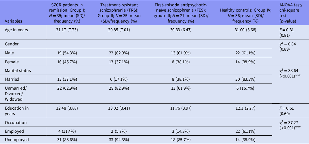
The mean age of participants of all the four study groups was close to 30 years, and the mean duration of education varied from 11.76 years to 13.02 years. In all the schizophrenia groups, the majority of the participants were currently single and unemployed (Table 1). No significant difference was seen on the demographic variables between the different schizophrenia groups. Compared to healthy controls, participants with schizophrenia were more often single and unemployed.
Comparison of the socio-demographic profile of all the four study groups

χ2: Chi-square value; F: ANOVA value; SD: standard deviation; *p ≤ 0.05; *** p ≤ 0.001.
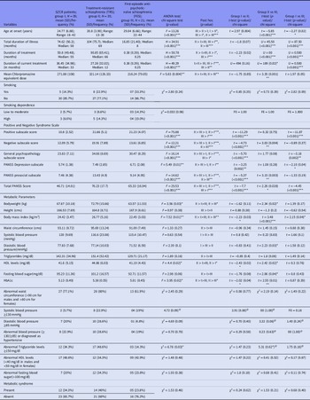
The participants with FES differed significantly from the other two groups, for the variables of the age of onset, and duration of illness (Table 2). When the study participants of TRS (Group II) and those with SZCR (Group I) were compared, compared to those in clinical remission, participants with TRS had lower age of onset, and longer duration of treatment (Table 2). None of the patients had co-morbid lifetime diagnosis of obsessive–compulsive disorder or major depression. Few patients in all the study groups had co-morbid tobacco dependence (Table 2). When participants’ TRS and those with FES were compared, participants with TRS had significantly higher negative symptoms, lower prosocial subscale score, and PANSS total score (Table 2). Compared to the participants with SZCR, participants with TRS had higher body mass index (BMI), higher high-density level lipoproteins (HDL) levels, and Hba1c levels. However, both these groups did not differ in terms of the prevalence of abnormal metabolic parameters, which are components of metabolic syndrome and prevalence of metabolic syndrome. However, compared to participants with the FES, participants with TRS had significantly higher body weight, higher BMI, higher HDL levels, higher fasting blood glucose levels, higher Hba1c levels, higher prevalence of abnormal diastolic blood pressure, and higher prevalence of abnormal triglyceride levels. When participants with FES were compared with participants with SZCR, it was seen that compared to the participants with FES, participants with SZCR had significantly higher BMI (Table 2).
Comparison of clinical profile of participants with schizophrenia

χ2: Chi-square value; t: T-test; SD: standard deviation; U: Mann–Whitney value; FE: Fisher Exact value; H: Kruskal–Wallis value; @: Chi-square value with Yate’s correction; F: ANOVA value; *p ≤ 0.05; **p ≤ 0.01; ***p ≤ 0.001.
Four participants (11.1%) in the control group had tobacco dependence with low to moderate dependence as per FTND scale.
Comparison of OCT findings of participants with schizophrenia with healthy controls
All schizophrenia subjects vs healthy controls
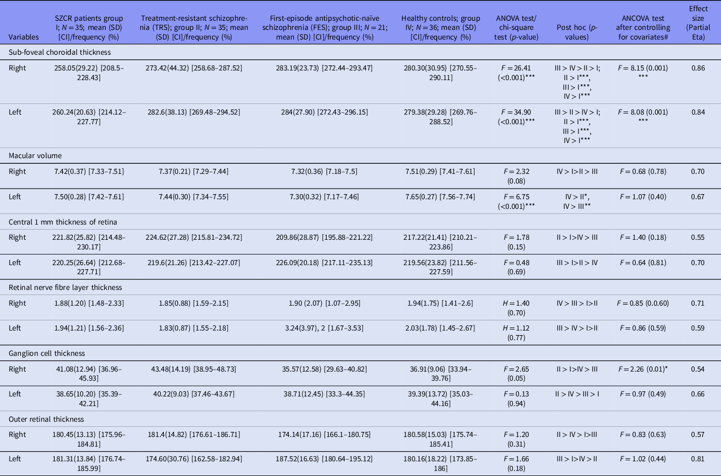
Before covariate analysis: Compared to healthy controls, patients with psychotic illness (FES, SZCR & TRS groups) had lower sub-foveal choroidal thickness. Additionally, patients with first-episode antipsychotic-naïve schizophrenia and TRS had significantly lower macular volume.
After covariate analysis : After controlling for various potential confounders [gender, duration of psychiatric illness, Fagerstrom Test for Nicotine Dependence (FTND) scores, current medications (CPZ equivalents), BMI, waist circumference, systolic blood pressure and diastolic blood pressure values, FBS, HbA1c, presence /absence of metabolic syndrome] by covariate analysis, the significant finding of lower sub-foveal choroidal thickness among patients with psychotic illness (FES, SZCR & TRS groups) persisted with a large effect size (0.84–0.86) (Table 3).
Comparison of OCT measurements among all groups

SD: standard deviation; CI: confidence interval; H: Kruskal–Wallis value; F: ANOVA & ANCOVA value; *p ≤ 0.05.
# Covariates – age, gender, duration of treatment, FTND scores, current medications (CPZ equivalents), body mass index, waist circumference, blood pressure (systolic and diastolic values), FBS, HbA1c, metabolic syndrome(present or absent).
Individual schizophrenia groups vs healthy controls
SZCR vs healthy controls
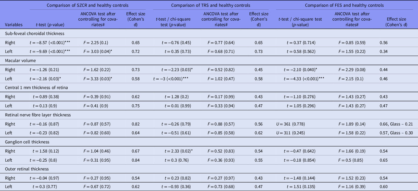
Before covariate analysis : Participants with SZCR had significantly lower thickness for the sub-foveal retinal thickness and left macular volume. SZCR group had higher central 1 mm thickness of retina, ganglion cell thickness, but lower thickness for RNFL and outer retinal thickness (left).
After covariate analysis: Only significant findings were lower left sub-foveal choroidal thickness and left macular volume (Table 4).
Comparison of OCT measurements among schizophrenia participants with healthy controls

t: T-test; SD: standard deviation; CI: confidence interval; F: ANCOVA value; Glass: Glass’s delta value; *≤0.05; **≤0.01; ***≤0.001.
# Covariates – age, gender, duration of psychiatric illness, duration of treatment, FTND scores, current medications (CPZ equivalents), body mass index, waist circumference, blood pressure (systolic and diastolic values), FBS, HbA1c, metabolic syndrome (present or absent).
TRS vs healthy controls
Before covariate analysis: Participants with TRS had significantly lower macular volume as compared to healthy controls. Further, participants with TRS had lower sub-foveal choroidal thickness, RNFL thickness but had higher thickness for central 1 mm thickness of retina ganglion cell thickness.
After covariate analysis: No significant differences were noted (Table 4).
FES vs healthy controls
Before covariate analysis: When participants with FES were compared with healthy controls, participants with FES had significantly lower thickness for macular volume.
After covariate analysis: No significant differences were noted (Table 4).
Within schizophrenia group comparisons
SZCR vs TRS
Before covariate analysis: On comparing SZCR and TRS groups, patients in SZCR group had significantly lower sub-foveal choroidal thickness.
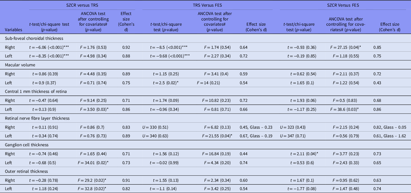
After covariate analysis: Only significant finding was found in the OCT of lower left ganglion cell thickness in SZCR group (Table 5).
Comparison of OCT measurements between different schizophrenia groups

t: T-test; SD: standard deviation; CI: confidence interval; F: ANCOVA value; Glass: Glass’s delta value; *≤0.05; **≤0.01; ***≤0.001.
# Covariates – age, gender, duration of psychiatric illness, duration of treatment, FTND scores, current medications (CPZ equivalents), body mass index, waist circumference, blood pressure (systolic and diastolic values), FBS, HbA1c, metabolic syndrome (present or absent).
SZCR vs FES
Before covariate analysis: Compared to participants with FES, participants in SZCR group had significantly lower sub-foveal retinal thickness, and left macular volume.
After covariate analysis : Only significant finding was lower left RNFL thickness in the SZCR group (Table 5).
TRS vs FES
Before covariate analysis: On comparing TRS group with FES groups, patients in the FES group had significantly lower right ganglion cell thickness.
After covariate analysis : TRS group had significantly lower right sub-foveal choroidal thickness and left central 1 mm thickness of retina (Table 5).
Discussion
Increasing evidence from literature sources of the last decades suggests noticeable changes in the thickness and/or volume of various retinal structures in patients with schizophrenia. Available studies in patients with schizophrenia have focused on evaluating the RNFL thinning, which is represented in the form of reduction in the ganglion cell axons and macular volume, which indicates the thinning of the fovea and surrounding tissue. Currently, available evidence suggests that compared to healthy controls, there is a reduction in the RNFL thickness in patients with schizophrenia (Ascaso et al., Reference Ascaso, Rodriguez-Jimenez, Cabezón, López-Antón, Santabárbara, De la Cámara, Modrego, Quintanilla, Bagney, Gutierrez, Cruz, Cristóbal and Lobo2015, Reference Ascaso, Laura, Quintanilla, Gutiérrez Galve, López-Antón, Cristóbal and Lobo2010; Cabezon et al., Reference Cabezon, Ascaso, Ramiro, Quintanilla, Gutierrez, Lobo and Cristobal2012; Celik et al., Reference Celik, Kalenderoglu, Sevgi Karadag, Bekir Egilmez, Han-Almis and Şimşek2016; Chu et al., Reference Chu, Kolappan, Barnes, Joyce and Ron2012; Lee et al., Reference Lee, Tajunisah, Sharmilla, Peyman and Subrayan2013; Schönfeldt-Lecuona et al., Reference Schönfeldt-Lecuona, Kregel, Schmidt, Kassubek, Dreyhaupt, Freudenmann, Connemann, Gahr and Pinkhardt2019; Topcu-Yilmaz et al., Reference Topcu-Yilmaz, Aydin and Ilhan2019; Yılmaz et al., Reference Yılmaz, Küçük, Ülgen, Özköse, Demircan, Ulusoy and Zararsız2016). Additionally, some of the studies suggest that there is also a reduction in the macular volume (Ascaso et al., Reference Ascaso, Rodriguez-Jimenez, Cabezón, López-Antón, Santabárbara, De la Cámara, Modrego, Quintanilla, Bagney, Gutierrez, Cruz, Cristóbal and Lobo2015; Lee et al., Reference Lee, Tajunisah, Sharmilla, Peyman and Subrayan2013; Miller et al., Reference Miller, Zemon, Nolan-Kenney, Balcer, Goff, Worthington, Hasanaj and Butler2020; Schönfeldt-Lecuona et al., Reference Schönfeldt-Lecuona, Kregel, Schmidt, Kassubek, Dreyhaupt, Freudenmann, Connemann, Gahr and Pinkhardt2019; Topcu-Yilmaz et al., Reference Topcu-Yilmaz, Aydin and Ilhan2019; Yılmaz et al., Reference Yılmaz, Küçük, Ülgen, Özköse, Demircan, Ulusoy and Zararsız2016). The studies that have compared the retinal changes in male and female patients with schizophrenia suggest some differences in the findings of male and female patients (Jerotic et al., Reference Jerotic, Ristic, Ignjatovic and Maric2020). However, one of the major problems with the existing literature is the confounding factors such as systematic diseases (such as hypertension and diabetes mellitus), chronic smoking, use of other substances, use of antipsychotic medications, obesity, and demographic variables such as gender and age have not been controlled consistently in various studies. Possibly due to this, the results are inconsistent with the thickness of RNFL and correlates of RNFL. Keeping these issues in mind, the present study aimed to evaluate the retinal layer changes in patients with schizophrenia with SS-OCT and compared it with healthy controls.
The present study included four groups of participants, that is, FES, SZCR, TRS, and a healthy control group. These four groups were selected considering the effect of antipsychotics and the acute symptoms on the RNFL. The patients with FES and those with TRS had acute symptoms, whereas the third group of patients, that is, SZCR group patients, were in clinical remission. TRS was also defined by using criteria proposed by Howes et al (Reference Howes, McCutcheon, Agid, de Bartolomeis, Citrome, Daskalakis, Davidson, Drake, Dursun, Ebdrup, Elkis, Falkai, Fleischacker, Gadelha, Gaughran, Glenthøj, Graff-Guerrero, Hallak, Honer, Kennedy, Kinon, Lawrie, Lee, Leweke, MacCabe, McNabb, Meltzer, Möller, Nakajima, Pantelis, Reis Marques, Remington, Rossell, Russell, Siu, Suzuki, Sommer, Taylor, Thomas, Üçok, Umbricht, Walters, Kane and Correll2016), which ensured proper categorisation of patients to the TRS group and also the SZCR group.
Age, gender, and level of education-matched healthy control group were selected to overcome the impact of these variables on the RNFL. Further, the study was limited to patients with schizophrenia, aged 18–45 years. This was done to minimise the effect of older age on the retina. Similarly, other conditions which can influence the retinal findings were excluded. In addition to this, all the metabolic parameters, such as fasting blood glucose level, Hba1c levels, triglyceride levels, high-density lipoprotein levels, very low-density lipoprotein levels, waist circumference, BMI, and metabolic syndrome, were taken as covariates while evaluating the differences in the RNFL thickness. Additionally, total duration of treatment and chlorpromazine dose equivalent of antipsychotic medications were also used as a covariate. The severity of tobacco dependence was also used as a covariate in the analysis. Accordingly, the present study can be considered as a methodological advancement, and the findings obtained can be considered as a true reflection of differences and similarities between patients with schizophrenia and healthy controls. Further, the similarities and differences in different group of patients with schizophrenia could be a reflection of clinical differences in the participants in the different groups.
In the present study, when patients with psychotic illness (first-episode drug-naïve schizophrenia, SZCR & TRS groups), currently in clinical remission were compared with healthy controls, patients had significantly lower sub-foveal choroidal thickness (both left and right eye). Additionally, patients with FES and TRS groups had significantly lower macular volume (left eye). However, these significant differences persisted for sub-foveal choroidal thickness (left eye) only when the various confounders were taken into account. The effect size of the difference between the patients with SZCR, when compared to healthy controls, was 0.72 for the sub-foveal choroidal thickness (left eye) and for the macular volume (left eye) was 0.58. When the patients with TRS were compared with healthy controls, initially significant differences were observed for the macular volume (left and right), and the ganglion cell thickness (right eye) but these differences disappeared after controlling for the various covariates. Similarly, when the patients with FES, currently in the acute episode, were compared with healthy controls, a significant difference was noted in the macular volume (left and right), but these differences disappeared after controlling for the various covariates.
Our findings are in concordance with the majority of the previous studies and suggest thinning of the retina in patients with schizophrenia when compared to the healthy controls (Ascaso et al., Reference Ascaso, Rodriguez-Jimenez, Cabezón, López-Antón, Santabárbara, De la Cámara, Modrego, Quintanilla, Bagney, Gutierrez, Cruz, Cristóbal and Lobo2015, Reference Ascaso, Laura, Quintanilla, Gutiérrez Galve, López-Antón, Cristóbal and Lobo2010; Cabezon et al., Reference Cabezon, Ascaso, Ramiro, Quintanilla, Gutierrez, Lobo and Cristobal2012; Celik et al., Reference Celik, Kalenderoglu, Sevgi Karadag, Bekir Egilmez, Han-Almis and Şimşek2016; Joe et al., Reference Joe, Ahmad, Riley, Weissman, Smith and Malaspina2018; Lee et al., Reference Lee, Tajunisah, Sharmilla, Peyman and Subrayan2013; Miller et al., Reference Miller, Zemon, Nolan-Kenney, Balcer, Goff, Worthington, Hasanaj and Butler2020; Samani et al., Reference Samani, Proudlock, Siram, Suraweera, Hutchinson, Nelson, Al-Uzri and Gottlob2018; Schönfeldt-Lecuona et al., Reference Schönfeldt-Lecuona, Kregel, Schmidt, Kassubek, Dreyhaupt, Freudenmann, Connemann, Gahr and Pinkhardt2019; Topcu-Yilmaz et al., Reference Topcu-Yilmaz, Aydin and Ilhan2019; Yılmaz et al., Reference Yılmaz, Küçük, Ülgen, Özköse, Demircan, Ulusoy and Zararsız2016). However, our study findings do not support the studies which have reported lack of difference between patients with schizophrenia and healthy controls (Chu et al., Reference Chu, Kolappan, Barnes, Joyce and Ron2012; Silverstein et al., Reference Silverstein, Paterno, Cherneski and Green2018). The lack of difference between patients with TRS and those with FES from healthy controls after controlling for confounders could be due to the effect of the symptoms per se on the retinal findings. Some of the authors have suggested that acute psychosis itself is associated with acute inflammatory response, which increases the retinal thickness (Ascaso et al., Reference Ascaso, Rodriguez-Jimenez, Cabezón, López-Antón, Santabárbara, De la Cámara, Modrego, Quintanilla, Bagney, Gutierrez, Cruz, Cristóbal and Lobo2015). Findings of the present study also possibly support the same.
The significant difference in the thickness of macular volume between the healthy controls and patients with SZCR, currently in remission, even after controlling for various covariates reflects that retinal changes are seen in patients with schizophrenia and support the exiting studies (Ascaso et al., Reference Ascaso, Rodriguez-Jimenez, Cabezón, López-Antón, Santabárbara, De la Cámara, Modrego, Quintanilla, Bagney, Gutierrez, Cruz, Cristóbal and Lobo2015; Joe et al., Reference Joe, Ahmad, Riley, Weissman, Smith and Malaspina2018; Lee et al., Reference Lee, Tajunisah, Sharmilla, Peyman and Subrayan2013; Miller et al., Reference Miller, Zemon, Nolan-Kenney, Balcer, Goff, Worthington, Hasanaj and Butler2020; Schönfeldt-Lecuona et al., Reference Schönfeldt-Lecuona, Kregel, Schmidt, Kassubek, Dreyhaupt, Freudenmann, Connemann, Gahr and Pinkhardt2019; Topcu-Yilmaz et al., Reference Topcu-Yilmaz, Aydin and Ilhan2019; Yılmaz et al., Reference Yılmaz, Küçük, Ülgen, Özköse, Demircan, Ulusoy and Zararsız2016). However, presence of these changes in the remission phase and absence of the same in the acute phase possibly suggests that retinal changes in patients with schizophrenia are ‘state’ markers. Accordingly, it can be said that future studies must attempt to compare the patients with acute symptoms and those in remission and longitudinally follow up the patients to evaluate the retinal changes during the different phases of illness to improve the understanding. Further, the present study suggests that among the various parameters, the macular volume appears to be the most important measurement. Hence, besides focusing on other measurements, future studies must focus on the macular volume by carrying out single-layer analysis to improve clarity on the topic.
However, one of the important findings of the present study is that although we did not find significant differences for various retinal parameters in the comparison statistics, effect sizes for various comparisons between the schizophrenia groups and healthy controls were medium to large, with the largest sizes (0.54–0.82) for the comparisons of patients with SZCR group and the healthy controls. For the comparisons of TRS and the healthy control group, the effect sizes varied from 0.43 to 0.73, and that for patients with FES and healthy controls varied from 0.34 to 0.66. Few of the previous studies have evaluated the effect sizes and have also reported medium to large effect sizes (Cabezon et al., Reference Cabezon, Ascaso, Ramiro, Quintanilla, Gutierrez, Lobo and Cristobal2012; Silverstein et al., Reference Silverstein, Paterno, Cherneski and Green2018). These medium to large effect size differences between patients with schizophrenia in different phases of illness and healthy controls suggest that retinal changes do occur in patients with schizophrenia.
When patients with TRS were compared with patients with SZCR, the initial comparison revealed thinning of the sub-foveal choroidal thickness (both right and left eye), but this disappeared when the covariates were included in the analysis. However, on the inclusion of covariate, the ganglion cell thickness, which was not significant in the initial analysis, appeared to be significant (left side). Only one previous study has compared patients with treatment-refractory schizophrenia and healthy controls, and this study suggests that global RNFL thickness ganglion cell thickness and internal plexiform layer thickness are lower in patients with treatment-refractory schizophrenia, compared to treatment-responsive schizophrenia. Our findings of significant difference in ganglion cell thickness after controlling for other variables support this study (Celik et al., Reference Celik, Kalenderoglu, Sevgi Karadag, Bekir Egilmez, Han-Almis and Şimşek2016). However, in the present study, we did not find the difference for other parameters, after controlling for the covariates. This difference in our study and the previous study could be attributed to the differences in the severity of psychopathology and the method of analysis.
In the present study, when the patients with SZCR were compared with the patients of FES, after controlling for the covariates, patients in clinical remission were found to have significant thinning of the RNFL thickness (left side). As none of the previous studies have compared these groups, it is difficult to make any conclusion about this difference between the two groups.
In the present study, patients with TRS and those with FES differed significantly for the thickness of sub-foveal choroidal thickness (right side) and central 1 mm thickness of the retina (left side), after controlling for the covariates. As none of the previous studies have compared these groups, it is difficult to make any conclusion about this difference between the two groups. These retinal changes may be a marker of the TRS. However, there is a need to validate the same in future studies.
Another important fact, which emerged from the present study, is that when the patients with schizophrenia with different phases of illness were compared, effect sizes between the different groups were mostly large (i.e. >0.8). This finding suggests that there are differences in the retinal thickness between the subjects with different phases of illness and this requires further evaluation by controlling for various covariates by using more stringent selection criteria.
The present study has certain limitations, which must be kept in mind. These include limited sample size, convenient sampling, cross-sectional assessment, lack of evaluation of the association of retinal changes with the brain changes by using neuroimaging, lack of assessment of functional retinal changes, and lack of evaluation of the association of retinal changes with cumulative antipsychotic dose exposure in the lifetime. The study also did not consider the evaluation of acute inflammatory markers. Future studies must attempt to overcome these limitations. In the present study, the mean age of the participants with FES was higher than that noted in some of the studies evaluating patients with first-episode schizophrenia.
To conclude, the present study suggests that compared to healthy controls, patients with SZCR (non-TRS), currently in clinical remission, show thinning of the retina, especially in the sub-foveal choroidal thickness (left eye), and macular volume (left eye). However, similar changes are not in patients in the acute phase of illness, either TRS or FES. These findings possibly suggest that retinal changes in patients with schizophrenia correspond with the brain changes marked by atrophy, which is more apparent only in remission phase. However, during the acute phase of illness, these changes are not visible due to neuro-inflammation-related oedema.
Author contributions
All the authors contributed to the concept of the study. AK collected the data. AK, SG and SS analysed the data. AK and SG drafted the manuscript. All the authors contributed to the revising of the manuscript and approved the final version of the manuscript.
Financial support
This research received no specific grant from any funding agency, commercial or not-for-profit sectors.
Conflict of interest
None.