Introduction
Advanced maternal age in mammals is associated with reduced fertility. The age-related decline in human female fertility begins by the age of 35 years and fecundity is virtually lost by the age of 45 (Szamatowicz & Grochowski, Reference Szamatowicz and Grochowski1998). The cause of this decline in fertility in older mammals has been the subject of many studies and controversies. The decline is the result of several factors that contribute to overall reproductive failure. These factors include decreasing oocyte number, oocyte quality, and endometrial receptivity (Fitzgerald et al., Reference Fitzgerald, Zimon and Jones1998).
In older women, reduced fertility was most likely to be due to a diminishing pool of ovarian follicles that are available for development (Faddy et al., Reference Faddy, Gosden, Gougeon, Richardson and Nelson1992). The number of oocytes obtained, even after hormone stimulation, declines progressively with ageing in women (Fitzgerald et al., Reference Fitzgerald, Zimon and Jones1998). However, it has been reported that age-related decline is attributable to poor oocyte quality (Navot et al., Reference Navot, Bergh, Williams, Garrisi, Guzman, Sandler and Grunfeld1991). It has been well recognized that chromosomal aneuploidy of oocytes and embryos is increased significantly with advancing age in humans (Pellestor et al., Reference Pellestor, Andréo, Arnal, Humeau and Demaille2003). Most aneuploidies that are associated with increased maternal age are due to non-disjunction and meiotic errors that occur during meiosis I (Pellestor et al., Reference Pellestor, Andréo, Arnal, Humeau and Demaille2002; Lamb et al., Reference Lamb, Yu, Shaffer, Feingold and Sherman2005).
Although age-related decline in fertility has also been found in mice, previous data on age-related decreases in oocyte number and age-related increases in aneuploidy in mice have been contradictory. The number of GV oocytes (Eppig & O'Brien, Reference Eppig and O'Brien1995), MII oocytes (Fujino et al., Reference Fujino, Ozaki, Yamamasu, Ito, Matsuoka, Hayashi, Nakamura, Ogita, Sato and Inoue1996; Pan et al., Reference Pan, Ma, Zhu and Schultz2008) or fertilized eggs (Sakurada et al., Reference Sakurada, Ishikawa and Endo1996) declines gradually with ageing in mice. However, several reports have demonstrated that there is not an age-related decrease in the number of ovulated oocytes in many laboratory mouse strains (Gosden, Reference Gosden1975; Ishikawa & Endo, Reference Ishikawa and Endo1996). Moreover, the aneuploidy of ovulated oocytes does not increase significantly during female ageing in majority of mice (Golbus, Reference Golbus1981; Zuccotti et al., Reference Zuccotti, Boiani, Garagna and Redi1998). Although low but significant increase in the incidence of aneuploidy has been found in aged oocytes and embryos of some mouse strains (Sakurada et al., Reference Sakurada, Ishikawa and Endo1996; Eichenlaub & Boll, Reference Eichenlaub-Ritter and Boll1989), the incidence of aneuploidy does not increase as steeply as it does in humans (Hansmann & Pabast, Reference Hansmann and Pabst1992). These observations suggest that there should be a suitable mouse strain for addressing the problem of age-related oocyte infertility in human.
A more direct indicator of predisposition to aneuploidy in oocytes seems to be the degree of order with which chromosomes are alignment in the metaphase II spindles. Increased aberrations in spindle formation and chromosome alignment are associated with ageing in humans (Battaglia et al., Reference Battaglia, Goodwin, Klein and Soules1996; Volarcik et al., Reference Volarcik, Sheean, Goldfarb, Woods, Abdul-Karim and Hunt1998), and mice (Liu & Keefe, Reference Liu and Keefe2002; Pan et al., Reference Pan, Ma, Zhu and Schultz2008). In addition, a close relationship exists between ageing and premature chromatid segregation during meiosis in human (Pellestor et al., Reference Pellestor, Andréo, Arnal, Humeau and Demaille2003; Vialard et al., Reference Vialard, Petit, Bergerem, Gomes, Martek-Petit, Lombroso, Ville, Gerard and Selva2006).
As human oocytes are not readily available for studies on maternal age effect and use of human oocytes is restricted for ethic considerations, it is important to have an animal model in which age effects are present and larger numbers of oocytes are available for chromosomal, developmental and biological analysis. Such model would provide insight into reproductive ageing in women.
Kunming (KM) mouse, an outbred strain, was introduced into China from the Haffkine institute of India in 1946. After being bred for several decades, the KM mouse has been a native breed, making up 70 per cent of all mice used in research in China.
Our intention is to determine the relationship between maternal ageing and reproductive loss in KM mouse, and to seek possible factors involved in maternal ageing. We collected oocytes from young (6–8 weeks), middle-aged (9 months) and aged (12 months) females of KM mouse. Oocyte number, segregation and alignment of meiotic chromosome, and developmental competence were compared. Our experiments showed that the KM mouse exhibited a significant decline in fertility at 9 months of age, and reduced number of oocytes, chromosome misalignment and premature chromatid segregation were associated with ageing. In addition, we observed age-related 1 h delay in pronuclear formation after in-vitro fertilization (IVF). The present study suggests KM mouse is a suitable model for age-related decline in female fertility in human.
Materials and methods
Animals
Kunming (KM) mice were purchased at 4 weeks of age from the Institute of Laboratory Animal Science, Chinese Academy of Medical Science of PLA, and were maintained under a 12 h light and 12 h dark with a room temperature between 21°C and 23°C, relative humidity of 50 ± 5% and free access to water and food. Virgin female mice were bred up to 6–8 weeks, 9 months and 12 months, respectively, setting up three different age groups (young group, 6–8 weeks; middle-aged group, 9 months; and aged group,12 months). Animal care and handling were conducted in accordance with policies on the care and use of animals promulgated by the ethical committee of the Institute of Genetics and Developmental Biology, Chinese Academy of Sciences, China.
Fertility of the mice
Female mice of different age group were paired with young adult KM males of proven fertility at 22:00 h. Matings were confirmed by the presence of a vaginal plug before 10:00 h the following morning (day 1 of pregnancy). The female mice with vaginal plugs were bred individually. Pregnant time of each mouse, number of pups in one litter and development of pups were recorded.
Oocyte collection
Female mice were injected i.p. with 5 iu pregnant mare serum gonadotrophin (PMSG; Sigma, St Louis, MD, USA). GV oocytes (including oocytes surrounded by and free of cumulus cells) were collected by puncturing the ovarian follicles at 44–48 h post injection into M2 medium (Sigma). The number of ovarian oocytes collected from each mouse was counted.
Female mice were injected i.p. with 5 iu PMSG followed 48 h later by 5 iu of human chorionic gonadotrophin (hCG, Sigma). MII oocytes were collected from the oviducts 15–16 h after the hCG injection into M2 medium. The cumulus cells were removed by brief incubation in 0.3 mg/ml hyaluronidase (Sigma) in M2 medium. The number of ovulated oocytes collected from each mouse was counted. The denuded MII oocytes were used for cytogenetic analysis and immunocytochemistry. MII oocytes with cumulus cells were used for in-vitro fertilization (IVF).
All in-vitro manipulations were carried out at 36–37°C on a heated stage.
Histological study of ovaries
Female mice were injected i.p. with 5 iu PMSG and killed 48 h later by cervical dislocation. The ovaries of the animals were fixed in Bouin's solution, serially sectioned at 8 μm, and stained with haematoxylin and eosin. The numbers of follicles were counted.
In-vitro maturation (IVM) and IVF
GV oocytes (including oocytes surrounded by and free of cumulus cells) were cultured in human tubal fluid (HTF) medium (Irvine Scientific, Santa Anna, CA, USA) supplemented with 10% fetal calf serum (FCS; HyClone, Logan, UT, USA) under mineral oil at 37°C in an atmosphere of 5% CO2 in humidified air. Maturation of the oocytes was evaluated at 16–18 h of culture.
In vivo-matured MII oocytes enclosed by cumulus cells were fertilized in vitro as described (Hogan et al., Reference Hogan, Constantini, Lacey, Nagy, Gertsenstein, Vintersten and Behringer1986). Spermatozoa were collected from the cauda epididymides of male mice and capacitated in IVF medium that contained 15 mg/ml BSA (Sigma) for 1.5 h (Hogan et al., Reference Hogan, Constantini, Lacey, Nagy, Gertsenstein, Vintersten and Behringer1986). Oocytes were incubated with the spermatozoa in IVF medium with 15 mg/ml BSA for 4 h. The oocytes were then transferred to HTF medium with 10% FCS at 37°C, 5% CO2 for in-vitro culture. The presence of pronuclei was monitored in 4, 5, 6 and 7 h of IVF, respectively. Early embryonic development of the fertilized oocytes was recorded every 24 h for 5 days.
Cytogenetic analysis
Ovulated MII oocytes were submitted to cytogenetic analysis as described (Tarkowski, Reference Tarkowski1966). The oocytes were transferred into a 1% sodium citrate hypotonic solution for 10 min and then fixed with methanol: acetic acid (3:1) on a clean glass slide. Fixed oocytes were stained with Giemsa to determine the number and construction of chromosomes.
Immunocytochemistry
Ovulated MII oocytes were fixed in 3.7% paraformaldehyde (Sigma) in PBS for 40 min, and then permeabilized in PBS containing 0.1% Triton X-100 (Sigma) for 30 min at room temperature. They were washed subsequently for 1 h in PBS containing 5% BSA. Afterwards, the oocytes were incubated with β-tubulin mouse monoclonal antibody (1:150; ZYMED Laboratories Inc.) overnight at 4°C, then washed, and incubated with fluorescein isothiocyanate (FITC)-conjugated goat anti-mouse IgG (1:200; Sigma) at room temperature for 2 h. Hoechst 33258 (Sigma, 5 μg/ml) in PBS was included to localize chromosomes.
FITC localization patterns were observed using a confocal fluorescence microscope. Hoechst 33258 fluorescence was obtained simultaneously, and optical sections were collected and reproduced on a SPARC workstation. Paired images were digitally reproduced to examine the co-localization of tubulin and chromosomes.
Data analysis
All experiments were repeated three to five times and the results were analysed using chi-squared test or t-test with significance determined at a P-value < 0.05.
Results
Fertility of KM mice
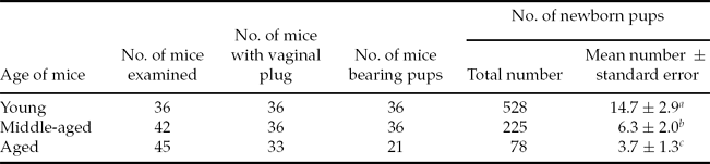
The number of pups born by mice from different age groups is summarized in Table 1. The mean number of pups born per mouse decreased significantly from 14.7 ± 2.9 in young mice to 6.3 ± 2.0 in middle-aged mice, and to 3.7 ± 1.3 in aged mice (P < 0.01). Pups from the young mice group were born on day 19 or 20 of pregnancy. All pups were alive at birth.
Effect of female ageing on fertility of KM mice

a > b > c P < 0.01 (t-test).
Of 225 pups from middle-aged females, 216 were born on days 19 or 20 of pregnancy, of which 12 died at birth. Nine pups were born on day 21 of pregnancy and all died at birth. Of 78 pups from aged females, only 30 were born on days 19 or 20 of pregnancy; the rest were born on days 21, 22, 23, 24, or 25 of pregnancy (3, 15, 3, 18 and 9, respectively) and all died at birth. It was found by anatomy that ovarian cyst, tumour of the uterus or abdomena tumour occurred in 12 aged females that did not bear pups on day 30 of pregnancy.
Number of oocytes retrieved from ovaries and oviducts of mice
As shown in Table 2, the maternal age had a striking effect on the mean number of oocytes retrieved from ovaries and oviducts per mouse. The mean numbers of both ovarian GV oocytes and ovulated MII oocytes decreased significantly as the age increased from the young to the middle-aged, and to the aged (P < 0.01).
Effect of female ageing on number of oocytes retrieved from ovaries and oviducts
a > b > c P < 0.01(t-test). GV, germinal vesicle.
Histology of mouse ovaries
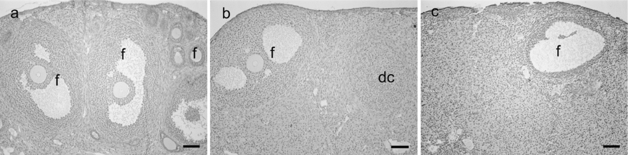
In one microscopic field of a section of ovaries from young females, there were a great number of primordial follicles, primary follicles, secondary follicles and mature follicles, and a small number of corpus luteum and atresic follicles (Fig. 1a). However, only one or a few follicles were observed in that from middle-aged females at the same magnification (×100), and the follicles were mainly large secondary follicles or mature follicles (Fig. 1b). The ovarian cortex consisted mainly of degenerated corpus luteum and atresic follicles, which were mostly replaced by connective tissue and left a broad outline of them. The number of follicles in ovaries from aged females was the least. There were one or two follicles that were mainly large secondary follicles or mature follicles in one microscopic field of a section of the ovaries (Fig. 1c). Degenerated corpus luteum and atresic follicles were almost replaced by connective tissue and their outline died out.
Sections of ovaries from young (a), middle-aged (b) and aged (c) mice. f, follicles; dc, degenerated corpus luteum. Bar = 100 μm.
IVM of GV oocytes
Table 3 shows in-vitro maturation of GV oocytes (surrounded by and free of cumulus cells) from mice of different ages. In 16–18 h of culture, maturation rate of oocytes from young mice was significantly lower than those of oocytes from middle-aged and aged mice (P < 0.01). In contrast, the rate of oocytes resting at GV stage was significantly higher for young mice, compared with middle-aged and aged mice (P < 0.01).
Effect of female ageing on in-vitro maturation of GV oocytes
Values with the same superscripts are not significantly different from each other.
(P > 0.05) a > b, f < g P < 0.01; c > d P < 0.05 (χ 2 test). GV, germinal vesicle; MI, metaphase I; MII, metaphase II.
Cytogenetic analysis of MII oocytes
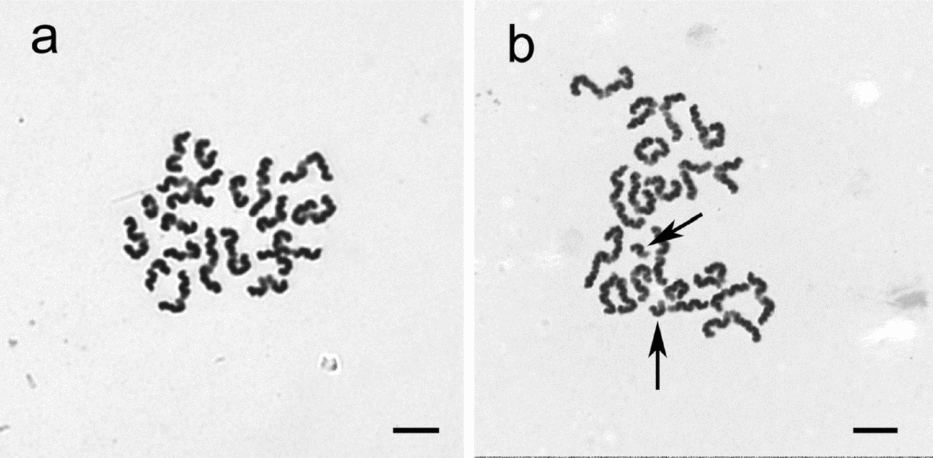
The results of cytogenetic analysis of MII oocytes are shown in Table 4. In total, 147 out of 443 matured oocytes were fixed successfully for cytogenetic study. The percentage of oocytes displaying 20 chromosomes (Fig. 2a) decreased slightly, but there was no significant difference with increase in age. Hyperploid oocyte (with more than 20 chromosomes) was not observed in mice of three groups.
Effect of female ageing on cytogenetics of MII oocytes
Values with the same superscripts are not significantly different from each other (P > 0.05).
b < c P < 0.01 (χ 2 test).
Cytogenetic analysis of oocyte from young mouse showing 20 chromosomes (a) and oocyte from aged mouse showing 20 chromosomes (b), one of which has split into two unassociated chromatids (arrows). Stained with Giemsa. Bar = 10 μm.
Interestingly, 1–4 chromosomes split into 2–8 distinct chromatids were found in some matured oocytes (Fig. 2b). Furthermore, the percentage of oocytes with 20 chromosomes was nearly identical whether the cells were derived from young, middle-aged or aged individuals, a correlation between premature chromatid segregation and maternal age was observed. The rate of premature chromatid separation in oocytes from aged mice was significantly higher than that from young mice (P < 0.01). The rate of chromatid separation in oocytes from the middle-aged mice was high (23.8%), but not significantly different from that of other two groups (Table 4).
Chromosome and spindle organization of MII oocytes
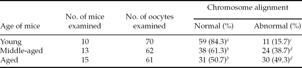
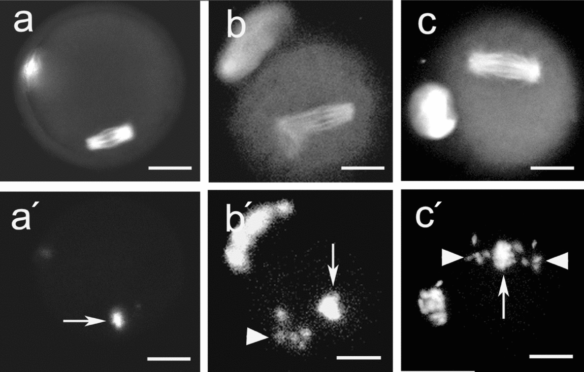
Confocal microscopy analysis (Table 5; Fig. 3) showed that most MII oocytes (84.3%) of young mice exhibited chromosome alignment in the mid-region of MII spindles, and the MII spindles were uniform in shapes. In contrast, oocytes of aged mice showed a significantly high rate (49.3%) in chromosome misalignment and dispersal over the metaphase plate, and some oocytes were either abnormally shaped or disordered with regard to microtubule placement. The rate (38.7%) of chromosome organization abnormality in oocytes from middle-aged mice was also significantly higher than that in oocytes from young mice, but comparable with that in oocytes from aged mice. The chromosome misalignment and dispersal were found over both intact and abnormal spindles.
Effect of females ageing on chromosome alignment in MII oocytes
Normal = chromosomes alignment in the middle region of metaphase spindles.
Abnormal = chromosomes misalignment and dispersal along intact or disrupted spindles.
Values with the same superscripts are not significantly different from each other (P > 0.05).
a > b, c < d P < 0.01 (χ2 test).
Immunofluorescence images of spindles and chromosomes of metaphase (M)II oocytes from young (a, a′), middle-aged (b, b′) and aged (c, c′) mice. (a–c) Meiotic II spindles stained by anti-β-tubulin and FITC-conjugated second antibody; (a′–c′) Chromosomes stained by Hoechst 33258. Arrows indicate chromosome accumulation at the plate. Arrowheads indicate misalignment of chromosomes. Bar = 30 μm.
Pronuclear formation and development of MII oocytes after IVF
Pronuclear formation of oocytes from mice of different ages in 4–7 h of IVF is shown in Table 6. Pronuclear formation was accomplished in 7 h of IVF and the rates (84.6–88.9%) of oocytes forming pronuclei were consistent among mice of three groups. However, the percentage of oocytes forming pronuclei in 5 h of IVF / the total oocytes forming pronuclei was 88.1% in young mice, but 35.5% and 42.3% in middle-aged and aged mice, respectively. The percentage of oocytes that form pronuclei in 6 h of IVF /the total oocytes forming pronuclei was 91.3% and 88.6% in middle-aged and aged mice respectively. The results demonstrated that pronuclei formed in oocytes from middle-aged and aged mice 1 h later than that from young mice.
Pronuclear formation and development of MII oocytes after IVF
Values in [] are percentages of oocytes forming pronuclei at certain time of IVF / the total oocytes forming pronuclei.
Values with the same superscripts are not significantly different from each other (P > 0.05).
a > b, c > d, e > f P < 0.01 (χ 2 test). MII, metaphase II; IVF, in-vitro fertilization.
Early embryonic development of oocytes after IVF is also shown in Table 6. The rates of oocytes developing to 2-cell, 4-cell, morula and blastocyst decreased slightly but not significantly from that of young to middle-aged or aged P > 0.05.
Discussion
The present study demonstrates that the KM mouse exhibits earlier significant decline in fertility at 9 months of age, and further significant decline at 12 months of age. Decreased litter size, prolongation of delivery and death of most pups at birth were observed in aged mice. Ageing of the female mice is associated with: (1) reduced number of ovarian follicles, ovarian GV oocytes and ovulated MII oocytes; (2) reduced percentage of oocytes exhibiting a normal distribution of chromosomes in the metaphase II plate; (3) increased percentage of oocytes displaying premature chromatid segregation; and (4) prolongation of duration of pronuclear formation after IVF.
Despite no relationship between oocyte number and maternal age in many laboratory mouse strains (Gosden, Reference Gosden1975; Ishikawa & Endo, Reference Ishikawa and Endo1996), the KM mouse exhibits an age-related decrease in oocyte pool. Aged KM mice produce much fewer GV oocytes and much fewer MII oocytes after hormone stimulation compared with young mice. Rare follicles were observed and ovarian cortex was almost displaced by connective tissue on sections of ovaries from aged mice. Our results are in agreement with studies in human (Fitzgerald et al., Reference Fitzgerald, Zimon and Jones1998; Tarin et al., Reference Tarin, Pérez-Albalá and Cano2001), a finding that suggested that the primary cause for fertility decline is the gradual disappearance of oocytes from the stock of oocyte that is formed during fetal development (Faddy et al., Reference Faddy, Gosden, Gougeon, Richardson and Nelson1992).
It has been suspected that the reproductive failure can be attributed to the functional and structural qualities of the oocytes (Navot et al., Reference Navot, Bergh, Williams, Garrisi, Guzman, Sandler and Grunfeld1991). It seems that the decline in oocytes number retrieved with age may be of less importance than the decline in oocyte quality (Lim & Tsakok, Reference Lim and Tsakok1997). The present study shows that the age-related decline in fertility of the mice is not only due to a reduced oocyte pool, but also the result of a decrease in oocyte quality. Chromosome misalignment and premature chromatid segregation are the most common abnormalities in MII oocytes of aged KM mice, which suggests that these factors play a role in the age-related decline in oocyte quality and female fertility.
A feature that is correlated with ageing, possibly, with aneuploidy induction, is the degree of order of chromosome alignment in the metaphase II spindles. It is reported that one or more chromosomes were displaced from the metaphase plate during the second meiotic division in the most oocytes from older women, leading to the high prevalence of aneuploidy (Battaglia et al., Reference Battaglia, Goodwin, Klein and Soules1996). The results that chromosome disorder and displacement are most pronounced in aged SAM mice are consistent with findings of an increased rate of aneuploidy in the mice (Liu & Keefe, Reference Liu and Keefe2002). Our results demonstrate the direct relationship between maternal age and chromosome misalignment in KM mouse oocytes. Because comparisons between oocytes from young and aged were confined to the metaphase II, we cannot accurately extrapolate what may be occurring during meiosis I or other phases of meiosis. However, disorder of chromosomes in the spindle of aged MII oocytes may reflect a disturbance in chromosome segregation during meiosis I, or indicate that these oocytes are predisposed to errors in chromatid segregation during meiosis II (Eichenlaub, Reference Eichenlaub-Ritter1998). Instead of numerical aberration of whole metaphase II chromosomes, we found two or several single chromatids in cytogenetic study of MII oocytes in KM mice. The presence of multiple chromatids is significantly higher in oocytes of the aged mice as compared with the young mice, which coincides with prominent chromosome misalignment as described above. Similar results were observed in the study on ageing in human (Vialard et al., Reference Vialard, Petit, Bergerem, Gomes, Martek-Petit, Lombroso, Ville, Gerard and Selva2006). It was observed that conventional whole chromosome non-disjunction and premature chromatid separation were the major types of numerical abnormalities after careful cytogenetic analysis of 1397 unfertilized oocytes in human (Pellestor et al., Reference Pellestor, Andréo, Arnal, Humeau and Demaille2003). A positive relationship was found between maternal age and these two types of non-disjunction, but the most significant correlation was observed with chromatid separation resulting in the presence of free chromatid in metaphase II oocytes. The data revealed that chromatid separation was an essential factor in the age-dependent occurrence of aneuploidy in human oocytes (Pellestor et al., Reference Pellestor, Andréo, Arnal, Humeau and Demaille2003). An increased incidence was found of disjoined single chromatids in MII oocytes of OX mice, which have a high incidence of chromosomal aneuploidy in the offspring, and it is suspected that such an anomaly might contribute to embryonic aneuploidy (Sakurada et al., Reference Sakurada, Omoe and Endo1994).
As single chromatids were identified in the oocyte and in its first polar body, it was suggested that the chromatid separation must have occurred during anaphase I, not in metaphase II (Eichenlaub, Reference Eichenlaub-Ritter1998). The presence of single chromatids in metaphase II signals that cohesion resolved precociously during prophase I and chromatids in functional univalents oriented amphitelically and underwent an equational rather than reductional segregation at anaphase I (predivision) (Angell, Reference Angell1997). It has also been shown that loss or reduction of cohesion between chromatids during the prolonged prophase I arrest with increasing maternal age results in premature chromatid separation, thus generating aneuploidy (Liu & Keefe, Reference Liu and Keefe2008). Cohesion components play an important role in coordinating reductional division and chromosome fidelity (Hodges et al., Reference Hodges, Revenkova, Jessberger, Hassold and Hunt2005).
Effect of maternal age on mouse oocyte aneuploidy has been studied previously and no conclusions have been reached, while such an effect does exist for mid-gestation mouse fetuses (Golbus, Reference Golbus1981; Fabricant & Schneider, Reference Fabricant and Schneider1978). In our study, in contrast with rate of premature chromatid separation, the percentage of MII oocytes with 20 chromosomes did not remain different among three age groups, and hyperploidy was not observed. This situation may be due to the small number of oocyte examined because the incidence of aneuploidy is very low in mouse. In the next study, we intend to examine more oocytes and various pre-implantation and early post-implantation stages of mouse development for aneuploidy incidence.
The aetiological factors responsible for the reduced quality of oocytes and their inability to separate chromosomal configurations faithfully in meiosis with increasing age of the females are still poorly understood. Theories that have been proposed relate chromosome aberrations to various disturbances and different critical events in oocyte development (Martin, Reference Martin2008). The understanding of the nuclear–cytoplasmic interactions necessary for normal meiosis of oocytes is also limited. We studied previously meiosis and nuclear–cytoplasmic interrelationships by GV exchange between young and aged females of KM mice and found transfer of GV to ooplasm of young mice could not rescue ageing-associated chromosome misalignment in meiosis of oocytes from aged mice (Cui et al., Reference Cui, Huang and Sun2005). Other research have also shown that nuclear or nuclear-associated defects appear to be a major factor that contributes to metaphase chromosome misalignment and meiotic abnormality in reproductive ageing of old SAM mice (Liu & Keefe, Reference Liu and Keefe2004). It has been suggested that the expression of oocyte genes, in a variety of major functional categories including cell cycle regulation, cytoskeletal structure, energy pathways, transcription control, and stress responses, were influenced by maternal age (Steuerwald et al., Reference Steuerwald, Bermúdez, Wells, Munné and Cohen2007). In addition, age-dependent changes in hormonal feedback mechanisms, especially in aged individuals with a depleted pool of oocytes, may lead to a predisposition to aneuploidy in the mammalian oocytes.
It has been reported that female ageing is associated with a decreased ability of oocytes to mature in vitro (Wu et al., Reference Wu, Zhang and Wang2000; Tarin et al., Reference Tarin, Pérez-Albalá and Cano2002). In the present study, however, significantly more oocytes from ovaries of young than aged mice were arrested in meiosis I, which might be as a consequence of more growth follicles in ovaries of the young as compared with the aged mice.
We also observed that pronuclear formation was delayed in oocytes of middle-aged and aged females (35.5 and 42.3% respectively in 5 h of IVF) as compared with young females (88.1%). The prolongation in the time required for pronuclear formation in middle-aged and aged females may be associated with chromosome misalignment and premature chromatid separation of MII oocytes, and thus may result in prolongation of delivery and death of most pups at birth in the aged females. However, the mechanism remains unknown.
Fertilization failure does not appear to be a factor in the decline in female fertility of KM mice because fertilization rates for young (88.9%), middle-aged (89.0%) and aged (84.6%) mice are similar. In addition, the rates of oocytes developing to 2-cell, 4-cell and blastocyst were comparable among the mice of three age groups. These results suggest that oocytes from aged mice are competent to be fertilized and undergo normal cleavage despite the decrease of their quality. It has been shown that women aged > 40 years can achieve similar fertilization rates as their younger counterparts (Oehninger et al., Reference Oehninger1995), and that oocytes displaying premature chromatid segregation from aged women still may possess the potential for fertilization and may undergo normal cleavage, although pregnancy rates consistently have been much lower in these women (Lim & Tsakok, Reference Lim and Tsakok1997). The oocytes with misaligned chromosomes and premature chromatid segregation in the aged mice undergo meiotic division and are fertilized without arrest, suggesting that the spindle assembly checkpoint for meiosis is inefficient or has reduced stringency when the KM mice are aged (Vogt et al., Reference Vogt, Kirsch-Volders, Parry and Eichenlaub-Ritter2008).
In conclusion, the KM mouse exhibits an age-related decline in female fertility. Significant reduction in the number of GV and MII oocytes and significant increase of metaphase chromosome misalignment and premature chromatid segregation after meiotic maturation of oocytes, similar to the aged women, presumably contribute to the decline in aged mice. Taken together, these results provide further support that the mouse is a suitable model with which to study age-related decline in female fertility in human.
Acknowledgements
This work was supported partly by grants from the State key Basic Research Program of China (Grant Nos. 2007CB948101 and 2006CB944004), Knowledge Innovation Program of the Chinese Academy of Sciences (Grant No. KSCX2-YW-N-017), the National Natural Science Foundation of China (Grant No. 30430390) and the Natural Science Foundation of Shandong Province in China (Grant Nos. YD2008D30 and ZR2011CQ003).