Implications
These swine that we construct are useful for future studies of human diseases. We can use these pigs as a tool just as the Mx1-Cre reporter mouse to knock out the target gene of human diseases. Many other human diseases in the fields of internal medicine, surgery, radiology, ophthalmology, dermatology, body-temperature regulation, nutrition, metabolism and protein and vitamin deficiencies can also be investigated in experimental porcine models. It is hoped that the study of porcine genes can especially be used to identify effective treatments for currently incurable diseases such as pathophysiologic obesity, malignant hyperthermia and various cancers.
Introduction
Gene disruption is a powerful technique for studying gene function in vivo l (Doetschman et al., Reference Doetschman, Gregg, Maeda, Hooper, Melton, Thompson and Smithies1987). A common technique for ablating genes is the use of the Cre-loxP recombination system, ‘Cre’ being an abbreviation for the phrase ‘causes recombination’. The Cre recombinase of bacteriophage P1 catalyzes recombination between two loxP recognition sequences (Sauer, Reference Sauer1998). As any DNA fragment that is flanked by loxP sites can be excised by Cre recombinase (Gu et al., Reference Gu, Zou and Rajewsky1993; Kühn et al., Reference Kühn, Schwenk, Aguet and Rajewsky1995; Schmidt et al., Reference Schmidt, Taylor, Prigge, Barnett and Capecchi2000; Li et al., Reference Li, Robinson, Lesche, Martinez-Diaz, Jiang, Rozengurt, Wagner, Wu, Lane, Liu, Hennighausen and Wu2002; Chen et al., Reference Chen, Wolfe, Wang, Chang, Yeh and Radovick2009), the Cre-loxP system provides a powerful means to delete specific targeted genes in cells or tissues of interest.
Swine are the optimal model species for the investigation of a large number of human diseases and have made valuable contributions to almost every field of human medicine. Porcine models are better for human disease research than a number of other laboratory animal model (Hammer et al., Reference Hammer, Pursel, Rexroad, Wall, Bolt, Ebert, Palmiter and Brinster1985; Miller et al., Reference Miller, Bolt, Pursel, Hammer, Pinkert, Palmiter and Brinster1989; Schwenk et al., Reference Schwenk, Kuhn, Angrand, Rajewsky and Stewart1998; Forsberg et al., Reference Forsberg, Strelchenko, Augenstein, Betthauser, Childs, Eilertsen, Enos, Forsythe, Golueke, Koppang, Lange, Lesmeister, Mallon, Mell, Misica, Pace, Pfister-Genskow, Voelker, Watt and Bishop2002; Lai et al., Reference Lai, Kolber-Simonds, Park, Cheong, Greenstein, Im, Samuel, Bonk, Rieke, Day, Murphy, Carter, Hawley and Prather2002; Raffai and Weisgraber, Reference Raffai and Weisgraber2002; Tucker et al., Reference Tucker, Belcher, Moloo, Bell, Mazzulli, Humar, Hughes, McArdle and Talbot2002; Thomas et al., Reference Thomas, Palm, Broers, Zezafoun and Desmecht2006; Fujimura et al., Reference Fujimura, Murakami, Kurome, Takahagi, Shigehisa and Nagashima2008). In this study, we combined the specificity of the Cre-loxP recombination system with the research model characteristics of swine. We constructed transgenic swine expressing Cre recombinase, using a gene construct that contains a porcine origin for the Cre recombinase gene, under the control of the porcine Mx1 promoter. This construct has previously been shown to be induced significantly by poly I/C to express the Cre recombinase in the porcine fibroblasts. These transgenic swine are likely to have a profound impact on the study of human diseases.
Material and Methods
Construction of the vector and transduction into fetal-derived fibroblasts
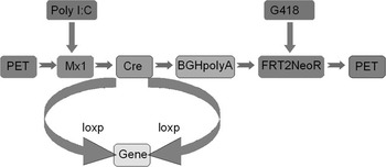
The vector was constructed, and transgenic porcine fetal fibroblast cells that inducibly express Cre recombinase were produced as follows. In our vector, the Cre recombinase gene is regulated by the Mx1 promoter, its expression can be induced by poly I/C, followed by FRT (Flp recognition target)-flanked cassette containing the neo-gene and transcriptional termination sequences. The final vector was named PET28a-Mx1-Cre (Figure 1). Cells were transfected using Lipofectamine 2000™ reagent (Invitrogen Diagnostics, CA, USA) using the manufacturer’s protocol. The cells were diluted 1 : 20 into fresh growth medium 24 h after transfection, and after 48 h, they were cultured in selection medium containing 300 μg/ml of G418 antibiotic (no. 2006B044; Amresco, Solon, OH, USA) for an additional 7 days. The surviving cell colonies were picked and propagated in a fresh 24-well dish (no. 3003; Becton Dickinson, NJ, USA), and then, the cells were frozen to be used as donor cells.
Construction of the vector PET28a-Mx1-Cre. In this vector, the Cre gene is under the control of the Mx1 promoter, its expression can be induced by poly I/C, followed by FRT-flanked cassette containing the neo-gene and transcriptional termination sequences.
Construction of transgenic pigs by nuclear transplantation
A positive cell clone was used to construct a transgenic pig by nuclear transfer (NT). Porcine NT was performed as described by Lai et al. (Reference Lai, Kolber-Simonds, Park, Cheong, Greenstein, Im, Samuel, Bonk, Rieke, Day, Murphy, Carter, Hawley and Prather2002). Briefly, ovaries were collected from pre-pubertal gilts at a local abattoir and transported to the laboratory at 30°C to 35°C. After cumulus–oocyte complexes were aspirated from the antral follicles, they were washed three times in maturation medium, and the oocytes were matured for 40 h at 39°C. After oocyte maturation, the cumulus cells were removed. Metaphase II oocytes were enucleated by removal of the polar body and the associated metaphase plate. An electrical pulse of 95 v for 45 μs from an ElectroCell Manipulator 200 (Genetronics, San Diego, CA, USA) was used to fuse the membranes of the donor cell and oocyte, forming a cytoplasmic hybrid. After the donor nuclei were transferred into the enucleated oocytes, the reconstructed oocytes were activated electrically in order to initiate subsequent development. After activation, healthy embryos that were cultured for 18 to 22 h were surgically transferred into the oviduct of a surrogate.
DNA extraction and PCR analysis
Genomic DNA from the umbilical cord was extracted for PCR analysis. The genomic DNA was used for the PCR amplification (35 cycles) of a 1044-bp fragment of the Cre recombinase gene using the primers 5′-AGTACTCCACCATGGCCAATCTCCTGACCGTCCACCAGAA-3′ and 5′-GGCAACTAGAAGGCACAGTCAATCGCCATCTTCCAGCAGG-3′. The PCR protocol consisted of 30 cycles at 98°C for 10 s, 68°C for 1.5 min and an extension at 72°C for 10 min. PCR products were analyzed by gel electrophoresis (1.0%; w/v).
Integration site analysis
The transgenic pig genome was extracted from the umbilical cord. The integrated sites were analyzed by high-efficiency thermal asymmetric interlaced PCR (hiTAIL-PCR) (Li et al., Reference Li, Robinson, Lesche, Martinez-Diaz, Jiang, Rozengurt, Wagner, Wu, Lane, Liu, Hennighausen and Wu2002). Primers used for hiTAIL-PCR were as follows: GSP1, GSP2 and GSP3; they are specific for transgenic vectors:
GSP1: 5′-GCGAACAGAAGCGAGAAGCGAACT-3′
GSP2: 5′-ACGATGGACTCCAGTCCGGCCAGGGTCATTTTCAGGTCCTTGGGG-3′
GSP3: 5′-CGCATCTGGGGACCATCTGTTCTT-3′
Longer arbitrary degenerate primers (LAD)
LAD1: 5′-ACGATGGACTCCAGAGCGGCCGC (G/C/A) N (G/C/A) NNNGGAA-3′
LAD2: 5′-ACGATGGACTCCAGAGCGGCCGC (G/C/T) N (G/C/T) NNNGGTT-3′
LAD3: 5′-ACGATGGACTCCAGAGCGGCCGC (G/C/A) (G/C/A) N (G/C/A) NNNCCAA-3′
LAD4: 5′-ACGATGGACTCCAGAGCGGCCGC (G/C/T) (G/A/T) N (G/C/T) NNNCGGT-3′
AC1: 5′-ACGATGGACTCCAGAG-3′
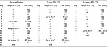
Pre-amplification reactions (20 μl) were prepared, each containing 2.0 μl PCR buffer, 200 μm each of dATP, dCTP, dGTP and dTTP (dNTPs), 1.0 μm of any one of the LAD primers (in the cases in which two LAD primers were used in single reactions, each was in 1.0 μm), 0.3 μm GSP1, 0.5 U Ex Taq (Extension Taq polymerase), and 20 to 30 ng transgenic swine DNA. Each 25-μl primary TAIL-PCR contained 2.5 μl PCR buffer, 200 μm each of dNTPs, 0.3 μm AC1 and GSP2, 0.6 U Ex Taq and 1 μl 40-fold diluted pre-amplified product. Each secondary 25-μl TAIL-PCRs contained 2.5 μl PCR buffer, 200 μm each of dNTPs, 0.3 μm AC1 and GSP3, 0.5 U Ex Taq and 1 μl 10-fold diluted primary TAIL-PCR product. When necessary, the primary or secondary TAIL-PCRs were scaled up to 50 μl. The PCRs were performed using a PCT-100 PCR cycler (MJ Research, Waltham, MA, USA) with thermal conditions shown in Table 1. The amplified products were analyzed on 1% agarose gels, and single fragments were recovered from the gels and purified using a DNA purification kit. The amplified products were analyzed on 1% agarose gels, and single fragments were recovered from the gels and purified using a DNA purification kit and sequencing. The resultant sequences were analyzed using the online BLAST database for pig genomic DNA.
Thermal conditions of high-efficiency thermal asymmetric interlaced PCR

Induction of the Mx1-Cre-transgenic swine
The Mx1-Cre transgene was injected intraperitoneally with 6 mg polyinosinic–polycytidylic acid (Poly I/C; Sigma-Aldrich; St Louis, MO, USA) three times at 2-day intervals. The day for the first injection was defined as day 1. The liver was examined histologically 2 days after the last injection.
Reverse transcriptase (RT)-PCR analysis
Total RNA was purified for RT-PCR 2 days after infection. RT-PCR was performed using the Simply P Total RNA Extraction kit (Bioer Technology, Beijing Co., Ltd, China) and the BioRT one-step RT-PCR kit (Bioer Technology), according to the manufacturer’s protocols. The RT-PCR products were separated by electrophoresis in 1% agarose gels and visualized by staining with ethidium bromide. The primers used for the RT-PCR were specific to the Cre sequence.
Immunohistochemistry analysis of Cre recombinase expression
Liver tissues were fixed in 10% paraformaldehyde, dehydrated, embedded in paraffin, sectioned and analyzed by strept actividin–biotin complex (SABC; Boster, Wuhan Co., Ltd, Beijing Co., Ltd, China) staining. For the Cre immunohistochemistry study, paraffin sections were incubated with the primary Cre mAb (Sigma-Aldrich), followed by horseradish peroxidase-conjugated goat anti-mouse IgG. After incubation for 30 min at room temperature, the specimens were visualized with 3.3′-diaminobenzidine in H2O2 and counterstained with hematoxylin solution. Monoclonal antibodies were used at dilutions of 1 : 100.
Results
Generation of a pig with inducible Cre expression
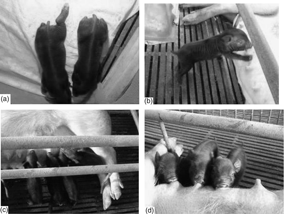
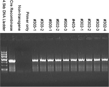
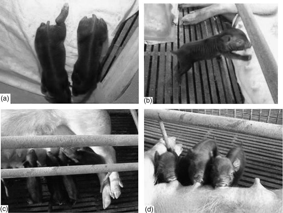
We transferred 210, 230, 250 and 215 embryos to the oviducts of four surrogate gilts, 4 days after estrus. Four litters were delivered: one on day 116 (two piglets, litter no. 303), a second on day 115 (three piglets, litter no. 505), the third on day 113 (one piglet, litter no. 403), and the fourth on day 118 (four piglets, litter no. 302) (Figure 2). All 10 piglets were males and alive, and PCR confirmed that they carried the Cre gene (Figure 3). The average birth weight was 0.78 kg.
Nuclear transfer piglets from four litters. The piglets were photographed on the day of birth (a), and 1 day after birth (b, c and d).

Amplification of the Cre gene of piglets. All 10 piglets were positive for the Cre recombinase gene. PMD18 T plasmid with Cre was used as the positive control; non-transgenic pig DNA and water served as negative controls.

Activating Cre expression in the transgenic pigs with pI-pC
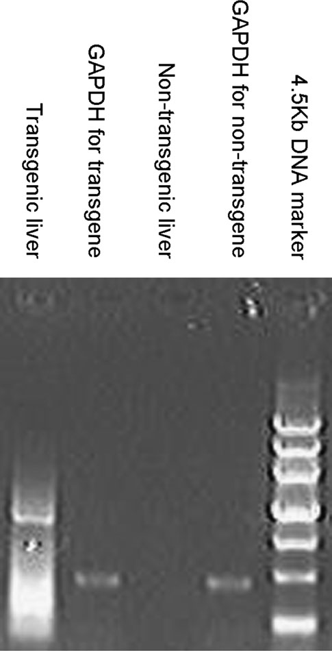
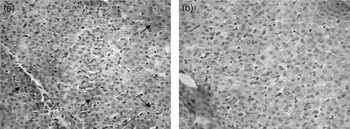
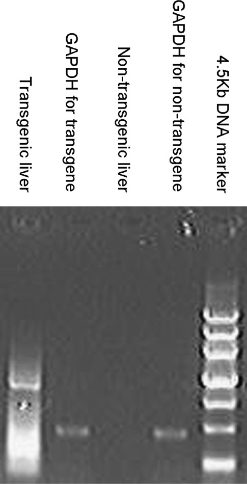
Treatment of transgenic pig litter no. 303-1 with pI-pC was effective in inducing Cre expression in the liver. The transgenic pig was induced by pI-pC treatment to express Cre recombinase, as judged by RT-PCR (Figure 4). Immunohistochemistry revealed that Cre recombinase was expressed in the hepatocyte cytoplasm (Figure 5a). A non-transgenic pig (Figure 5b) was used as the negative control and the housekeeping gene, GAPDH (glyceraldehyde 3-phosphate dehydrogenase II), was used as a positive control in RT-PCR.
Reverse transcriptase (RT)-PCR. The 1044-bp Cre fragment was amplified from the total RNA of liver tissues. The 480-bp GAPDH was used as a positive control for RT-PCR. A non-transgenic pig served as a negative control for the amplification of the Cre fragment.
Immunohistochemistry (×400). (a) The Cre recombinase expresses in the hepatocyte cytoplasm of Mx1-Cre transgenic pigs; (b) the Cre recombinase was not expressed in non-transgenic pigs.

Discussion
Transgenic pigs are of great value for research and commercial applications (Forsberg et al., Reference Forsberg, Strelchenko, Augenstein, Betthauser, Childs, Eilertsen, Enos, Forsythe, Golueke, Koppang, Lange, Lesmeister, Mallon, Mell, Misica, Pace, Pfister-Genskow, Voelker, Watt and Bishop2002; Fujimura et al., Reference Fujimura, Murakami, Kurome, Takahagi, Shigehisa and Nagashima2008) and could serve as models for human disease. The Cre-loxP system is a powerful tool for establishing human disease models and for manipulating DNA both in vitro and in vivo.
To study human diseases using animal models by the Cre-loxP system, transgenic swine with inducible expression of Cre recombinase were generated. In this study, we showed that Mx1-Cre swine harbor the Cre gene in genomic DNA using PCR. PCR tests showed that the foreign gene had been integrated, and integration site analysis revealed that the fragments were randomly integrated into the intron region of the chromosome 13 and chromosome 6 (unpublished results). Cre recombinase expression was studied in transgenic pigs by using RT-PCR and immunohistochemistry. We found that Cre recombinase is expressed in the cytoplasm of hepatocytes of Mx1-Cre transgenic pigs. Poly I/C doses may not be enough for the high expression of Cre recombinase. RT-PCR analysis confirmed Cre transcription. In future, this will allow the knock out of genes in Mx1-Cre swine that correspond to human diseases. This animal model promises to contribute substantially to an increased understanding of individual gene function in development and pathogenesis. In conjunction with inducible systems for the control of Cre expression and function, these transgenic swine are likely to have a profound impact on developmental biology and in the generation of useful animal models of human disease.
We believe that our findings hold promise for the future use of nuclear transplantation technology and the induced expression of Cre recombinase in creating porcine models of human disease.
Acknowledgments
The authors thank Professor Stefano Casola of the IFOM-Fondazione Istituto FIRC di Oncologia Molecolare, Milan, Italy; Dr Annabelle Decreux and Professor Daniel Desmecht of Liège University, Liege, Belgium, for their help with construction of the vector described in this report; Professor James B. Campbell University of Toronto for his help in editing the manuscript; and Chen Zhenwen from Capital Medical University School of Basic Medical Sciences, Beijing, for his help in the isolation of porcine embryonic fibroblasts.