Introduction
Subtotal petrosectomy is an effective surgical technique for treating chronic suppurative otitis media (CSOM) when the affected ear has no serviceable hearing.Reference Coker, Jenkins and Fisch1,Reference Prasad, Roustan, Piras, Caruso, Lauda and Sanna2 It involves complete exenteration of all air cells of the temporal bone (middle ear and mastoid), closure of the Eustachian tube, and closure of the external auditory canal with a blind sac. Finally, the middle ear and tympanomastoid cavity are obliterated.
Obliteration provides a relatively safe and secure closure of the surgical defect in the temporal bone, and eliminates problems associated with an open mastoid cavity, such as the need for regular cleaning, infection, and possible vertigo after exposure to water or wind. Usually, abdominal fat is used for this purpose.Reference Coker, Jenkins and Fisch1,Reference Bartels and Sheehy3 However, there is a considerable risk of complications (ranging from 10–40 per cent) when using fat for obliteration, such as infection or fat necrosis, which may necessitate revision surgery.Reference Yung4
In general, patients requiring subtotal petrosectomy are compromised because of prior surgery or co-morbidities, and therefore might have a higher risk of complications such as infections. However, patient burden and considerable additional hospitalisation costs can be avoided if infection rates can be lowered.
Bioactive glass might be a candidate to reduce infection rates after obliteration. S53P4 bioactive glass (BonAlive Biomaterials, Turku, Finland) has been successfully used in the treatment of bone and bone cavity infections.Reference Vos, de Veij Mestdagh, Colnot, Borggreven, Orelio and Quak5–Reference Silvola9 S53P4 consists of 53 per cent silicon dioxide (silica) and 4 per cent phosphorus pentoxide, which has antibacterial properties. Furthermore, it is readily available; hence, no extra time or abdominal incision is necessary for its harvesting. It cannot be contaminated with diseased cells as opposed to autologous material, and has osteoconductive and osteostimulative properties.Reference Jones10,Reference van Gestel, Geurts, Hulsen, van Rietbergen, Hofmann and Arts11
Several long-term studies have already shown successful mastoid cavity obliteration with S53P4 bioactive glass for patients with cholesteatoma, troublesome radical cavities or CSOM.Reference Vos, de Veij Mestdagh, Colnot, Borggreven, Orelio and Quak5,Reference de Veij Mestdagh, Colnot, Borggreven, Orelio and Quak12–Reference Skoulakis, Koltsidopoulos, Iyer and Kontorinis14 This case series describes the follow up of two patients after subtotal petrosectomy and obliteration of the tympanomastoid cavity with S53P4 bioactive glass in chronically discharging ears. With these cases, we would like to illustrate our experience with S53P4 bioactive glass as an obliteration material in subtotal petrosectomy, an alternative to the widely used autologous abdominal fat.
Cases
Patient A was a 59-year-old male well known at the department of otorhinolaryngology because of a history of pain and discharge of the right ear caused by CSOM, complicated by profound perceptive hearing loss. He had already been surgically treated with a mastoidectomy twice, and subsequently extensively treated conservatively with topical and systemic antibiotics. However, chronic disabling otorrhoea continued.
Patient B was a 76-year-old male patient with a history of diabetes type 2 and CSOM of the right ear with cholesteatoma since the age of 59 years. He had already been surgically treated with a radical modified mastoidectomy and subsequent revisions, including a bony obliteration, complicated by a profound perceptive hearing loss. Furthermore, both local and systemic antibacterial therapies were unsuccessful, and chronic fetid otorrhoea persisted.
In order to control the chronic ear infection, we decided to perform a subtotal petrosectomy with S53P4 bioactive glass obliteration of the cavity in both patients. Total follow-up duration for patient A and patient B was 84 months and 18 months, respectively. No complications occurred peri-operatively and complete control of the CSOM with pain relief was obtained for both patients. In both cases, follow up consisted of diffusion-weighted magnetic resonance imaging.
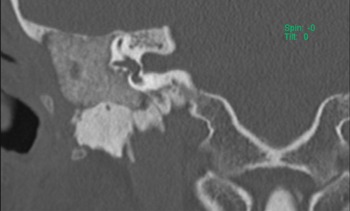
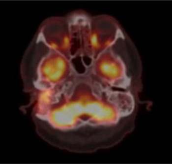
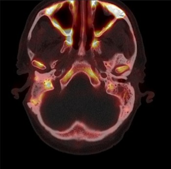
One year post-operatively, patient A noticed a feeling of pressure in the right temporal region, but no signs of an (ear) infection such as otorrhoea or pain were observed. Computed tomography (CT) showed a homogeneous obliterated cavity (Figure 1). Magnetic resonance imaging showed no signs of diffusion restriction. The former imaging could not explain the complaints; therefore, it was decided to perform a fluorine-18 (18F) fluorodeoxyglucose (FDG) positron emission tomography (PET)/CT (18F-FDG-PET/CT), which showed FDG uptake in the obliterated cavity only (Figure 2). The level of FDG uptake seemed too low for an infection, and was more likely to reflect (reactive) metabolic activity in the obliterated mastoid cavity. However, a distinction between new bone formation, a foreign body reaction or scar tissue could not be made based on this 18F-FGD-PET/CT scan. Three years post-operatively, an 18F sodium fluoride (NaF) PET/CT (18F-NaF-PET/CT) was performed to attempt to visualise new bone formation, which showed increased uptake of 18F-NaF at the cavity border (Figure 3). This was considered as osteoblast activity induced by the S53P4 bioactive glass, indicating new bone formation.
Coronal computed tomography scan of the right middle ear of patient A, showing homogeneous distribution of S53P4 in the surgical cavity of the middle ear and epitympanic space.

Axial fluorine-18 fluorodeoxyglucose (FDG) positron emission tomography/ computed tomography (18F-FDG-PET/CT) scan, revealing increased FDG uptake in the obliterated area of the right middle ear and mastoid, without FDG uptake in the contralateral mastoid.

Axial fluorine-18 (18F) fluorodeoxyglucose positron emission tomography/ computed tomography scan, showing increased uptake of 18F-sodium fluoride on the edges of the obliterated cavity of the right middle ear and mastoid, indicating active bone formation.

Discussion
The use of abdominal fat to obliterate the tympanomastoid cavity in subtotal petrosectomy has a long history and is well accepted by the otologist community; hence, it is surprising that high rates of post-operative complications persist.Reference Yung4,Reference Yan, Reddy, Isaac, Nguyen, McRackan and Meyer15 Abdominal fat tends to shrink (or become atrophic), reversing the desired effect. There are risks of infection and fat necrosis, necessitating antibiotic treatment or revision surgery, and its harvesting involves a superfluous abdominal incision. These issues have prompted the search for a safer and more effective obliteration material that is ideally accessible at any time and in any amount, is easy to handle, and maintains a stable size and shape over time.
In recent years, there has been an increasing interest in the potential properties of bioactive glass.Reference Vos, de Veij Mestdagh, Colnot, Borggreven, Orelio and Quak5–Reference van Gestel, Geurts, Hulsen, van Rietbergen, Hofmann and Arts11 Bioactive glass has significant advantages associated with two main features: bacterial growth inhibition and stimulation of new bone formation. Various mechanisms have been proposed regarding these features’ modes of action. These include: the leak of ions resulting in bactericidal action against micro-organisms; an increase of environmental pH and osmolarity in the surrounding environment; and ‘needle-like’ sharp glass debris that could potentially damage bacterial cell walls and facilitate the penetration of antimicrobial agents in the microbial cytoplasm.Reference Gubler, Brunner, Zehnder, Waltimo, Sener and Stark16–Reference Begum, Johnson, Worthington and Martin18
In this case series, both patients showed that obliteration with bioactive glass resulted in complete elimination of CSOM, with no post-operative wound infection. Therefore, bioactive glass granules might be viable as an alternative filler material to abdominal fat, to reduce the risk of post-operative infections. Moreover, by using synthetic bioactive glass, there is no need to harvest abdominal fat using a transverse midline incision below the umbilicus, thereby eliminating the risk of infection and fat necrosis, and resulting in a shorter operating time. Furthermore, bioactive glass stimulates various biological responses, prompting osteostimulation and osteoconduction. Osteostimulation refers to osteoblast cell recruitment and differentiation, as well as osteoblast activation to produce new bone in a bony environment. When immersed in physiological fluids, bioactive glass causes a leak of ions, culminating in the development of a bone-like hydroxyapatite layer on the granules’ surface, starting the process of osseointegration. Osteoconduction by bioactive glass occurs by providing latticework or scaffolding for the bone to grow along in vivo.Reference Jones10,Reference Zhang, Leppäranta, Munukka, Ylänen, Viljanen and Eerola17,Reference Drago, Toscano and Bottagisio19
The 18F-NaF PET/CT scans in this case series showed increased uptake of 18F on the edges of the obliterated cavity. Timmermans and colleagues, in 2019, showed that 18-NaF-PET/CT is suitable for detecting new bone formation.Reference Timmermans, Quak, Hagen and Colnot20 The mechanism of 18F-NaF uptake in bones is based on the exchange of 18F ions with hydroxyl ions (OH−) on the surface of the hydroxyapatite layer to produce fluorapatite. The uptake of 18F is higher in new bone because of the higher availability of binding sides; therefore, nearly all causes of increased new bone formation have increased 18F localisation.Reference Timmermans, Quak, Hagen and Colnot20 Thus, in addition to its antibacterial property, bioactive glass S53P4 also induces new bone formation.
Despite the promising results, we need to acknowledge that bioactive glass is a costly material, which adds to the overall expenses related to the procedure. However, it facilitates wound healing, reduces wound infection and obviates the need, and associated time taken, to harvest abdominal fat. Furthermore, we realise that this is explorative research conducted on a very small population. Consequently, further research with higher case numbers and a longer follow-up period is required to confirm our findings and expand our knowledge.
Conclusion
S53P4 bioactive glass is a promising effective and safe obliterating material for use following subtotal petrosectomy, and elimination of CSOM can be achieved with this technique. It has comparable outcomes to autologous abdominal fat, with the advantages of antibacterial properties, the potential for complete osseointegration and no donor site morbidity. Avoiding revision surgery for infection of obliteration material reduces burden and hospital costs.
Competing interests
None declared