Heart development is a complex process during which the heart needs to evolve from a single myocardial tube towards a fully septated heart with two atria, two ventricles, and a separated outflow tract. Figure 1 shows the cardiac events that must occur to achieve a normal circulation. These developmental processes do not occur sequentially, but largely overlap in time. It is therefore not surprising that maldevelopment during one timepoint might affect multiple processes. Correct remodelling of the heart most likely requires normal haemodynamics. Studies on chick and zebrafish demonstrate that reduced blood flow during heart development leads to impaired looping, incomplete septation, and abnormal valvulogenesis;Reference Hove, Koster, Forouhar, Acevedo-Bolton, Fraser and Gharib1, Reference Broekhuizen, Hogers, DeRuiter, Poelmann, Gittenberger-de Groot and Wladimiroff2 therefore, abnormal blood flow through the cardiovascular system during early embryonic stages can result in structural heart disease.Reference Vermot, Forouhar and Liebling3, Reference Slough, Cooney and Brueckner4
Schematic overview of the time span of development of the different cardiac components. Modified after Jongbloed et al.Reference Jongbloed, Mahtab, Blom, Schalij and Gittenberger-de Groot87

Congenital heart disease affects four to eight newborns per 1000 live births and is the most frequently encountered congenital malformation.Reference Allan, Benacerraf and Copel5 Congenital heart malformations are responsible for majority of infant deaths in the first year of life.Reference Allan, Benacerraf and Copel5 Second trimester ultrasound screening programmes are routine pregnancy care in most developed countries. In the last 2 decades, ultrasound systems have improved rapidly, allowing the visualisation of the first trimester heart in detail. First trimester echocardiography is defined as an attempt to visualise foetal heart anatomy at a gestational age before 14 completed weeks (13+6). The use of first trimester echocardiography to detect foetal anomalies at this early gestational age is increasingly gaining popularity, driven by the urge to detect foetal anomalies at very young stages. These examinations are conducted mainly in high-risk pregnancies, for example, in cases with increased nuchal translucency or in patients with a positive family history for cardiac malformations.Reference Carvalho6 At the same time, the desire to recognise complex anatomy already in these early developmental stages stresses the need for a thorough knowledge of basic developmental processes as well as recognition of cardiac compartments based on their morphology.
Despite several studies that stated that foetal heart examination could be incorporated in the first trimester examinations, its use is currently still limited to a few specialised centres. In the current paper, we aim to provide a comprehensive overview of the use first trimester echocardiography and the correlation with morphology. In addition, we give an overview of the current possibilities and limitations, and define the indications in which first trimester echocardiography could be offered.
Visualisation of the foetal heart before 10 weeks of gestational age: ultrasound versus morphology
Organogenesis occurs in the first 8 weeks after conception. In this relatively short period, which corresponds to 10 completed weeks of gestation, all major organ systems develop. At the end of this period, most organs have reached a size that can be visualised by ultrasound.
The cardiovascular system begins to develop within the lateral intra-embryonic mesoderm at 4 weeks gestational age.Reference Gittenberger-de Groot and Poelmann7 At this time, the primitive heart tube is formed and starts pulsating after 5 completed weeks of gestation, that is, day 22 after conception.Reference Larsen8 This single tube propulses the blood initially in a peristaltic manner. The primitive myocardial heart tube undergoes a process of looping, remodelling, and septation, thereby transforming the single lumen into the four chambers of the definitive heart with a separated outflow tract. This forms the basis for the separation of the pulmonary and systemic circulations at birth.Reference Larsen8 The final closure of the ventricular septum and ultimate formation of the atrioventricular valves is finished at 10 completed weeks of gestation.Reference Schoenwolf, Bleyl, Brauer and Francis-West9, Reference Cook, Yates and Anderson10 After development, the different cardiac compartments can be distinguished based on their morphological characteristics.Reference Anderson and Yen Ho11
Ultrasonographic studies on the foetal heart, before a gestational age of 10 completed weeks, have to take into account that heart morphology is not definitive yet. Therefore, examinations before this gestational age are studies on the developing embryo, rather than a diagnostic tool. Timor-Tritsch et al described embryonic development in 38 well-dated pregnancies.Reference Timor-Tritsch, Farine and Rosen12 He observed that foetal heartbeats could be visualised from 5+5 weeks gestational age. In a similar study, the ventricular septum was seen at 9+1 weeks and the four chambers were identified at 14 weeks.Reference Timor-Tritsch, Peisner and Raju13 With the use of 7.5 MHz transvaginal probes, more detailed structures of the foetal heart could be identified.Reference D’Amelio, Giorlandino and Masala14 The atrial and ventricular walls were visualised at the end of week 8, and the atrioventricular valves at the end of week 10 by Blaas et al. In this study, reference curves for the diameter of the foetal heart at this early gestation were constructed.Reference Blaas, Eik-Nes, Kiserud and Hellevik15 Allan et al compared the results of echocardiographic studies at 5–12 weeks gestational age, using transvaginal ultrasonography, with the anatomy of microdissectioned hearts.Reference Allan, Santos and Pexieder16 Before a gestational age of 9 weeks, cardiac morphology could not be visualised by ultrasound. The results in the study by Johnson et al, agree with these findings.Reference Johnson, Sharland, Maxwell and Allan17 At 9 weeks gestational age, Allan found the foetal heart to be centrally positioned in the foetal chest, directionally opposite the foetal spine.Reference Allan, Santos and Pexieder16 The pulmonary trunk could be visualised in the B-mode, in contrast to the ascending aorta, which could only be identified with colour Doppler. At 10 weeks gestational age, they found the heart’s position changing to a more left-sided orientation, as is typical later in pregnancy. Figure 2 shows an example of first trimester echocardiography and the correlation to morphology.
Normal echocardiography versus morphology. The transverse plane of the three-vessel view is depicted by the square (a). First trimester ultrasound (b) and a histological (c) section of the three-vessel view clearly show the superior vena cava (SVC), aorta (Ao), pulmonary trunk (PT), ductus arteriosus (DA), descending aorta (AoD). Plane (d) depicts a normal foetal heart, corresponding to ultrasound (e), and histological (f) sections of the four-chamber view showing the left ventricle (LV), right ventricle (RV), left atrium (LA), and right atrium (RA). Spine (S) marks the dorsal side of the foetus on the ultrasound plane.
Echocardiography in late first trimester foetuses
From 10 completed weeks of gestation onwards, the foetal heart has reached its definitive anatomy for foetal life. The final closure of the foramen ovale and the occlusion of the ductus arteriosus occurs after birth and results in the definitive separation of the systemic and pulmonary circulation.
In the ultrasonographic examination of the foetal heart from 10 weeks gestational age onwards, a predefined strategy can be applied, similar to the way mid-gestational cardiac examination is conducted. Owing to the increased axial (0.3–0.4 mm) and lateral resolution of the modern broadband probes, the foetal heart and its detailed structures up to 1 mm can be readily visualised. Initially, the majority of early first trimester echocardiographic studies were conducted using transvaginal probes. More recent studies showed that modern high-frequency abdominal probes could reach a sufficient level of imaging quality.Reference Carvalho, Moscoso and Ville18–Reference Bellotti, Fesslova and De23 Transabdominal scanning has the advantage that the imaging plane can be acquired in more angles compared with transvaginal ultrasound. In a review by Rasiah et al,Reference Rasiah, Publicover, Ewer, Khan, Kilby and Zamora24 it was stated that regarding specificity, transabdominal approach appears superior to transvaginal approach, but Gembruch et al stated that at lower gestational age transvaginal approach has better feasibility.Reference Gembruch, Shi and Smrcek25 Considering these limitations it seems advisable to master both methods, to be able to individualise the approach according to the goal of the ultrasound, detecting or excluding/reassuring, and technical factors such as patient stature and size of the foetus).
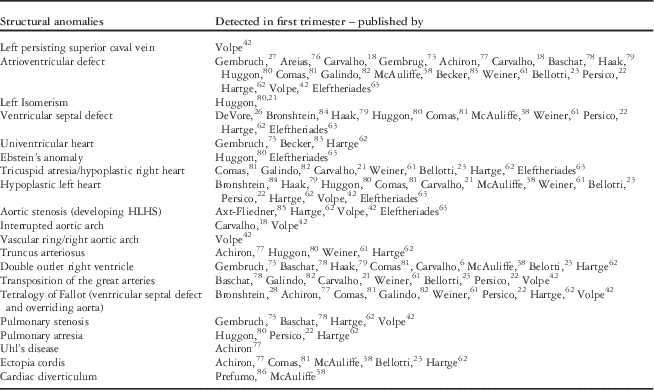
The first published studies on the diagnosis of heart malformations in the first trimester foetus were case reports,Reference DeVore, Steiger and Larson26–Reference Bronshtein, Siegler, Yoffe and Zimmer28 published up to 4 years before Dolkart and ReimersReference Dolkart and Reimers29 systematically described the possibilities of examining the normal foetal heart in this early period of gestation. Following these case reports, larger series of diagnosed malformations were published by several authors. Table 2 provides an overview of the structural heart defects that have been described to be accurately diagnosed in the first trimester.
The four-chamber view
The transverse section through the foetal heart at the inflow level, called “the four-chamber view”, is incorporated in routine second trimester obstetric scanning in nearly every centre. In this ultrasonographic plane, the size and position of the heart, two equally sized ventricles and atria, the opening and closure of the mitral and tricuspid valves, and an intact ventricular septum up to the cardiac crux are evaluated (Fig 2e). Allan et al and later Copel et al described that a normal “four-chamber view” ruled out the majority of foetal heart malformations in mid-gestational foetuses.Reference Allan, Crawford, Chita and Tynan30, Reference Copel, Pilu, Green, Hobbins and Kleinman31
An attempt to visualise the four-chamber view in first trimester foetuses proved feasible in several articles, as reviewed by Haak et al.Reference Haak and Van Vugt32 In the past few decades, the visualisation of this plane has increased to 90% of the examinations from 12 weeks gestational age onwards. Furthermore, a shift towards earlier gestational ages in which the four-chamber view can be visualised is observed. These improvements in early foetal echocardiography are probably because of the development of new transvaginal and transabdominal probes. Higher frequencies are used nowadays, and the increased variety in post-processing possibilities produce a very high image quality, leading to more detailed visualisation of small structures.Reference Votino, Jani and Verhoye33, Reference Timor-Tritsch, Fuchs, Monteagudo and D’alton34 The most frequently reported congenital heart defects in first trimester echocardiography are those with an abnormal four-chamber view, such as atrioventricular defects or defects with asymmetrical or disproportional ventricles. In most cases, follow-up ultrasounds are necessary to determine additional details and definitive diagnosis of the abnormalities and state the correct diagnosis, or sporadically conclude that there is a normal cardiac anatomy.Reference Carvalho6, Reference Persico, Moratalla, Lombardi, Zidere, Allan and Nicolaides22
Outflow tracts
During development, the outflow tract of the heart needs to develop from a single tube to a situation where the aorta and pulmonary trunk are separated and connect to the proper ventricle. During this process, a lengthening of the pulmonary part of the outflow tract will occur, whereas the aorta will stay relatively short.Reference Scherptong, Jongbloed and Wisse35 After normal development, the great arteries are located ventrally, wedged in between both atrial appendages. In the normal heart, the aorta has a right posterior position with respect to the pulmonary trunk (Fig 3a).
Echocardiography and morphology of a foetal heart with transposition of the great arteries. The aorta has, compared with the normal posterior position (a), in transposition of the great arteries, a right anterior position (panel (b) and enlargement (c)). On a transverse section through the foetal chest at the level of the “three-vessel view” (d), only the aorta (Ao) and superior vena cava (SVC) can be observed, owing to the caudal position of the pulmonary trunk. Spine (S) marks the dorsal side of the foetus on the ultrasound plane.
Although the four-chamber view is a potent plane to screen for major heart defects, it is not sufficient to rule out heart defects concerning the outflow tracts. To detect outflow tract anomalies with a normal four-chamber view, several planes have to be added to the examination.Reference Sklansky, Berman, Pruetz and Chang36 The position and size of the ascending aorta is the first plane cranial to the four-chamber view. This is a relatively difficult plane to achieve, because the probe has to be rotated and slightly tilted.
Just above the four-chamber view, above the outflow of the aorta, both the pulmonary trunk and the aorta can been seen in their spatial relationship in a transverse plane through the foetal chest. The superior caval vein is visible next to the aorta. This “three-vessel view” is not difficult to acquire and easy to teach to ultrasonographers (Fig 2a–c). It is generally accepted to be of great benefit in screening programmes, as it is abnormal in several heart malformations with abnormal position of the great vessels.
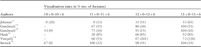
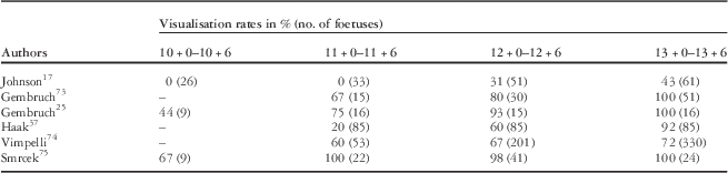
In the first trimester foetuses, the ascending aorta and pulmonary trunk measure about 1–2 mm at the valve annulus.Reference Gembruch, Shi and Smrcek25, Reference Haak, Twisk and Van Vugt37 Haak et alReference Haak and Van Vugt32 observed an increase in the visualisation of the outflow tracts in the past few decades up to 95% at gestational age of 13 weeks, confirmed by later studies.Reference McAuliffe, Trines, Nield, Chitayat, Jaeggi and Hornberger38Table 1 provides an overview of the visualisation rates of both the outflow tracts and the four-chamber view.
Visualisation of both four-chamber view as well as the outflow tract, obtained by different studies.
Reports on diagnosis of malformations solely affecting the outflow tracts, before 14 weeks gestational age, are increasingly being published, but mainly in the last decade. This suggests that these types of defects are more difficult to diagnose in the first trimester. Table 2 summarises several examples of outflow tract malformations detectable by echocardiography. Possibly, the most important of these is transposition of the great arteries, which is one of the most frequently missed severe heart defects, and could possibly benefit the most from prenatal detectionReference Blyth, Howe, Gnanapragasam and Wellesley39 (Fig 3, transposition of the great arteries is further described below).
Overview of heart anomalies detected by first trimester sonography (up to 14 weeks of gestational age).
HLHS=hypoplastic left heart syndrome
Sensitivity and specificity of echocardiography in first trimester foetuses
As discussed, echocardiography in first trimester foetuses has been proven feasible. More recent studies have assessed the reliability and accuracy for detecting or excluding congenital heart disease. In a systematic review by Rasiah et alReference Rasiah, Publicover, Ewer, Khan, Kilby and Zamora24 a high sensitivity (85%) and specificity (99%) were reported in a pooled group of foetuses predominantly with a high risk for congenital heart defects such as increased nuchal translucency, previous child with cardiac defect, and cardiotoxic medication. These studies had all been conducted by specialised obstetricians in tertiary care hospitals. Large population-based studies concerning second trimester screening of low-risk pregnancies have demonstrated the limited accuracy regarding the detection of congenital heart defects. Specialised tertiary centres markedly showed higher detection rates.Reference Sharland40 It is therefore probable that considering first trimester echocardiography in population-based screening programmes will achieve lower reliability. A large randomised control trialReference Westin, Saltvedt and Bergman41 demonstrated a low detection rate in a 12-week scan policy (11%). This did not differ significantly from an 18-week scan policy (15%), likely reflecting the level of training and experience of the ultrasonographers in this programme. Evidence regarding first trimester screening for congenital heart defects of low-risk mothers is scarceReference Volpe, Ubaldo and Volpe42 and does not assess cardiac anatomy only, but focuses on first trimester detection of structural defects in general.Reference Grande, Arigita, Borobio, Jimenez, Fernandez and Borrell43–Reference Rissanen, Niemimaa, Suonpaa, Ryynanen and Heinonen45 The results of these pioneering studies are conflicting and no systematic reviews are available yet.
Examples of cardiac malformations detectable with first trimester ultrasound; ultrasound versus morphology
The following section describes three examples of congenital malformations in the first trimester.
In transposition of the great arteries, there is a concordant atrio-ventricular connection in combination with ventriculo-arterial discordance. The aorta connects to the morphological right ventricle and the pulmonary trunk connects to the morphological left ventricle. There is often a parallel course of the great arteries, with a right anterior position of the aorta with respect to the pulmonary orificeReference Bartelings and Gittenberger-de Groot46 (Fig 3b–c). There is usually a fibrous continuity of the mitral valve and pulmonary valve, whereas the aortic valve is separated from the tricuspid valve by a muscular infundibulum. In transposition of the great arteries, both the aortic valve and the pulmonary valve are often situated at the same level, in contrast to normal hearts where the aortic valve is situated at a lower level than the pulmonary valve. In about 50% of the cases of transposition of the great arteries, a ventricular septal defect is present.Reference Liebman, Cullum and Belloc47
Transposition of the great arteries is frequently missed during routine second trimester anomaly scans.Reference Pinto, Keenan, Minich, Puchalski, Heywood and Botto48 This is probably owing to the normal four-chamber view in this defect. In transposition of the great arteries the vessel that arises from the left ventricle branches in a transverse plane. This is called “laterally branching”, which identifies the pulmonary artery. The vessel that arises from the right ventricle is the aorta, which gives rise to the neck vessels (superiorly branching). Easier to recognise is the fact that the “three-vessel view” described above cannot be achieved in transposition of the great arteries. In transposition of the great arteries, the vessel that arises from the right ventricle produces a “two-vessel view”. These two vessels are the aortic arch and the superior caval vein. Figure 3d illustrates that it is possible to diagnose transposition of the great arteries, even at an early gestational age. The images are comparable to those encountered in the second trimester transposition of the great arteries.
Another example of a congenital malformation that may be detected during first trimester echocardiography is atrioventricular septal defect. Atrioventricular septal defects cover a spectrum of heart malformations resulting in a common atrioventricular junction and common atrioventricular valve (Fig 4). The extent of the incomplete development of the atrioventricular septum varies and is used to categorise atrioventricular septal defects. A complete atrioventricular septal defect includes an ostium primum defect of the atrial septum, as well as a defect in the inflow portion of the ventricular septum. In incomplete or partial atrioventricular septal defect, the common atrioventricular annulus and valve are combined with an atrial septal defect – only shunting at the atrial level – or, less commonly, a ventricular septal defect – shunting only at the ventricular level. Extreme disbalance of the ventricles may result in hypoplastic right heart, in cases of left ventricular predominance, or hypoplastic left heart, if the right ventricle is predominant).Reference Christensen, Andersen and Garne49–Reference Yildirim, Gungorduk and Yazicioglu51
Echocardiography and morphology of a foetal heart with an atrioventricular septal defect. The common valve between the atria (a) and ventricles (RV and LV) can be recognised on the ultrasound four-chamber view (A; printed with permission of Ultrasound in Obstetrics and Gynaecology. Please note that this figure displays an oblique plane through the thorax. Owing to persistent foetal position, a correct four-chamber view was not obtainable) and histological section (b). On plane C, the common valve is seen from the left ventricle. IL=inferior bridging leaflet; SL=superior bridging leaflet.
In echocardiography, atrioventricular septal defects can be recognised in the four-chamber view (Fig 4a) by the absence of the crux and a common atrioventricular valve. As the four-chamber view is the most basic plane to screen for heart defects, atrioventricular septal defects, especially complete atrioventricular septal defects with moderate to large ventricular septal defects, are usually not missed, even in the first trimester scanning.
As balanced complete atrioventricular septal defects carry a risk of around 60–80% for Down’s syndrome, karyotyping has to be offered. A careful examination of other organs is furthermore warranted, as 75% of cases with atrioventricular septal defects have anomalies in other organ systems.Reference Christensen, Andersen and Garne49 A high prevalence of atrioventricular septal defects is also observed in patients with heterotaxy syndrome.Reference Peoples, Moller and Edwards52, Reference Irving and Chaudhari53
The final example is the hypoplastic left heart, in which aortic atresia or aortic stenosis is combined with either mitral atresia or mitral stenosis. The left side of the heart is underdeveloped, with a left ventricle of about 1/3 or less of its regular size, and there is hypoplasia of the ascending aorta and left atrium.
When inflow of blood into the heart exceeds the outflow, a profound intimal thickening called fibro-elastosis can be observed. Whereas this intimal thickening can be readily observed during later development stages, this is usually not observed during first trimester echocardiography yet. A profound disbalance of the ventricles can be visible already during first trimester echocardiography (Fig 5). However, this disbalance may also occur only later in development, so that the observation of equal-sized ventricles does not always rule out the hypoplastic left heart. Follow-up studies are mandatory.
Echocardiography and morphology of a hypoplastic left heart. On ultrasound at 12 weeks gestational age (a), disbalance between the right ventricle (RV) and left ventricle (LV) can be observed. Foetal specimens of 13 (b) and 25 (c) weeks also show hypoplasia of the ascending aorta. Right atrium (RA), left atrium (LA), aorta (Ao), pulmonary trunk (PT). *Shows pericardial fluid.
Limitations for foetal cardiac evaluation during first trimester
The most important disadvantage and therefore the major limitation of first trimester echocardiography is the intrauterine development of congenital heart disease at later gestation.Reference Allan54, Reference Allan55 Several malformations, such as mild pulmonaryReference Todros, Presbitero, Gaglioti and Demarie56, Reference Rice, McDonald and Reller57and aortic stenosis or coarctationReference Allan, Crawford and Tynan58 or even the hypoplastic left heartReference Allan, Sharland and Tynan59 can develop in second or third trimester foetuses. Furthermore, some heart lesions such as cardiac rhabdomyoma or cardiomyopathy may not be present in the first or second trimester and can evolve in later gestation or even after birth.Reference Yagel, Weissman and Rotstein60 Therefore, it is important to clarify to parents that some lesions could be missed in early pregnancy, even after detailed examination, and that, however rarely, a serious defect can develop after the mid-trimester. This statement is also supported by reports of heart defects, being missed in the first trimester ultrasonography.Reference Carvalho6, Reference McAuliffe, Trines, Nield, Chitayat, Jaeggi and Hornberger38, Reference Volpe, Ubaldo and Volpe42, Reference Weiner, Weizman, Beloosesky, Goldstein and Bombard61–Reference Eleftheriades, Tsapakis, Sotiriadis, Manolakos, Hassiakos and Botsis63 Thus, after the first trimester echocardiography, a follow-up examination on the second trimester of pregnancy should always be conducted. The knowledge that ventricular septal defects usually remain undetected in second trimester echocardiography,Reference Garne, Stoll and Clementi64, Reference Jaeggi, Sholler, Jones and Cooper65 probably because the size of these defects are beyond the resolution of the currently available ultrasound devices, makes it a neglectable aspect of first trimester examination, at which time this technical limitation is even more severe.
Another disadvantage of the first trimester echocardiography is the possible detection of defects that could resolve spontaneously in later pregnancy, such as muscular ventricular septal defects.Reference Paladini, Palmieri, Lamberti, Teodoro, Martinelli and Nappi66 This could result in unnecessary anxiety of the parents.
If the first trimester echocardiography is performed transvaginally, the direction of probe manipulation is limited. If the foetal position is consistently unfavourable, the technique does not provide sufficient images of the foetal heart. Transvaginal scanning, however, produces images with detail comparable to the second trimester abdominal scanning. In our opinion, transvaginal scanning should therefore be the preferred approach, despite the fact that specific training is needed.
Finally, probably the most significant reason that first trimester echocardiography (abdominal or transvaginal) is still a rarely applied method is the need for highly skilled staff with several years of experience in second trimester echocardiography, and the use of high-end ultrasound equipment with high-frequency (transvaginal) probes.Reference Rustico, Benettoni and D'Ottavio67
Conclusion
Although the advantages of first trimester transvaginal echocardiography seem of considerable value for couples at risk for having offspring with cardiac defects, this method is nowadays still limited to a few specialised centers. The numerous reports on the poor performance of mid-gestational sonography, regarding the detection of cardiac abnormalities, as well as the aforementioned limitations, could be the reason that centres are discouraged to extend echocardiography to the first trimester. Furthermore, high-end ultrasound facilities are needed, as well as operators that are experienced and have a particular interest in echocardiography. These factors, as well as the need for a follow-up examination at 20 weeks, are probably responsible for the limited utilisation of first-trimester echocardiography, and its restricted use in foetuses at a high risk of cardiac abnormalities.
In short, it seems clear that the capacity of high-frequency transvaginal probes is sufficient to perform echocardiographic examination in the late first trimester of pregnancy. However, the capability to detect and correctly diagnose heart defect is highly dependent on the operator, as is the case in the second trimester echocardiography.
Acknowledgements
E.C. is supported by a grant of the Willem Alexander Kinderfonds. M.J. is supported by a ZonMw/NWO personal research grant (project number 90700368).